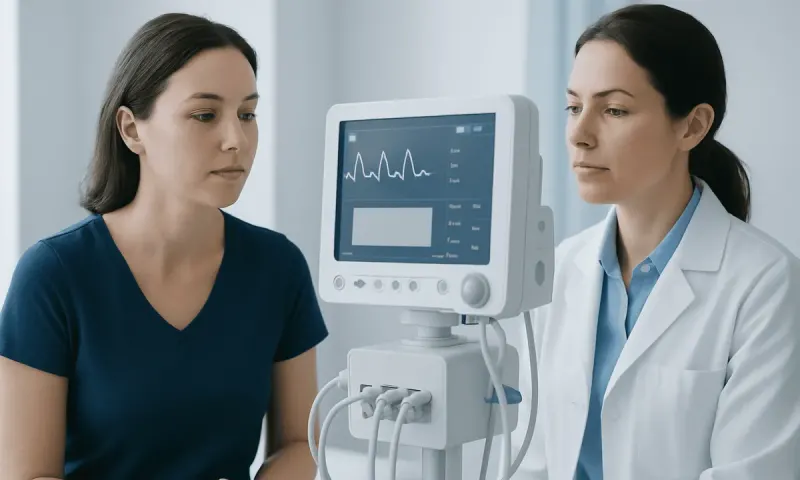
PET Scan Prep: What to Do Before Your Scan
PET scan prep helps the imaging team obtain clear pictures of how tissues and organs are functioning. Most people need to fast for several hours, drink plain water, avoid strenuous exercise and tell the team about medicines, diabetes or possible pregnancy before the appointment.
PET scan prep: what to expect
Pet scan prep is the set of steps taken before positron emission tomography (PET) imaging to help produce accurate, useful pictures. PET is a nuclear medicine scan that shows metabolic activity in the body. It is often paired with computed tomography (CT) or magnetic resonance imaging (MRI), allowing areas of altered activity to be viewed alongside detailed anatomical images.
For the most common type, an FDG PET scan, a small amount of a glucose-like radioactive tracer is injected into a vein. Cells take up the tracer at different rates, and a scanner detects its signal. The scan can help assess some cancers, heart conditions, brain disorders, inflammation and infection, but it does not diagnose a condition on its own.
Instructions differ depending on the body area being assessed, the tracer used and a person’s health needs. The imaging center should provide personalized guidance in advance. People should contact the center before changing prescribed medicines, especially insulin, diabetes tablets, steroids or medicines for anxiety.
How PET imaging works and who may need it
PET imaging measures the distribution of a radiotracer after it enters the bloodstream. FDG is the most frequently used tracer and behaves similarly to glucose. Areas with higher or lower tracer uptake may provide important clinical clues, although uptake can also occur with normal body processes, healing tissue and inflammation.
A clinician may request PET imaging to investigate an abnormal finding, stage or monitor certain cancers, assess response to treatment, evaluate recurrent disease, or examine selected heart and neurological conditions. In oncology, it may complement other tests used for conditions such as cancer treatment. The reason for the scan determines the most appropriate preparation and the way results are interpreted.
PET is suitable for many adults and children when clinically indicated. However, patients should tell the care team if they are pregnant, might be pregnant or are breastfeeding. They should also mention diabetes, kidney disease, recent surgery, infection, vaccination, chemotherapy, radiation therapy, allergies, difficulty lying flat or claustrophobia, as these factors can affect planning or interpretation.
What are the best foods to eat the day before a PET scan?
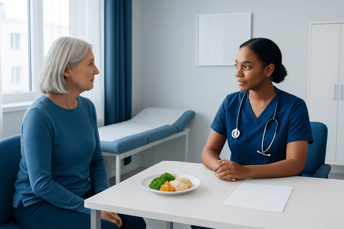
The best foods depend on the planned examination. For a typical FDG PET scan, most people can follow their usual balanced meals the day before, then begin fasting at the time instructed by the imaging center. Plain water is usually encouraged because good hydration can support comfort and tracer clearance.
People are commonly asked to avoid alcohol and large amounts of sugar in the period before fasting begins. Sugary drinks, sweets and high-carbohydrate meals may affect blood glucose and can interfere with FDG distribution. However, a special low-carbohydrate, high-fat preparation may be recommended for certain cardiac PET examinations, so patients should not adopt a restrictive diet unless their own team has advised it.
During the fasting window, many centers allow only plain water. Gum, candy, mints, juice, milk, coffee and tea may not be permitted because they can alter metabolism or stimulate muscles. Diabetes requires individualized planning: patients should ask the imaging center well ahead of time for instructions about meals, glucose monitoring and medicines.
What not to do 24 hours before a PET scan?
For many FDG PET scans, strenuous exercise should be avoided for at least 24 hours and sometimes longer if instructed. Activities such as running, intense gym sessions, heavy lifting and vigorous cycling can increase muscle uptake of the tracer, which may make images harder to interpret. Gentle everyday movement is generally acceptable unless the team says otherwise.
Patients should not eat or drink anything other than permitted plain water during the stated fasting period, which is often several hours before the scan. They should also avoid smoking, vaping, chewing gum and using mints if the center instructs them to fast completely. These actions can affect glucose metabolism or activate muscles in the head and neck.
It is important not to stop routine medication without medical advice. The team needs an up-to-date list of prescription medicines, over-the-counter products and supplements. People with diabetes should not guess how to adjust insulin or tablets; their appointment may need to be timed carefully so blood glucose is in an appropriate range.
PET scan procedure: step by step
On arrival, staff confirm the patient’s identity, medical history and preparation. They may check blood glucose, particularly for FDG imaging. The patient should wear comfortable clothing without metal where possible and may need to change into a gown. Metal objects and some jewelry may need to be removed for the CT or MRI portion of a combined scan.
A radiotracer is injected through a small intravenous line. This usually feels similar to a routine blood draw or injection. The patient then rests quietly, often for about 30 to 60 minutes, while the tracer circulates and is taken up by tissues. Remaining warm, relaxed and still helps reduce unwanted muscle activity.
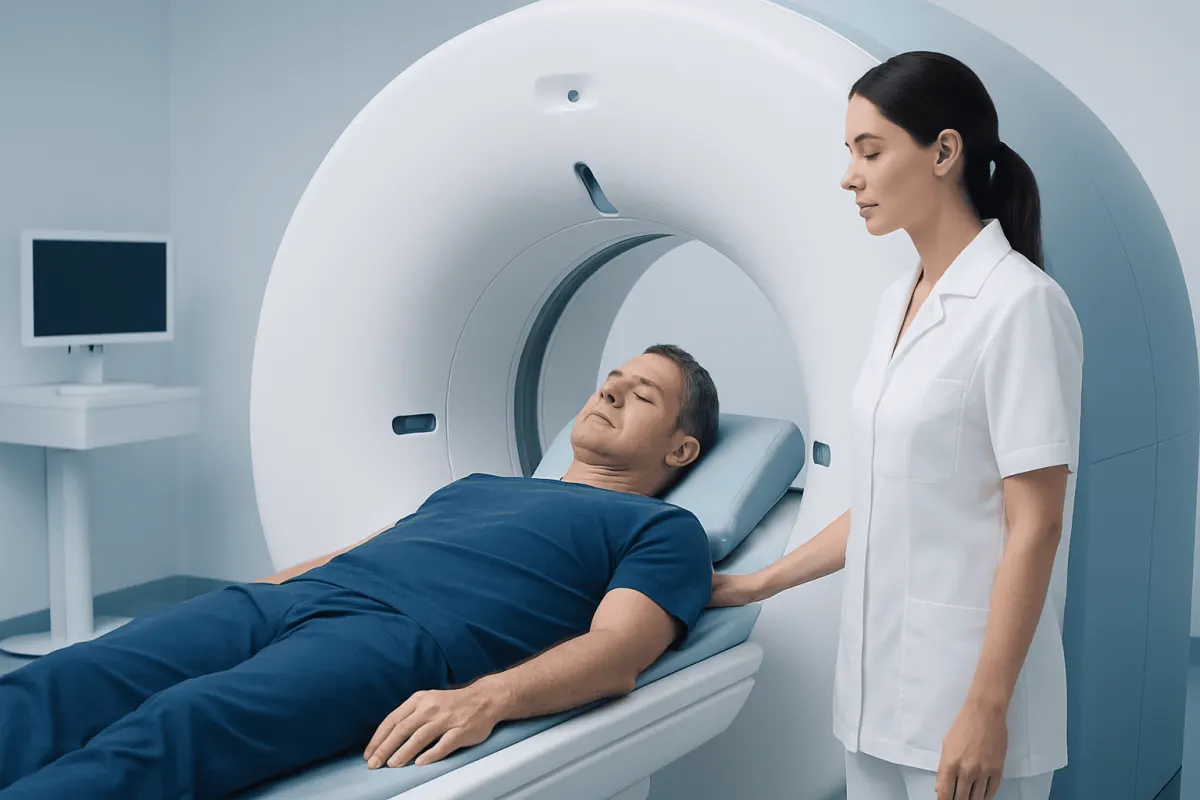
For the scan, the patient lies on a padded table that moves slowly through the scanner. The imaging itself commonly takes around 20 to 40 minutes, although the full visit may last two to three hours because of check-in, tracer uptake and scanning. The machine is open compared with many MRI scanners, and the imaging staff can communicate throughout the examination.
Depending on the clinical question, the appointment may include CT with contrast, which has separate preparation and safety considerations. Patients should report any prior contrast reaction or kidney problem. PET imaging may be part of a wider diagnostic plan that guides oncology treatment or other specialist care.
How to get the best results from a PET scan
The best way to get reliable PET scan results is to follow the appointment-specific instructions exactly and share complete health information with the team. Arriving on time, fasting as directed, drinking permitted water and avoiding strenuous activity can all help. Bringing previous scan reports or images, if requested, may also help specialists compare findings over time.
Before the scan, patients should mention recent infections, fever, surgery, biopsies, injuries and vaccinations. These can cause temporary inflammation that may show tracer uptake. They should also tell staff about any recent cancer treatment, including chemotherapy or radiotherapy, because the timing of PET imaging after treatment can influence interpretation.
During the tracer uptake period and scan, remaining comfortable, warm and as still as possible is helpful. Patients can ask staff for a blanket, positioning support or help managing anxiety. If they feel unwell, are in pain or need assistance, they should tell the staff rather than trying to continue silently.
A radiologist or nuclear medicine physician reviews the images alongside the reason for the test and other available medical information. A result may be described as normal, abnormal, indeterminate or requiring correlation with other tests. An abnormal area does not automatically mean cancer, and a normal scan does not rule out every medical condition.
Recovery timeline, safety and aftercare
Most people can return to usual daily activities shortly after a PET scan. The injected tracer contains a small amount of radiation and loses its activity naturally over time. Drinking extra water, unless a clinician has advised fluid restriction, and urinating regularly can help the tracer leave the body.
People are often advised to limit very close, prolonged contact with pregnant people, infants and young children for several hours after the scan. The exact period depends on the tracer and local protocol, so the imaging staff should provide the relevant advice before discharge. Breastfeeding instructions also vary by tracer and should be discussed individually.
Why do you have to flush the toilet twice after a PET scan? The tracer is mainly removed in urine, so double flushing reduces the small amount of radioactive material left in the toilet for other users. Good handwashing after using the bathroom is also advised. These simple precautions are temporary and are intended to reduce unnecessary exposure to others.
Risks from PET imaging are generally low. The injection can cause brief discomfort, bruising or rarely infection at the needle site. Allergic reactions to the radiotracer are uncommon; if CT contrast is used, it has its own small risk of reaction. The benefit is that PET can provide information that helps clinicians select, plan or monitor appropriate care.
When to seek medical care
Patients should contact the imaging center before their appointment if they cannot follow fasting instructions, have an acute illness, have significantly high or low blood glucose, are pregnant or breastfeeding, or need advice about medicines. It is better to clarify these issues early than to arrive unprepared and risk needing to reschedule.
After the scan, urgent medical attention is appropriate for signs of a severe allergic reaction, such as trouble breathing, swelling of the face or throat, widespread hives, severe dizziness or fainting. Although these reactions are rare, they require prompt assessment. Increasing redness, swelling, warmth or pain at the injection site should also be discussed with a healthcare professional.
Results are usually sent to the referring clinician after specialist review, and timing varies by facility and urgency. Patients should arrange follow-up to discuss the report rather than trying to interpret it alone. Acıbadem Health Point’s multidisciplinary specialists and JCI-accredited hospitals can support diagnostic evaluation and treatment planning for international patients when PET imaging is needed.
Frequently asked questions
01How long should a person fast before a PET scan?
For many FDG PET scans, fasting is required for several hours before the appointment, often around four to six hours. Requirements vary by the tracer and reason for imaging, so the instructions from the imaging center should be followed exactly. Plain water is commonly allowed, but patients should confirm this with their own team.
02Can a person take medication before a PET scan?
Many regular medicines can be taken, often with a small amount of water, but this depends on the test and the medicine. People should not stop prescription medication without advice. Diabetes medicines and insulin need particular planning because glucose levels can affect FDG PET images.
03Can a person drink coffee before a PET scan?
Coffee is commonly restricted during the fasting period for an FDG PET scan, even if it is black. Caffeine, sweeteners, milk and other additives may affect preparation or scan interpretation. The imaging center can confirm what drinks are permitted for the specific examination.
04Will a PET scan make a person radioactive?
A small amount of radiotracer remains in the body for a short time after the scan. It decays naturally and is eliminated mainly through urine. Temporary hygiene and close-contact precautions may be advised, particularly around pregnant people and young children.
05How soon are PET scan results available?
The images need to be reviewed by a radiologist or nuclear medicine physician, who prepares a report for the referring clinician. Results may be available within a few days, but timing depends on the facility and clinical urgency. The referring clinician explains what the findings mean alongside symptoms, examination and other tests.
06Can PET scan results show cancer?
PET scans can identify areas of increased or reduced metabolic activity that may be associated with cancer, but they do not confirm cancer by themselves. Infection, inflammation and normal body processes can also produce uptake. Clinicians may use PET findings with CT, MRI, laboratory tests or biopsy results to make a diagnosis.
This article is for general information only and is not a substitute for professional medical advice. Please consult a qualified doctor about your individual situation.
Oncology care in Turkey — second opinion and treatment plan
JCI-accredited · board-certified surgeons · reply within 24h
Add us as a Preferred Source to see more of our trusted health content across Google Search, AI Overviews and Discover.