Open vs Closed Angle Glaucoma: How They Differ
Open-angle glaucoma usually develops gradually because fluid drains too slowly through an open drainage angle. Closed-angle, or angle-closure, glaucoma happens when the angle becomes narrow or blocked; it can sometimes cause a sudden emergency with pain and rapid vision changes.
Open vs Closed Angle Glaucoma at a Glance
Open vs closed angle glaucoma refers to the condition of the eye’s drainage angle, where the iris meets the cornea. In open-angle glaucoma, this angle appears open but fluid leaves the eye less efficiently over time. In closed-angle glaucoma, also called angle-closure glaucoma, the angle is narrowed or blocked, which may lead to a rapid rise in eye pressure.
| Feature | Open-angle glaucoma | Closed-angle glaucoma |
|---|---|---|
| Drainage angle | Open, but drainage does not work efficiently | Narrow or blocked by the iris |
| Typical onset | Usually gradual | May be gradual or sudden |
| Early symptoms | Often none | May be none if chronic; sudden attacks cause symptoms |
| Possible acute symptoms | Not typical | Severe eye pain, red eye, blurred vision, halos, headache, nausea |
| How it is confirmed | Eye pressure, optic nerve testing, visual field testing, gonioscopy | Gonioscopy and examination of the angle, alongside pressure and optic nerve tests |
| Usual approach | Pressure-lowering drops, laser treatment, or surgery when needed | Urgent pressure reduction for an acute attack, followed by laser or other treatment to prevent recurrence |
Glaucoma is a group of conditions that can damage the optic nerve, the structure that carries visual signals from the eye to the brain. Eye pressure is an important risk factor, but glaucoma can occur even when pressure readings are within the usual range. Regular comprehensive eye examinations are important because damage may begin before a person notices changes in sight.
Why the Drainage Angle Matters
The front of the eye continuously produces a clear fluid called aqueous humor. This fluid helps maintain the eye’s shape and supports internal tissues. It normally exits through a drainage system located at the angle between the clear cornea and the colored iris.
In primary open-angle glaucoma, the angle is physically open, yet the trabecular meshwork, the eye’s natural filter, does not drain fluid adequately. Pressure may increase gradually, placing stress on the optic nerve over time. The condition commonly affects both eyes, although it may progress differently in each eye.
In angle-closure glaucoma, the iris moves close to or against the drainage system. This may prevent fluid from leaving the eye. Some people have naturally narrow angles without glaucoma, but narrow angles can raise the risk of closure and need assessment by an ophthalmologist.
Angle closure may be chronic, developing slowly with few symptoms, or acute, when the angle closes suddenly. Acute angle closure is an eye emergency because pressure can rise quickly and threaten vision without prompt treatment.
Symptoms: Gradual Changes Versus Sudden Warning Signs

Open-angle glaucoma is often called a silent condition because central vision can remain clear until later stages. Early peripheral, or side, vision changes are usually subtle and may not be noticed in daily life. This is why relying on symptoms alone is not a safe way to screen for glaucoma.
As open-angle glaucoma progresses, a person may develop patchy blind spots in side vision. In advanced disease, vision may become noticeably narrowed. These changes are usually painless and do not cause eye redness or a sudden change in how the eye looks.
Acute angle closure usually causes symptoms that are much more obvious. These can include severe pain in or around one eye, eye redness, blurry or cloudy vision, rainbow-colored halos around lights, headache, nausea, and vomiting. Vision may worsen rapidly, and the pupil can appear larger or less reactive than usual.
Chronic angle closure may cause no symptoms or may lead to intermittent episodes of mild blur, headache, halos, or discomfort, particularly in dim light. These symptoms are not specific to glaucoma, but they should be evaluated, especially in someone known to have narrow angles.
How a Clinician Tells Them Apart
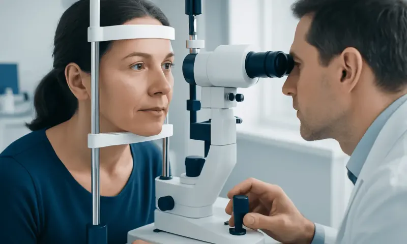
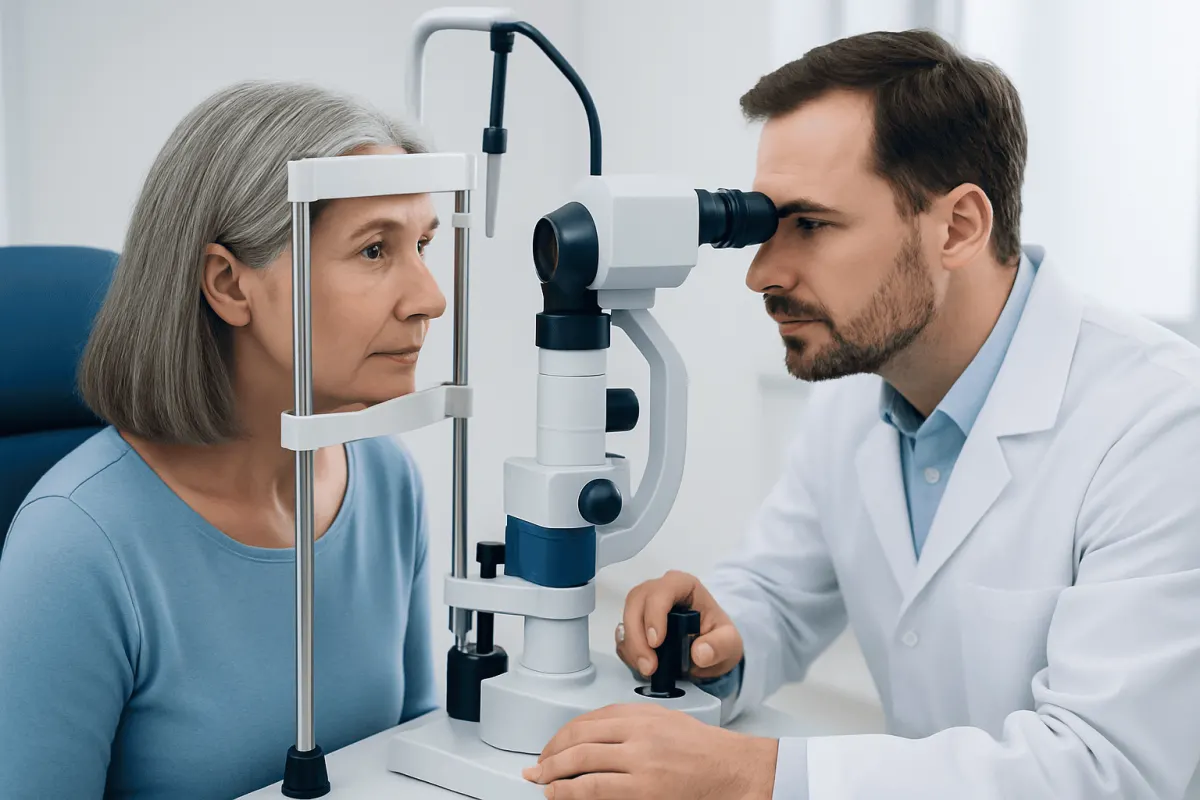
An ophthalmologist or suitably trained eye specialist identifies the glaucoma type through a complete eye examination rather than through symptoms alone. The most direct test is gonioscopy. After numbing drops are used, the clinician places a small mirrored lens gently on the eye to view the drainage angle and determine whether it is open, narrow, partly closed, or closed.
Eye pressure is measured with tonometry, but one reading cannot diagnose or exclude glaucoma. Pressure can change during the day, and optic nerve damage may occur at pressure levels that are not unusually high. The clinician also examines the optic nerve at the back of the eye for characteristic changes.
Additional tests commonly include optical coherence tomography, which measures layers of nerve tissue around the optic nerve, and visual field testing, which checks for areas of reduced peripheral vision. Corneal thickness may also be measured because it can influence the interpretation of pressure readings.
Imaging of the front part of the eye, such as anterior-segment optical coherence tomography or ultrasound biomicroscopy, may help assess narrow angles in selected cases. These tests complement, but do not always replace, gonioscopy. The full picture of angle anatomy, pressure, optic nerve health, and visual function guides diagnosis and follow-up.
Causes and Risk Factors
Primary open-angle glaucoma has no single cause. It is associated with age, family history, higher eye pressure, certain ethnic backgrounds, thin corneas, and medical conditions such as diabetes or high blood pressure. Previous eye injury, long-term corticosteroid use, and some eye conditions may also increase risk.
Angle closure is more likely when the eye has a naturally shallow front chamber or a narrow drainage angle. Risk tends to increase with older age, family history, farsightedness, and changes in the lens that occur over time. People of East or Southeast Asian heritage and Inuit ancestry have a higher population risk of angle closure, although it can occur in any group.
Some medicines can trigger angle closure in people with predisposed narrow angles. Examples may include medicines that dilate the pupil or affect fluid movement within the eye. A person should not stop prescribed medicine without medical advice, but should tell their doctor and eye specialist about all prescription drugs, over-the-counter products, and supplements they use.
Having narrow angles does not mean that a person will definitely develop an acute attack. However, it does mean an ophthalmologist should discuss individual risk, monitoring, and whether preventive treatment is appropriate.
What Doctors May Recommend for Each Type
For open-angle glaucoma, treatment aims to lower eye pressure to a level that reduces the chance of further optic nerve damage. Prescription eye drops are often the first option. Depending on the individual situation, laser treatment to improve drainage or glaucoma surgery may be considered if pressure remains above the target level or damage continues.
Follow-up is essential because glaucoma treatment usually controls risk rather than restoring vision already lost. A clinician monitors eye pressure, the optic nerve, and visual field results over time, then adjusts treatment when necessary. Using drops exactly as prescribed and attending planned appointments are important parts of care.
Acute angle closure requires urgent treatment to lower eye pressure and relieve the blockage. This may involve eye drops, oral or intravenous medicines, and laser treatment once the eye is stable. Laser peripheral iridotomy creates a small opening in the iris to help fluid move more freely and is commonly used when pupillary block contributes to angle closure.
When one eye has had angle closure, the other eye may also have a narrow angle and should be evaluated. Preventive laser treatment may be advised for the fellow eye in appropriate cases. Lens surgery or other procedures can be considered for selected people, depending on their anatomy, cataract status, pressure control, and overall eye health.
Protecting Eye Health and When to Seek Medical Care
There is no proven lifestyle measure that prevents every form of glaucoma, but routine comprehensive eye examinations allow earlier detection. Adults with a family history of glaucoma, known narrow angles, diabetes, prior eye injury, or long-term steroid treatment may need assessments more often. An eye specialist can recommend an interval based on individual risk.
People diagnosed with glaucoma should continue prescribed treatment even if their vision seems normal. They should also let other healthcare professionals know about their eye condition, particularly before starting a new medication. Regular physical activity and management of general health conditions can support overall wellbeing, but they do not replace glaucoma treatment.
Seek urgent medical care immediately for sudden severe eye pain, a red eye with blurred vision, halos around lights, severe headache, nausea, or vomiting. These symptoms may be caused by acute angle closure or another serious eye problem and should not be managed at home or delayed until a routine appointment.
Non-urgent eye assessment is appropriate for gradual side-vision changes, a strong family history of glaucoma, a previous finding of raised eye pressure, or advice that the drainage angles are narrow. Acıbadem Health Point’s multidisciplinary specialists and JCI-accredited hospitals diagnose and treat glaucoma for international patients, with care plans based on detailed ophthalmic assessment.
Frequently asked questions
01Is closed-angle glaucoma the same as angle-closure glaucoma?
Yes. Closed-angle glaucoma and angle-closure glaucoma are terms commonly used for glaucoma related to a narrowed or blocked drainage angle. It may be acute and sudden, or chronic and slowly progressive.
02Which is more common: open-angle or closed-angle glaucoma?
Primary open-angle glaucoma is the most common type of glaucoma in many populations. Angle-closure glaucoma is less common overall, but an acute angle-closure episode requires immediate medical attention.
03Can a person have glaucoma with normal eye pressure?
Yes. Some people develop optic nerve damage and visual field loss despite pressure readings within the usual range; this is often called normal-tension glaucoma. This is why clinicians assess the optic nerve and visual field as well as eye pressure.
04Can open-angle glaucoma turn into closed-angle glaucoma?
These conditions usually arise from different angle anatomies and mechanisms. However, a person with open-angle glaucoma can also develop narrowing of the angle for other reasons, so ongoing eye examinations remain important.
05Does laser treatment cure angle-closure glaucoma?
Laser peripheral iridotomy can relieve or prevent angle closure caused by pupillary block and can substantially reduce risk in suitable eyes. However, some people still need ongoing monitoring, eye-pressure treatment, or other procedures because angle closure can have more than one cause.
06What should someone do if they miss glaucoma eye drops?
They should follow the instructions given by their prescribing clinician or pharmacist and avoid using extra doses unless advised. If missed doses happen often, the person should discuss practical solutions with their eye specialist, as consistent treatment is important for protecting vision.
This article is for general information only and is not a substitute for professional medical advice. Please consult a qualified doctor about your individual situation.
Persistent digestive symptoms? Get evaluated in Turkey
JCI-accredited · board-certified surgeons · reply within 24h
Add us as a Preferred Source to see more of our trusted health content across Google Search, AI Overviews and Discover.