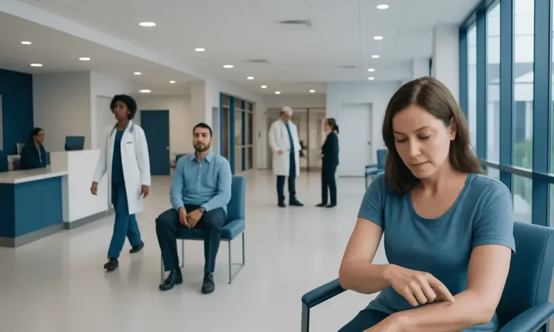
Nevus and Nevi: Monitoring Common Skin Moles
Nevus is the medical term for a mole, while nevi is the plural form. Most nevi are harmless and need no treatment, but regular awareness of their appearance can help people recognize changes that need a medical assessment.
Nevus Nevi: What the Terms Mean
Nevus nevi refers to moles. More precisely, nevus is the singular medical term and nevi is the plural. A mole is a localized area of skin with a higher concentration of pigment-producing cells called melanocytes, although some types of nevi involve other skin structures. They may be flat or raised and can range in color from skin-colored or pink to tan, brown, blue-gray, or black.
Most moles are benign, meaning they are not cancer. They commonly develop during childhood, adolescence, and early adulthood, and many people have several. A person’s number and appearance of moles are influenced by inherited traits, skin type, sun exposure, and normal hormonal changes. Having moles does not mean that a person has skin cancer, but it is sensible to become familiar with the skin’s usual pattern.
Some moles remain stable for decades, while others gradually become lighter, raised, or less noticeable over time. These changes can be part of normal mole evolution. However, a mole that changes noticeably over weeks or months, or a spot that looks distinctly different from a person’s other marks, deserves professional evaluation rather than self-diagnosis.
Common Types of Nevi
Melanocytic nevi are the most familiar type of mole. They can be classified by where melanocytes are located within the skin. Junctional nevi are usually flat and evenly pigmented. Compound nevi may be slightly raised and have pigment in both upper and deeper skin layers. Intradermal nevi are often raised, skin-colored, or lightly pigmented and may develop hairs.
Congenital melanocytic nevi are present at birth or appear shortly afterward. They vary greatly in size and appearance. Small congenital moles are relatively common and are usually monitored in the same way as other moles. Larger congenital nevi may need individualized follow-up because their care depends on size, location, appearance, symptoms, and a person’s overall circumstances.
Some nevi have distinctive appearances. Blue nevi often look blue-gray because pigment lies deeper in the skin. Spitz nevi may be pink, reddish-brown, or dark and are more often seen in children and young adults. Dysplastic or atypical nevi can be larger or less uniform in color and shape than ordinary moles. An atypical mole is not the same as melanoma, but people with numerous atypical nevi may benefit from regular skin examinations guided by a dermatologist.
Why Moles Develop and Who May Have More
Nevi form when melanocytes grow in small clusters rather than being spread evenly through the skin. Genetics has an important role: moles often run in families, and some people naturally develop more than others. Most acquired moles appear before about the third or fourth decade of life, although this timeline varies among individuals.
Ultraviolet radiation from sunlight and artificial tanning devices can contribute to the development of new moles and can damage skin cells. Intermittent intense sun exposure and sunburn are particularly relevant skin health concerns. Hormonal changes, including those during puberty and pregnancy, may cause existing moles to darken slightly or appear larger as the surrounding skin stretches. Still, a clearly changing mole during pregnancy should not automatically be attributed to hormones and should be assessed when appropriate.
People with fair skin, a history of substantial sun exposure or sunburn, many moles, a personal history of skin cancer, or a close relative with melanoma may have a higher need for professional skin surveillance. Risk is personal and cannot be determined from one feature alone. A clinician can recommend an appropriate review schedule based on the individual’s skin, medical history, and family history.
How to Check Moles: The ABCDE and “Ugly Duckling” Clues
A regular skin self-check can help a person recognize what is normal for them. It is useful to examine the front and back of the body, arms, legs, scalp, soles, spaces between the toes, and areas that are difficult to see with the help of a mirror or trusted person. Photographs may help track lesions over time, but they do not replace a clinical examination.
The ABCDE guide is a practical way to identify features that warrant medical attention:
- A — Asymmetry: one half does not closely resemble the other.
- B — Border: edges are irregular, blurred, scalloped, or poorly defined.
- C — Color: there are several colors or an uneven distribution of pigment.
- D — Diameter: the spot is larger than about 6 millimeters, although smaller melanomas can occur.
- E — Evolving: the mole changes in size, shape, color, surface, or symptoms.
Another helpful concept is the “ugly duckling” sign. This means a lesion looks noticeably unlike a person’s other moles, even if it does not meet every ABCDE feature. New spots in adulthood, a persistently scaly area, or a lesion that repeatedly itches, hurts, crusts, or bleeds should also be evaluated. These signs do not prove cancer, but they are good reasons to arrange a skin assessment.
How Doctors Diagnose a Concerning Nevus
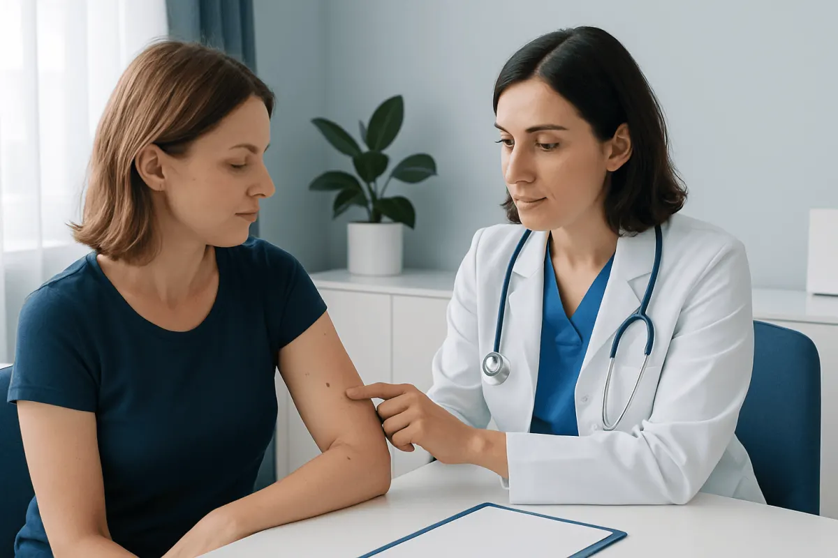
A dermatologist or other qualified clinician begins with a history and close visual examination. They may ask when the lesion appeared, whether it has changed, whether it causes symptoms, and whether there is a personal or family history of melanoma or other skin cancers. The clinician also considers the person’s age, skin type, pattern of sun exposure, and number of moles.
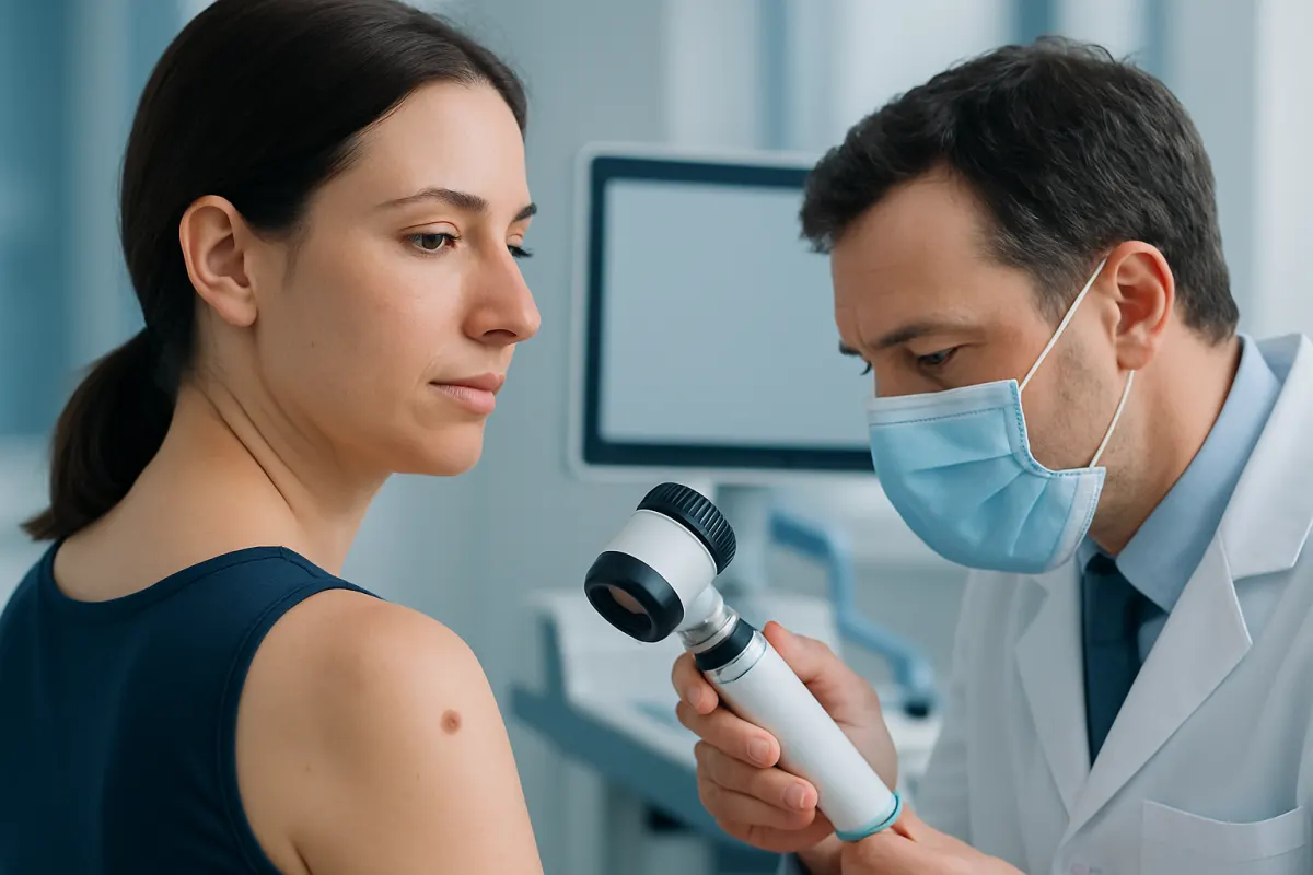
Dermoscopy is a painless examination using a handheld lighted device that magnifies skin structures not visible to the unaided eye. It can help clinicians decide whether a lesion appears benign, needs monitoring, or should be sampled. For people with many moles or elevated risk, total-body photography or digital dermoscopic monitoring may be considered to identify meaningful changes over time.
If a mole is suspicious or cannot be confidently classified through examination, the definitive way to assess it is a biopsy. A clinician removes all or part of the lesion under local anesthetic and sends the tissue to a laboratory for review by a pathologist. A biopsy is not performed simply because a person has ordinary moles; it is used when clinical findings make tissue confirmation appropriate.
Treatment and Removal Options
Most benign nevi do not need treatment. Removal may be considered if a mole is concerning, repeatedly irritated by clothing or shaving, located where it is easily injured, or unwanted for cosmetic reasons. The safest approach depends on the mole’s characteristics and the reason for removal. A clinician should examine a changing or unusual mole before any cosmetic procedure is considered.
Common medical techniques include surgical excision, in which the lesion is removed with a small margin of surrounding skin, and shave removal for selected raised lesions. The procedure is typically done with local anesthetic. When a lesion could be cancerous, complete excision or an appropriately chosen biopsy technique allows the tissue to be examined accurately. Removing a mole at home, cutting it, burning it, or using unregulated chemical products is not recommended because of risks such as infection, scarring, incomplete removal, and delayed diagnosis.
After removal, mild tenderness, redness, or a small scar can occur. The care team provides wound-care instructions and explains when pathology results are expected if tissue has been sent for analysis. A benign mole can occasionally recur if cells remain after a partial removal; any pigment returning in a scar should be reviewed by the treating clinician.
Prevention and Everyday Skin Care
Not every mole can be prevented, because genetics strongly influences mole development. However, reducing ultraviolet exposure is an important practical step for protecting skin. Broad-spectrum sunscreen, protective clothing, a wide-brimmed hat, sunglasses, and seeking shade during strong sunlight can all help. Sunscreen should be used as part of a broader sun-protection routine rather than as a reason to remain in intense sun for longer.
Avoiding indoor tanning devices is also advisable, as these expose the skin to ultraviolet radiation. Parents and caregivers can support healthy habits by protecting children’s skin from sunburn and teaching them that skin checks are a normal part of health awareness. People who work outdoors may need to plan additional protective measures with their daily routine.
Self-checks are most useful when performed consistently, such as once a month or at an interval recommended by a clinician. Rather than trying to identify every type of lesion independently, the goal is to notice new or changing marks. Keeping a record of important moles and attending recommended dermatology appointments can provide reassurance and support timely care.
When to Seek Medical Care
A person should arrange a medical appointment for a new mole appearing in adulthood, a lesion that changes in size, shape, color, or elevation, or one with irregular borders or multiple colors. Prompt review is also appropriate for a spot that bleeds without a clear injury, develops a persistent crust or sore, becomes painful, or causes ongoing itch. These symptoms often have non-cancerous explanations, but they should not be ignored.
People should seek more timely assessment if a lesion changes rapidly, looks very different from their other moles, or appears on the palms, soles, nail area, genitals, or scalp, where self-assessment can be more difficult. A person with a previous melanoma, many atypical moles, a large congenital nevus, or a strong family history of melanoma should ask a dermatologist about an individualized monitoring plan.
Acıbadem Health Point’s multidisciplinary specialists and JCI-accredited hospitals can assess concerning skin lesions and discuss diagnostic and treatment options for international patients. A professional examination is the most reliable next step when there is uncertainty about a mole.
Frequently asked questions
01Is a nevus the same as a mole?
In everyday use, a nevus is usually a mole. Nevus is the singular medical term, while nevi is the plural. Many nevi are made of clustered pigment-producing cells and are benign.
02Are all nevi harmless?
Most nevi are harmless and do not require removal. However, some lesions can look similar to one another, so a new or changing spot should be assessed by a qualified clinician. A medical examination is especially important if there is bleeding, persistent itching, pain, crusting, or uneven color.
03Can a normal mole turn into melanoma?
Melanoma can develop in or near an existing mole, but many melanomas arise as new spots on previously normal-looking skin. This is why both new lesions and changes in established moles matter. Monitoring the whole skin surface is more useful than focusing only on a single mole.
04What does the ABCDE rule mean for moles?
ABCDE stands for asymmetry, border irregularity, color variation, diameter, and evolving change. It is a screening guide that helps people notice potentially concerning features. A mole does not need to meet every ABCDE point to deserve a professional review.
05Should a mole that is itchy be removed?
Itching alone does not always mean a mole needs removal, as irritation can result from friction, dry skin, or inflammation. However, persistent itch, bleeding, pain, or visible change should be evaluated. A clinician can determine whether monitoring, biopsy, or removal is appropriate.
06Can moles be removed for cosmetic reasons?
Yes, some benign moles can be removed for cosmetic or comfort reasons after clinical assessment. The technique depends on the lesion's type, location, and whether there is any concern about cancer. It is important not to attempt mole removal at home because a suspicious lesion may require laboratory evaluation.
This article is for general information only and is not a substitute for professional medical advice. Please consult a qualified doctor about your individual situation.
Hair transplant in Turkey — costs, graft counts & a free assessment
JCI-accredited · board-certified surgeons · reply within 24h
Add us as a Preferred Source to see more of our trusted health content across Google Search, AI Overviews and Discover.