Neurofibromas: Benign Nerve Growths and Their Link to NF1
You notice a soft little bump under your skin. Maybe it has been there for years, maybe it is new. Maybe there are several. It is natural to wonder what it is and whether it matters.
Neurofibromas are growths that develop from cells surrounding peripheral nerves. They are commonly benign, meaning they are not cancer. What decides whether you simply need monitoring or actual treatment is where the growth sits, what symptoms it causes, how it changes over time and whether it is linked to neurofibromatosis.
What are neurofibromas?
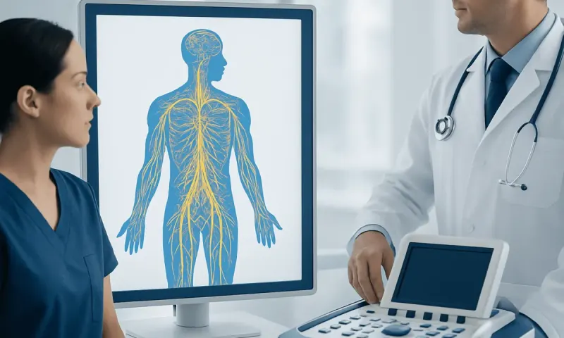
Neurofibromas are tumors that arise from the tissues surrounding peripheral nerves, which carry signals between the brain, spinal cord and the rest of the body. They are generally benign, so they do not behave like cancer or spread to distant organs. However, they can vary considerably in size, number, location and effect on daily life.
Some neurofibromas are small, soft bumps in or beneath the skin. Others grow more deeply along nerves and may involve larger areas of the body. A neurofibroma can occur as a single, isolated finding in an otherwise healthy person, or it can be one feature of neurofibromatosis type 1 (NF1), a genetic condition that can affect the skin, nerves, bones and other tissues.
One neurofibroma does not mean you have NF1. And if you do have NF1, it does not mean every tumor needs treating. Your care is shaped around your symptoms, how the growth looks, where it sits, how it changes over time and your overall health.
Types and how they may appear
Cutaneous neurofibromas develop in the skin. They often look like soft, flesh-colored, pink or slightly darker bumps and may range from a few millimeters to several centimeters. They can be sessile, meaning broad-based, or pedunculated, meaning attached by a small stalk. Many are painless, although itching, sensitivity or irritation from clothing can occur.
Subcutaneous neurofibromas develop under the skin and may feel firmer or more tender because they involve deeper nerves. A localized neurofibroma usually affects one small nerve. It may be noticed as a slow-growing lump, sometimes with discomfort or a tingling sensation when pressure is applied.
Plexiform neurofibromas are more extensive tumors that can grow along multiple branches of a nerve. They are most often associated with NF1 and may be present from childhood, although they can become more noticeable later. Their effects depend on location: a plexiform neurofibroma may be mainly cosmetic, or it may affect movement, vision, breathing, pain or the function of a nearby organ.
Not every lump under the skin is a neurofibroma. Cysts, lipomas, enlarged lymph nodes, skin tumors and other conditions can look much the same. So if a lump is new, changing or bothering you, have it looked at.
Why neurofibromas develop and who is affected

Neurofibromas form when cells associated with a peripheral nerve grow in an unregulated way. In NF1, changes in the NF1 gene reduce the activity of neurofibromin, a protein involved in controlling cell growth. NF1 is usually inherited in an autosomal dominant pattern, meaning a parent with the condition can pass it on to a child, but it can also result from a new genetic change in someone with no family history.
People with NF1 can develop multiple cutaneous, subcutaneous or plexiform neurofibromas over time. The number and size of skin neurofibromas can increase during adolescence, adulthood, pregnancy or other periods of hormonal change, although individual patterns differ greatly. These changes are not caused by poor hygiene, touching the bumps or a person’s lifestyle choices.
Solitary neurofibromas may occur without NF1 and often have no clear inherited cause. They may develop at any age. A clinician may consider an assessment for NF1 when a person has multiple neurofibromas, characteristic light-brown skin patches known as café-au-lait macules, freckling in skin folds, a family history of NF1 or other suggestive findings.
Let us clear up a common worry: neurofibromas are not contagious, and you cannot pass a benign neurofibroma to someone else through contact. Genetic counseling can help individuals and families understand inherited NF1, testing options and family-planning questions.
Symptoms and possible effects on health
Many neurofibromas cause no medical symptoms and are identified because of a visible or palpable bump. Skin lesions may affect confidence, body image or comfort, particularly when they are numerous or occur in highly visible areas. Those concerns are real, and you should feel free to raise them with your doctor.
Symptoms are more likely when a tumor presses on a nerve or nearby structure. Depending on its location, a neurofibroma may cause localized aching, burning pain, tenderness, numbness, tingling, weakness, reduced range of movement or changes in sensation. Larger plexiform neurofibromas can cause asymmetry or interfere with nearby tissues.
Most neurofibromas remain benign. In people with NF1, however, a small proportion of deeper nerve-sheath tumors may undergo malignant change into a malignant peripheral nerve sheath tumor. This is uncommon, but persistent new pain, rapid growth, increasing firmness, weakness or a change in a previously stable tumor should be evaluated without delay.
Children and adults with NF1 may also need monitoring for health features that are separate from neurofibromas, such as learning differences, blood pressure concerns, skeletal changes, vision-related issues or other tumors. Joined-up follow-up looks after the whole person, not just the bumps you can see.
How neurofibromas are diagnosed
Diagnosis often begins with a careful medical history and physical examination. The clinician will ask when the lump appeared, whether it has changed, and whether it is painful or associated with numbness, weakness or other symptoms. They may examine the skin and assess nerve function, strength, reflexes and sensation when appropriate.
A typical small cutaneous neurofibroma may be recognized clinically. If the diagnosis is uncertain, if a lesion is atypical, or if removal is being considered, a biopsy may be performed. In a biopsy, a small tissue sample or the entire lesion is examined by a pathologist under a microscope.
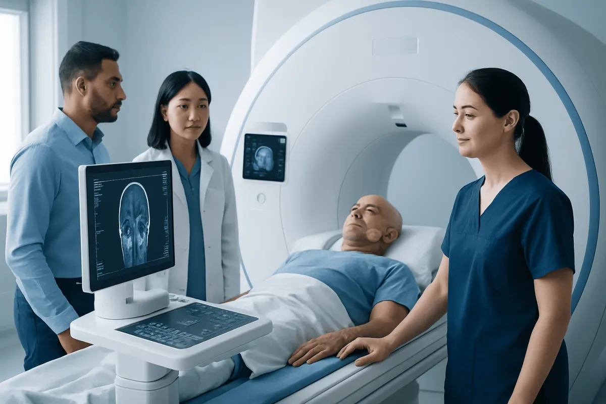
Ultrasound, magnetic resonance imaging (MRI) or other imaging may be used for a deep, large, painful or complex tumor. MRI is particularly useful for evaluating plexiform neurofibromas and their relationship to nerves, muscles, blood vessels and organs. Imaging can also help establish a baseline for future comparisons.
When NF1 is suspected, assessment may include a family history, skin examination, eye evaluation and referral to clinicians experienced in genetic conditions. Care Planning" class="ahp-ilk">Neuron Disease May Run in Families" class="ahp-ilk">Genetic testing can be useful in selected situations, but it is not always required to make a diagnosis when the clinical features of NF1 are clear.
Treatment options and ongoing care
Observation is appropriate for many neurofibromas that are small, stable and not causing significant symptoms. Follow-up may include periodic examinations, photographs for comparison or imaging for selected deeper tumors. Watching is not the same as ignoring. It means any decision to treat will be based on a real change in the tumor or in how you feel.
Surgical removal may be considered for a painful lesion, a tumor affecting nerve or organ function, a lump with concerning changes, or a neurofibroma causing substantial cosmetic or emotional distress. Complete removal can be possible for some localized tumors, while plexiform neurofibromas can be more difficult to remove because they may involve several nerve branches and surrounding tissues. Surgery also carries possible risks, including scarring, bleeding, nerve injury and recurrence.
Additional approaches may include treatment for pain, physical or occupational therapy for functional limitations, and psychological support when visible tumors affect quality of life. For some people with symptomatic, inoperable plexiform neurofibromas associated with NF1, specialist teams may consider targeted medicines. These treatments require careful selection, monitoring and discussion of potential benefits and side effects.
Care may involve dermatology, neurology, genetics, oncology, plastic surgery, pain medicine, radiology and rehabilitation specialists. Acıbadem Health Point’s multidisciplinary specialists and JCI-accredited hospitals assess and treat neurofibromas and related NF1 concerns for international patients, with plans guided by each person’s clinical needs.
Self-care and when to seek medical care
There is no proven home remedy that removes neurofibromas or prevents NF1-related tumors from developing. People can support comfort by reducing friction from clothing or jewelry, avoiding picking or cutting skin lesions, and using simple skin care if irritation occurs. Any persistent itching, pain or skin breakdown should be discussed with a clinician rather than treated aggressively at home.
Keeping a record of symptoms can be useful. A person may note when a lump first appeared, whether it is changing in size or texture, and whether it causes pain, altered sensation or difficulty with movement. For individuals with NF1, keeping scheduled follow-up appointments is an important part of preventive care.
Medical care should be sought promptly for a lump that grows quickly, becomes persistently painful, feels unusually hard, changes suddenly, or is associated with weakness, numbness, trouble walking, changes in bladder or bowel control, vision changes or breathing difficulty. These symptoms do not necessarily mean cancer, but they need timely assessment.
It is also reasonable to arrange a non-urgent appointment for any new unexplained lump, multiple skin bumps, or concerns about a family history of NF1. Early evaluation can provide clarity, address symptoms and identify whether specialist follow-up would be helpful.
Frequently asked questions
01Are neurofibromas cancer?
Most neurofibromas are benign nerve-sheath tumors and are not cancer. In people with NF1, a small proportion of deeper tumors can develop malignant change, which is why new pain, rapid growth or neurological symptoms should be assessed promptly.
02Can a person have a neurofibroma without neurofibromatosis?
Yes. A single localized neurofibroma can occur without neurofibromatosis type 1 or a family history of the condition. Multiple neurofibromas or other characteristic findings may lead a clinician to evaluate for NF1.
03Do neurofibromas go away on their own?
Neurofibromas generally do not disappear spontaneously. Small stable tumors that do not cause symptoms often do not need treatment, while troublesome or concerning lesions can be evaluated for removal or other management.
04Can neurofibromas be removed?
Some neurofibromas can be removed surgically, especially localized skin or subcutaneous lesions. The suitability of removal depends on the tumor’s size, location, connection to nerves, symptoms and the balance between expected benefit and risks such as scarring or nerve injury.
05Are neurofibromas inherited?
Neurofibromas themselves are not always inherited. However, neurofibromatosis type 1 is a genetic condition that can be inherited and commonly includes neurofibromas; it can also arise from a new genetic change in a person without an affected parent.
06What does a neurofibroma feel like?
A cutaneous neurofibroma may feel soft and movable within the skin, while a deeper tumor can feel firmer. Some are painless, but others may be tender or cause tingling, numbness or pain if they involve or press on a nerve.
This article is for general information only and is not a substitute for professional medical advice. Please consult a qualified doctor about your individual situation.
Joint and spine care in Turkey — expert assessment & treatment
JCI-accredited · board-certified surgeons · reply within 24h
Add us as a Preferred Source to see more of our trusted health content across Google Search, AI Overviews and Discover.