MRI for Back Pain or Numbness: When Spine Imaging Is Really Needed

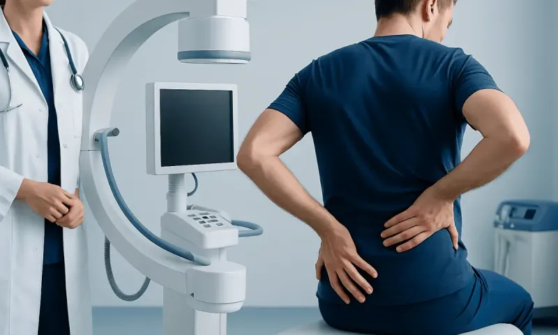
MRI for back pain can be very helpful, but it is not necessary for every episode of back pain or numbness. In most people, imaging is reserved for symptoms that suggest nerve involvement, serious underlying disease, or pain that does not improve with appropriate care.
Overview: What MRI Can and Cannot Show
MRI for back pain is a detailed imaging test that uses a magnetic field and radio waves to create pictures of the spine. It is particularly good at showing soft tissues such as spinal discs, nerves, ligaments, the spinal cord, and surrounding structures. This makes it more informative than an X-ray when a doctor suspects a problem involving nerve compression or other changes inside the spinal canal.
However, MRI is not the first step for every person with back pain. Many episodes of back pain are mechanical, meaning they are related to muscles, joints, or ligaments and improve with time, movement, and conservative care. In these cases, an early MRI often does not change treatment and may reveal age-related findings that are common even in people without symptoms.
When numbness, tingling, pain traveling into the leg or arm, or muscle weakness are present, MRI may be more useful. It can help identify whether a disc bulge, a narrowed spinal canal, inflammation, infection, or another issue is affecting the nerves. In this way, MRI is often used to answer a specific clinical question rather than simply confirm that pain exists.
When an MRI Is Usually Needed
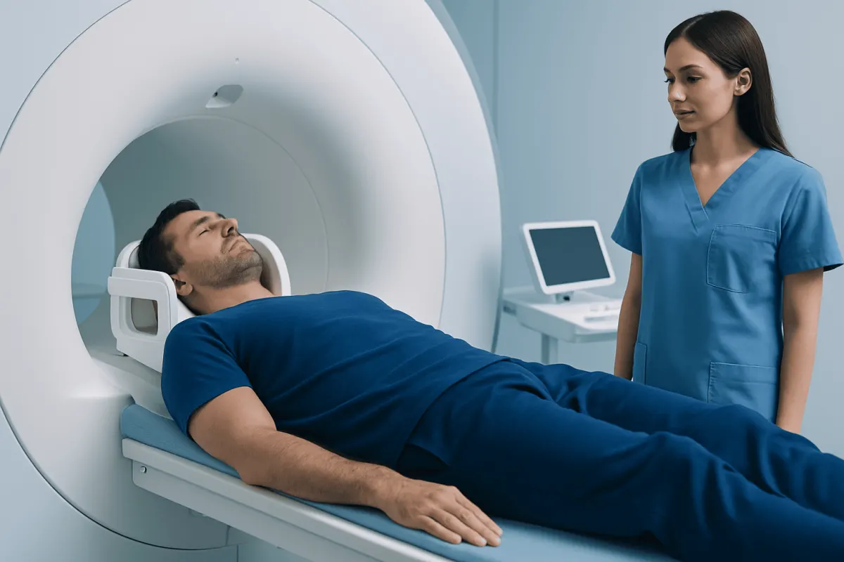
Doctors usually recommend a spine MRI when symptoms suggest that the pain may be coming from more than a simple strain. A common reason is persistent pain that radiates down the leg, especially when it is accompanied by numbness, tingling, or weakness. These features can point to irritation or compression of a spinal nerve, sometimes referred to as a herniated disc or another cause of nerve root pressure.
MRI is also considered when symptoms have lasted for several weeks despite appropriate treatment such as activity modification, physiotherapy, and pain management. If symptoms are getting worse rather than improving, imaging can help guide the next steps. This may be particularly relevant before procedures, injections, or surgery are being considered.
Another important reason for MRI is the presence of warning signs that raise concern for a more serious condition. These include a history of cancer, unexplained weight loss, fever, recent infection, significant trauma, immune suppression, or suspected fracture. MRI may also be used when a clinician suspects spinal stenosis or inflammation affecting the spine or spinal cord.
Symptoms and Red Flags That Need Prompt Assessment

Back pain with numbness can have many causes, but some patterns deserve urgent medical attention. Numbness that spreads, weakness in a leg or foot, difficulty walking, or a foot that begins to drag can signal increasing nerve compression. If these symptoms are new or worsening, a doctor may request urgent imaging.
Special concern arises when back pain is combined with bowel or bladder problems, numbness around the inner thighs or genital area, or severe weakness in both legs. These symptoms can suggest pressure on important nerve structures low in the spine and should be assessed immediately. Rapid evaluation is important because treatment decisions may need to be made quickly.
Other red flags include back pain with fever, chills, or a recent bloodstream or spinal infection; severe pain after a fall or accident; pain in someone with known osteoporosis; or pain that wakes a person repeatedly at night and is not explained by posture or movement. While these symptoms do not always mean something serious is present, they are reasons not to delay professional assessment.
- Radiating pain with numbness or tingling
- Muscle weakness or difficulty walking
- Loss of bladder or bowel control
- Numbness in the saddle area
- Fever, cancer history, recent infection, or major trauma
Why MRI Is Not Recommended for Every Episode of Back Pain
Most acute low back pain improves within a few weeks, even without imaging. Clinical guidelines generally recommend conservative treatment first when there are no red flags. This may include staying as active as tolerated, avoiding prolonged bed rest, using simple pain relief when appropriate, and starting guided exercise or physiotherapy.
One reason doctors avoid unnecessary MRI is that age-related changes are very common. Disc bulges, mild degeneration, and other findings often appear on scans even in people who have no pain at all. If these changes are not the true source of symptoms, they can create confusion and lead to anxiety or treatments that are not needed.
MRI also has practical limitations. Some people are unable to lie still because of pain or claustrophobia, and certain implanted devices may require special review before scanning. For these reasons, MRI is most valuable when the result is likely to affect treatment decisions or help clarify a concerning symptom pattern.
How Doctors Decide Whether Spine Imaging Is Appropriate
The decision to order MRI for back pain starts with a careful history and physical examination. A doctor will ask where the pain is felt, whether it radiates, what makes it better or worse, and whether numbness, weakness, balance changes, or bladder symptoms are present. Past medical history also matters, including cancer, osteoporosis, infections, inflammatory disease, or previous spine surgery.
During the examination, the clinician may assess strength, reflexes, sensation, walking, posture, and movements that reproduce symptoms. This helps determine whether the pain is likely muscular, joint-related, or linked to irritation of a nerve root. In many cases, this clinical evaluation is enough to begin treatment without immediate imaging.
If MRI is needed, the scan is often targeted to a region such as the lumbar or cervical spine. In some situations, contrast dye may be used, especially if infection, inflammation, tumor, or previous surgery needs closer evaluation. In patients whose symptoms suggest a structural problem that may need intervention, imaging may be paired with further consultation in neurosurgery or orthopedics and traumatology.
What MRI Findings May Mean
An MRI can detect disc herniation, nerve compression, spinal stenosis, fractures, inflammation, infection, tumors, and degenerative changes. It can also help evaluate the spinal cord in the neck or upper back, which is important when symptoms involve balance problems, hand clumsiness, or widespread numbness. In some people, the scan helps confirm a diagnosis and supports a clear treatment plan.
Even so, MRI findings do not always explain symptoms by themselves. A mild disc bulge may look dramatic to a patient but may not be the cause of pain. On the other hand, a person can have significant symptoms with relatively subtle imaging findings. This is why doctors interpret MRI results together with the history, examination, and sometimes other tests.
When MRI suggests a condition affecting the nerves or spinal cord, further treatment may range from rehabilitation to more specialized care. Depending on the cause, this may include physical therapy and rehabilitation or, in selected cases, spine surgery. The goal is not to treat the scan itself, but to treat the person and the symptoms that matter most.
Treatment After MRI: What Happens Next
Treatment depends on both the scan result and the severity of symptoms. Many people with disc-related pain or numbness improve without surgery, using a combination of activity modification, exercise, posture advice, and pain control under medical guidance. Physiotherapy is often central to recovery, especially when strength, flexibility, and movement patterns need attention.
If MRI shows significant nerve compression and symptoms are severe, persistent, or progressive, a specialist may discuss injections or surgery. Surgical treatment is generally considered when there is major weakness, loss of function, spinal cord compression, or pain that continues despite well-managed conservative care. The exact plan depends on the level of the spine involved and the impact on daily life.
For people with infection, inflammatory disease, fracture, or suspected tumor, treatment is directed at the underlying cause and may involve several specialists. Near the end of the care pathway, some international patients may choose centers such as Acıbadem Health Point, where multidisciplinary specialists in JCI-accredited hospitals diagnose and treat spine-related conditions in a coordinated setting.
Self-care, Prevention, and When to See a Doctor
Although not all back pain can be prevented, healthy movement habits can lower the chance of recurring symptoms. Regular physical activity, core and back strengthening, safe lifting technique, smoking avoidance, and maintaining a healthy weight can all support spinal health. Long periods of sitting may also worsen symptoms for some people, so frequent position changes can help.
At home, people with simple back pain are often advised to stay gently active rather than remain in bed for long periods. Short walks, gradual stretching, and guided exercise can be helpful when tolerated. It is best to avoid self-diagnosing based on online imaging terms, because many MRI findings are common and not necessarily harmful.
A doctor should be consulted if back pain is severe, lasts longer than expected, repeatedly returns, or is associated with numbness, tingling, weakness, balance problems, or pain traveling into the arms or legs. Urgent medical care is needed for bladder or bowel changes, saddle numbness, fever with back pain, or major weakness. These symptoms do not always mean an emergency is present, but they are important enough to check promptly.
Frequently asked questions
01Should everyone with back pain have an MRI?
No. Most short-term back pain improves without imaging, especially when there are no warning signs or nerve-related symptoms. MRI is usually reserved for cases where the result is likely to change diagnosis or treatment.
02Is numbness a reason to get a spine MRI?
Sometimes, yes. Numbness may suggest nerve irritation or compression, especially if it travels down an arm or leg, or occurs with tingling or weakness. A doctor will decide based on the pattern, duration, and severity of symptoms.
03What is the difference between MRI and X-ray for back pain?
X-rays are better for showing bone alignment and some fractures, but they do not show nerves, discs, or the spinal cord well. MRI provides much more detail about soft tissues and is more useful when nerve or spinal canal problems are suspected.
04Can an MRI show the exact cause of back pain?
Not always. MRI can reveal important abnormalities, but some findings are common in people without pain and may not be the true cause of symptoms. That is why scan results need to be interpreted alongside the medical history and physical examination.
05When is back pain with numbness an emergency?
Urgent assessment is needed if numbness occurs with new bowel or bladder problems, numbness around the groin or inner thighs, severe weakness, or trouble walking. Fever, major trauma, or rapidly worsening symptoms also need prompt medical attention.
06If an MRI shows a disc problem, does that mean surgery is needed?
No. Many disc-related problems improve with conservative treatment such as exercise, physiotherapy, and symptom management. Surgery is usually considered only when there is significant weakness, spinal cord or nerve compression, or symptoms that do not improve with appropriate non-surgical care.
This article is for general information only and is not a substitute for professional medical advice. Please consult a qualified doctor about your individual situation.
Joint and spine care in Turkey — expert assessment & treatment
JCI-accredited · board-certified surgeons · reply within 24h
Add us as a Preferred Source to see more of our trusted health content across Google Search, AI Overviews and Discover.