Melanoma Screening Moves From Skin Exam to Biopsy When Needed
You notice a mole on your shoulder that looks a little different than it did last summer. Is it worth a visit? Usually, yes.
Melanoma screening tests are used to identify suspicious skin lesions and determine whether further testing is needed. A clinical skin examination is usually the first step, while dermoscopy and biopsy help confirm or rule out melanoma.
Melanoma Screening Tests: How They Work
Melanoma screening tests aim to find suspicious skin changes before they become more advanced. The process usually begins with a visual examination of the skin by a dermatologist or another qualified clinician. They look for moles, freckles or other spots that have an unusual appearance or have changed over time.
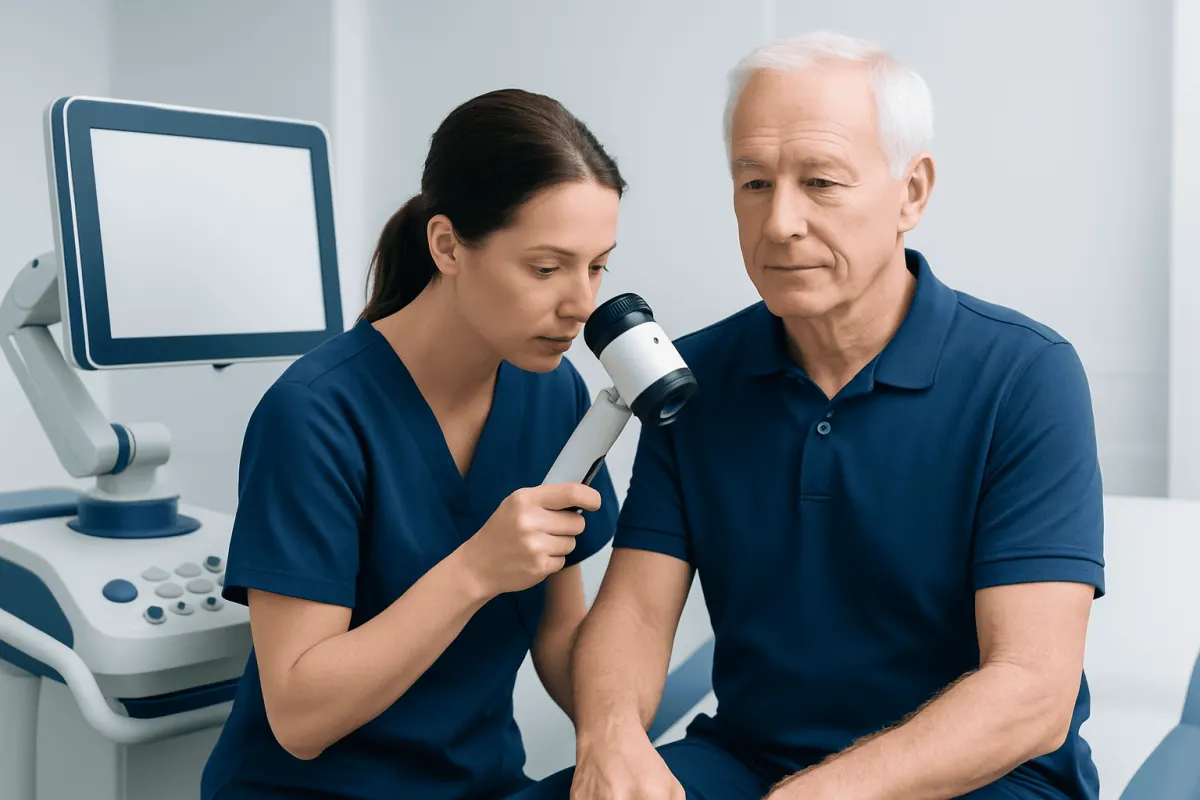
The most common tools are a full-body skin examination and dermoscopy, which uses a handheld lighted magnifier to inspect patterns beneath the surface of a lesion. If a spot appears concerning, the clinician may recommend a biopsy. A biopsy removes all or part of the area for laboratory examination and is the only test that can confirm melanoma.
Screening is especially useful if you have a personal or family history of melanoma, many moles, fair skin that burns easily, substantial ultraviolet exposure or a weakened immune system. That said, anyone can develop melanoma, including people with darker skin tones and those with no known risk factors at all.
Who May Benefit From a Melanoma Skin Check?
There is not one universal melanoma screening schedule for every adult. A clinician can help decide whether regular professional skin checks are appropriate based on individual risk factors, skin type and past medical history. People who have previously had melanoma generally need ongoing follow-up because they have a higher chance of developing another melanoma.
A person may benefit from a dermatologist-led examination if they have had intense intermittent sun exposure or sunburns, regularly used tanning beds, have many or atypical moles, or have a close relative with melanoma. Organ transplant recipients and people taking medicines that suppress the immune system may also need more frequent surveillance.
What you notice at home matters just as much as what we see in clinic. A monthly look over your own skin can catch a new lesion, or a change in an old mole, between appointments. A partner or family member can help inspect difficult-to-see areas, such as the scalp, back, buttocks and the backs of the legs.
What Happens During Melanoma Screening Tests?
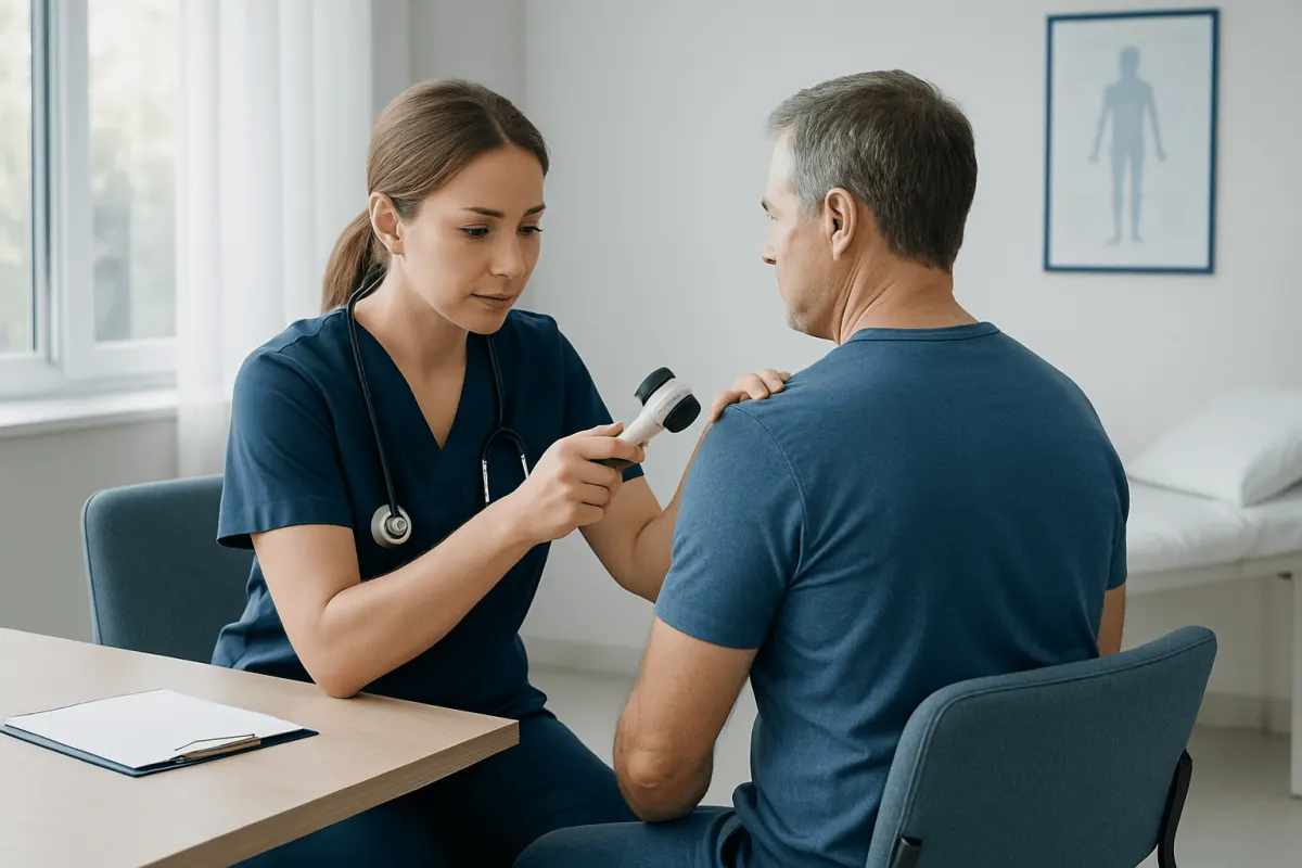
Before the examination, the clinician may ask about personal and family history of skin cancer, previous sunburns, tanning bed use, immune conditions and any lesions that have changed. It can be helpful to mention when a spot was first noticed and whether it has itched, bled, crusted, become painful or grown.
During a full-skin examination, the clinician checks the skin from the scalp to the soles of the feet. This may include the ears, lips, nails, between the fingers and toes, and genital skin when clinically appropriate. The examination is usually painless and takes only a short time, although the duration varies with the number of lesions that need assessment.
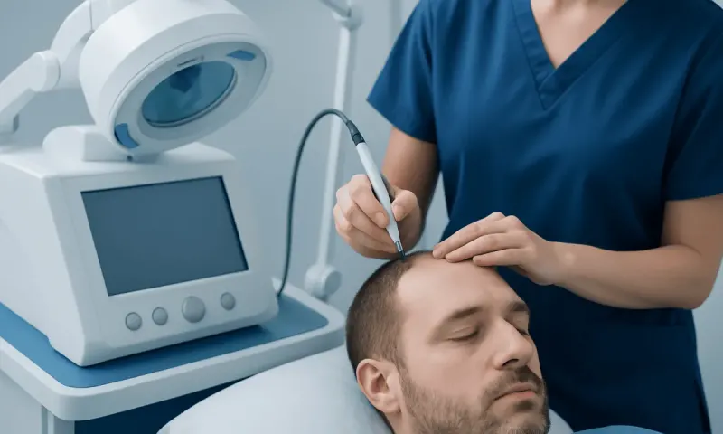
Dermoscopy may be used for selected moles. This non-invasive technique allows the clinician to assess pigment patterns, borders and structures that cannot be seen clearly with the naked eye. Some practices may also use total-body photography or digital mole monitoring to compare lesions over time, particularly for people with numerous moles.
If a lesion needs further investigation, a clinician may arrange a skin biopsy. Depending on its size and location, the lesion may be removed completely or sampled after local anesthetic is applied. The specimen is examined by a pathologist, and results help guide the next steps in melanoma care.
Understanding Screening Results and Biopsy Findings
A screening examination may find no concerning lesions, identify a spot that can be monitored, or reveal a lesion that should be biopsied. A recommendation for biopsy does not mean melanoma is confirmed. Many biopsied skin lesions prove to be harmless or represent non-melanoma skin conditions.
If melanoma is diagnosed, the pathology report provides important details, including the thickness of the melanoma, whether the skin surface is ulcerated and whether the cancer cells extend to the edge of the sample. These features help doctors estimate risk and plan treatment. Further assessment may be recommended in some cases to determine whether melanoma has spread.
Early melanoma is commonly managed with surgical removal of a wider margin of surrounding normal-looking skin. Depending on the diagnosis and stage, a care team may discuss melanoma treatment options, including surgery, lymph node assessment, immunotherapy, targeted therapy or radiotherapy. The appropriate approach depends on the individual diagnosis rather than on the screening test alone.
Benefits, Limits and Possible Risks of Screening
The main benefit of melanoma screening tests is that they can identify suspicious lesions at an earlier, more treatable stage. Skin examinations and dermoscopy are non-invasive, and routine self-checks can increase familiarity with an individual’s normal skin pattern.
Screening also has limitations. Not every melanoma has a typical appearance, and a lesion may be difficult to recognize in areas such as the scalp, nails, palms, soles or mucosal surfaces. Digital monitoring may be useful for selected people, but it does not replace medical review of a concerning new or changing lesion.
A biopsy can leave a small scar and may cause temporary soreness, bruising, bleeding or infection, although significant complications are uncommon when aftercare instructions are followed. Waiting for results can also be stressful. A clinician can explain why a biopsy is recommended and how the result will affect care.
There is no reliable blood test used as a routine melanoma screening test for the general population. And scans are not usually used to screen people without a confirmed diagnosis or a specific clinical reason for them.
What Is the 2 Week Rule for Melanoma?
The “2 week rule” is not a universal medical rule that can diagnose melanoma. It is commonly used as a practical reminder to seek clinical advice if a new skin spot, mole or sore does not settle, heal or improve within about two weeks, especially if it is changing or bleeding.
A lesion should not be watched at home for two weeks if it is rapidly changing, has multiple colors, has an uneven border, looks noticeably different from other moles, or is bleeding without a clear injury. In these situations, prompt medical assessment is more appropriate.
The ABCDE guide can be useful: asymmetry, border irregularity, color variation, diameter that is increasing, and evolution or change. Another useful sign is the “ugly duckling” concept: a lesion that looks distinctly different from a person’s other moles should be checked.
What Are the Signs Your Body Is Fighting Melanoma?
There are no reliable outward signs that show a person’s body is successfully fighting melanoma. The immune system can recognize and respond to cancer cells, but this response cannot be judged by whether a mole becomes itchy, inflamed, painful or changes in appearance.
Some melanomas may change, regress partially or develop inflammation, but these features do not confirm that the melanoma is improving. So please have any change in a mole or skin lesion looked at, rather than reading it as a good sign.
After melanoma has been diagnosed, doctors monitor response to treatment through examinations, pathology findings, imaging or other tests when appropriate. People should report new or persistent symptoms, such as unexplained lumps, ongoing cough, neurological symptoms or unexplained weight loss, but such symptoms have many possible causes and require medical assessment.
How Long Does It Take for Melanoma to Go From Stage 1 to 4?
There is no fixed timeline for melanoma to progress from stage 1 to stage 4. Melanoma behavior varies greatly between individuals and between tumor types. Some melanomas grow slowly over years, while others may become more invasive over a shorter period.
Staging is based on features of the melanoma at diagnosis, including tumor thickness, ulceration and whether cancer has reached lymph nodes or distant organs. It is not possible to accurately predict from appearance alone how quickly a particular lesion may progress.
That uncertainty is exactly why a suspicious spot is worth checking soon. Catching melanoma while it is still localized usually means simpler treatment than if it has spread. Acting quickly is not about assuming the worst; it is about getting a clear answer and the right advice.
Is Stage 1 Melanoma a Big Deal?
Stage 1 melanoma is an early form of invasive melanoma that is confined to the skin. It should be taken seriously because it is cancer, but it is generally more manageable than later-stage disease. Treatment often involves surgery to remove the melanoma and a margin of nearby skin.
Follow-up is still important after treatment. The care plan may include regular skin checks, examination of nearby lymph node areas and education about sun protection and self-examination. The exact follow-up schedule depends on the pathology findings and an individual’s overall risk profile.
Most people with stage 1 melanoma get back to their normal daily life once they have recovered from surgery. A diagnosis is worrying, of course. Going through the pathology report and the follow-up plan with your dermatologist or oncology team usually makes things feel a lot clearer.
When to Seek Medical Care
A person should arrange a medical skin assessment for a new lesion that persists, a mole that changes in size, shape or color, or a spot that repeatedly bleeds, crusts, itches or becomes tender. A lesion that looks unlike the person’s other moles also deserves professional review.
Urgent assessment is advisable for a rapidly changing pigmented lesion, a dark streak beneath a nail that is widening or irregular, or a non-healing sore without an obvious explanation. Melanoma can occur in less visible locations, including the scalp, palms, soles and under nails.
Sun protection remains an important preventive measure: seek shade when practical, wear protective clothing and broad-spectrum sunscreen, and avoid tanning beds. At Acıbadem Health Point, multidisciplinary specialists in JCI-accredited hospitals can assess suspicious skin lesions and coordinate melanoma diagnosis and treatment for international patients.
Frequently asked questions
01Can melanoma be detected with a blood test?
No blood test is currently recommended as a routine screening test for melanoma in people without symptoms. Clinical skin examination, dermoscopy and biopsy of suspicious lesions are the main methods used to assess melanoma.
02How often should a person have a melanoma skin check?
The right interval depends on personal risk factors, such as a history of melanoma, atypical moles, family history and immune suppression. A dermatologist can recommend an individualized follow-up schedule.
03Does a changing mole always mean melanoma?
No. Many moles and skin lesions change for non-cancerous reasons. However, a changing mole should be assessed by a qualified clinician because melanoma can also present with change.
04Is dermoscopy painful?
No. Dermoscopy is a painless, non-invasive examination using a handheld device with light and magnification. It does not involve needles, radiation or damage to the skin.
05What happens if a mole biopsy shows melanoma?
The doctor will review the pathology report and explain the recommended next steps. Treatment commonly includes surgery to remove a wider area of skin, while additional tests or treatments depend on the melanoma’s stage and characteristics.
06Can melanoma occur on skin that is not exposed to the sun?
Yes. Although ultraviolet exposure is an important risk factor, melanoma can develop on the soles, palms, under nails, scalp and other areas that receive little sun exposure. This is why full-skin awareness is important.
This article is for general information only and is not a substitute for professional medical advice. Please consult a qualified doctor about your individual situation.
Joint and spine care in Turkey — expert assessment & treatment
JCI-accredited · board-certified surgeons · reply within 24h
Add us as a Preferred Source to see more of our trusted health content across Google Search, AI Overviews and Discover.