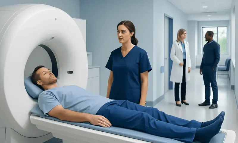
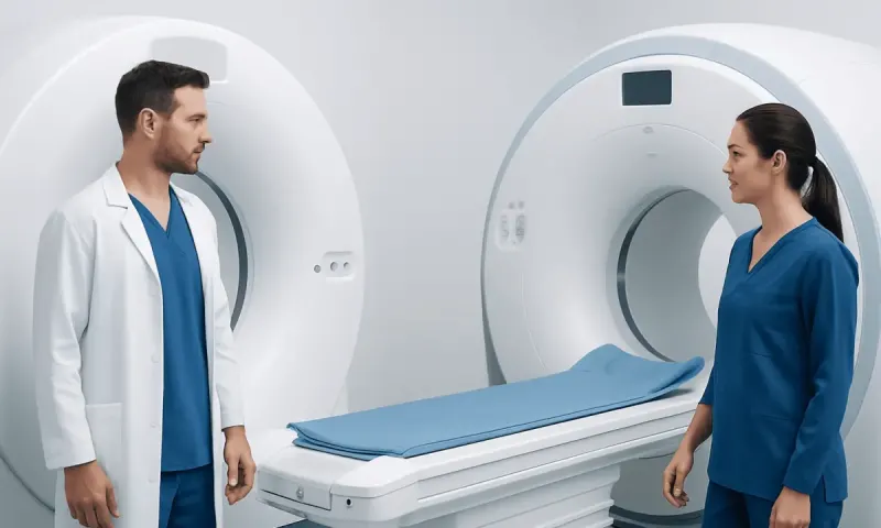
Head CT Scan vs MRI: Which Test Is Used When?
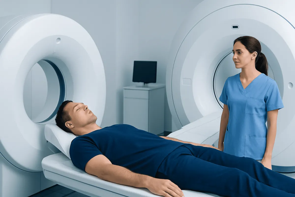
For ct scan head vs MRI, neither test is always better: a head CT is usually the quickest choice for emergencies, while brain MRI gives more detailed images of soft brain tissue. The most appropriate scan depends on the person’s symptoms, timing, medical history, and the clinical question being investigated.
CT Scan Head vs MRI: A Side-by-Side Comparison
When comparing a ct scan head vs mri, the main difference is the type of information each examination provides. A CT scan, also called computed tomography, uses X-rays to create cross-sectional images. An MRI, or magnetic resonance imaging scan, uses a powerful magnet and radio waves to produce highly detailed images of the brain and nearby structures.
A CT scan of the head and a CT scan of the brain are commonly used phrases for a similar examination. The scan may include the brain, skull, sinuses, and other head structures depending on the requested study. MRI can also assess these structures, but it is particularly valuable when doctors need a closer look at brain tissue, nerves, blood vessels, or areas at the base of the brain.
- Speed: CT is usually completed in minutes; MRI often takes longer and requires remaining still.
- Best for urgent concerns: CT is commonly the first test for acute trauma, suspected bleeding, or sudden severe neurological symptoms.
- Brain-tissue detail: MRI generally provides greater detail for many non-emergency brain conditions.
- Radiation: CT uses ionizing radiation; MRI does not use X-ray radiation.
- Bone detail: CT is especially effective for skull fractures and many sinus or facial bone problems.
- Practical factors: Some implants, severe claustrophobia, or the need for close monitoring may affect whether MRI is appropriate.
How a Clinician Tells CT and MRI Apart
The ct scan vs mri difference is not simply image quality. Each test captures different tissue characteristics, and the value of the result depends on the suspected condition. CT is especially good at detecting fresh blood, many fractures, calcification, and major swelling or pressure changes in the brain. It is also widely available in emergency settings and can be performed with monitoring equipment in place.
MRI separates soft tissues in greater detail. It can show subtle changes in the brain’s white and gray matter, the brainstem, the pituitary gland, cranial nerves, and areas near the spinal fluid spaces. Different MRI sequences highlight different processes, such as recent ischemic stroke, inflammation, scarring, infection, or certain patterns associated with seizures.
Both examinations may be performed with contrast material when it would help answer the clinical question. CT contrast is usually iodine-based, while MRI contrast is commonly gadolinium-based. Before contrast is given, the imaging team reviews factors such as kidney function, prior contrast reactions, pregnancy, and relevant medical history.
What Can Head MRI Show That CT Cannot?

Head MRI can reveal subtle abnormalities that may not be visible on CT, particularly in the early stages of some conditions or in areas where CT has less soft-tissue contrast. For example, MRI is often more sensitive for small or early ischemic strokes, especially in the posterior part of the brain, as well as for multiple sclerosis-type lesions, inflammation, infections, certain tumors, and some structural causes of seizures.
MRI may also give clearer information about the pituitary gland, optic pathways, inner structures near the skull base, cranial nerves, and certain abnormalities of the hippocampus and temporal lobes. These areas can be difficult to assess fully with CT because of nearby bone or because the tissue changes are too subtle.
However, MRI does not replace CT in every situation. A normal or unclear CT may be followed by MRI when symptoms persist or when the clinical concern involves brain tissue that MRI depicts more clearly. The choice is guided by the treating clinician rather than by a general rule that one scan is always superior.
Which Is More Accurate, CT or MRI of the Brain?
Whether CT or MRI is more accurate depends on what doctors are looking for and how quickly the scan is needed. MRI is generally more sensitive for many abnormalities within brain tissue, including small infarcts, demyelinating changes, some tumors, and inflammatory conditions. It may therefore be the more informative examination for ongoing neurological symptoms when the person is medically stable.
CT can be more accurate or more useful in urgent situations where rapid detection of acute bleeding, significant head trauma, skull fracture, or severe swelling is needed. A fast CT result can help clinicians make time-sensitive decisions. In suspected stroke, CT may be used first to rule out bleeding, while MRI may later clarify the type, location, or extent of brain injury in selected patients.
Image accuracy also depends on timing, the scanner protocol, whether contrast is used, motion during the scan, and the experience of the radiology team. A result should always be interpreted alongside symptoms, examination findings, and other tests rather than considered in isolation.
Why Do Doctors Choose CT Over MRI?
Doctors often choose CT over MRI when time is critical. A person with sudden severe headache, a recent head injury, confusion, reduced consciousness, or symptoms suggesting a possible brain bleed may need a CT because it is rapid and can identify urgent causes that require immediate treatment. CT is also useful for evaluating skull fractures and some complications of sinus disease.
CT may be preferred if an MRI would be difficult to complete safely or practically. For example, some implanted devices or metal fragments may make MRI unsuitable, although many modern implants are MRI-conditional and can be scanned under specific protocols. People who cannot lie still, need intensive monitoring, or have severe claustrophobia may also be better served initially by CT.
When a neurological emergency is suspected, imaging should not be delayed while deciding which test seems more comprehensive. Clinicians typically use the fastest suitable examination first, then arrange MRI or other tests if further detail would change management.
What to Do for Common Symptoms and Situations
For a recent blow to the head, worsening headache after injury, vomiting, drowsiness, confusion, or concern for a fracture, emergency clinicians commonly start with CT. A CT scan can rapidly check for bleeding, swelling, and bone injury. In some cases, MRI is later used if symptoms continue despite a normal CT or if a more subtle injury is suspected.
For new weakness, facial drooping, speech difficulty, vision loss, loss of balance, or numbness, urgent assessment is essential because these may be stroke symptoms. CT is frequently the first scan in emergency stroke pathways, and MRI may be added when it is available and clinically helpful. More information about warning signs and care pathways is available in stroke information.
For recurrent seizures, persistent unexplained headaches, gradual changes in memory or balance, possible pituitary symptoms, or suspected inflammatory neurological disease, MRI is often considered because of its detailed soft-tissue imaging. The clinician may also recommend blood tests, an eye examination, electroencephalography, or other assessments depending on the symptoms.
Before either scan, patients should tell the team about pregnancy, kidney disease, allergies or previous contrast reactions, implanted devices, hearing aids, surgical clips, metal injuries, and claustrophobia. They should also follow instructions about eating, drinking, and medications if contrast or sedation is planned.
When to Seek Medical Care
Emergency medical care is needed for sudden weakness or numbness on one side of the body, trouble speaking or understanding speech, sudden loss of vision, severe unexplained headache, a seizure lasting longer than expected, fainting, new confusion, or a significant change in consciousness. These symptoms can have several causes, but prompt assessment is important because some require immediate treatment.
After a head injury, urgent evaluation is advisable if there is repeated vomiting, worsening headache, increasing sleepiness, unusual behavior, weakness, seizure, fluid or blood from the nose or ears, or loss of consciousness. Children, older adults, and people taking anticoagulant medicines should receive individualized advice after head injury.
Non-urgent medical review is appropriate for persistent or recurrent headaches, new balance difficulties, ongoing dizziness, memory changes, unexplained seizures, or gradual neurological symptoms. A clinician can determine whether CT, MRI, or another assessment is the most appropriate next step. Acıbadem Health Point’s multidisciplinary specialists and JCI-accredited hospitals evaluate and treat neurological conditions for international patients.
Frequently asked questions
01Is a CT scan of the head the same as a CT scan of the brain?
The phrases are often used interchangeably in everyday clinical conversation. A head CT commonly includes the brain and skull, but the exact area and protocol can vary according to the reason for imaging. The ordering clinician and radiology team determine what needs to be assessed.
02Is MRI safer than CT for the head?
MRI does not use ionizing radiation, while CT uses X-rays. However, both tests are considered safe when appropriately recommended and performed. MRI may not be suitable for everyone, particularly with certain <a href="https://www.acibademhealthpoint.com/blog/mri-safety-with-metal-implants-what-patients-need-to-check-before-a-scan/" title="MRI Safety With Metal Implants: What Patients Need to Check Before a Scan" class="ahp-ilk">metal implants or foreign bodies, and contrast materials require individual safety checks.
03Can MRI detect a brain bleed?
Yes, MRI can detect bleeding in the brain and may provide helpful detail about the age or characteristics of blood products. In an acute emergency, CT is often used first because it is faster and is highly effective for identifying many fresh hemorrhages. The care team selects the test based on urgency and the suspected cause.
04Can a normal head CT still require an MRI?
Yes. A CT may be normal when symptoms are caused by subtle stroke, inflammation, small lesions, certain seizure-related abnormalities, or other soft-tissue changes that MRI can show more clearly. MRI is usually considered when symptoms, examination findings, or the suspected diagnosis warrant more detailed imaging.
05How long do CT and MRI scans take?
A head CT is often completed within a few minutes, although preparation and review can take longer. A brain MRI commonly takes longer because multiple image sequences are acquired, often ranging from around 20 minutes to an hour or more. The exact duration depends on the protocol and whether contrast is needed.
06Can a person choose MRI instead of CT?
The choice should be made with a qualified clinician because the scans answer different questions. In urgent situations, CT may be the safest and fastest first test even though MRI provides greater detail for some conditions. A clinician can explain why a particular examination is recommended and whether alternatives are appropriate.
This article is for general information only and is not a substitute for professional medical advice. Please consult a qualified doctor about your individual situation.
Joint and spine care in Turkey — expert assessment & treatment
JCI-accredited · board-certified surgeons · reply within 24h
Add us as a Preferred Source to see more of our trusted health content across Google Search, AI Overviews and Discover.