Haemangioma on the Spine: Causes, Symptoms, Diagnosis and Treatment

Key Takeaways
- Most spinal haemangiomas are benign and do not require treatment.
- Symptoms, when present, may include back pain, stiffness, numbness, or weakness.
- MRI and CT scans are the main tools used to confirm the diagnosis and assess risk.
- Treatment depends on symptoms and may include observation, pain control, vertebroplasty, embolisation, or surgery.
- New neurological symptoms should be assessed promptly to protect spinal function.
A haemangioma on the spine is usually a benign, slow-growing vascular lesion found within a vertebra. Many cause no symptoms and are discovered by chance, but some can lead to pain, nerve irritation, or spinal instability and need medical assessment.
Overview
A haemangioma on the spine is a benign growth made up of abnormal blood vessels inside a vertebra. It is often called a vertebral haemangioma, and in many people it is found incidentally on imaging done for another reason, such as an X-ray, CT scan, or MRI.
For most patients, the finding is reassuring rather than urgent. These lesions commonly stay small and stable, causing no symptoms at all. The main reason doctors take them seriously is not because they are usually dangerous, but because a small minority can become painful or affect nearby nerves if they grow in a way that weakens the bone or extends beyond the vertebra.
People who are planning care from another country often first hear the diagnosis during imaging for back pain, an injury, or another unrelated problem. In that setting, the important question is not simply what the lesion is called, but whether it is truly incidental, whether it explains the symptoms, and whether it needs monitoring or treatment.
Understanding the difference between a typical haemangioma and an aggressive one helps guide the next step. A careful review by a spine specialist or radiologist usually clarifies whether observation is enough or whether further evaluation is wise.
Symptoms
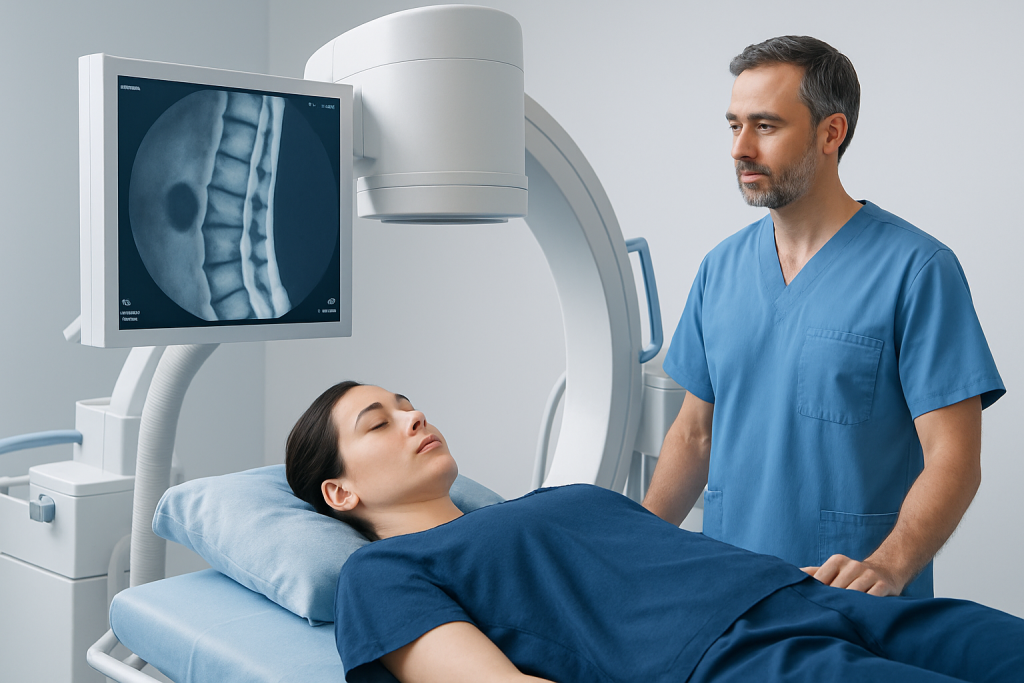
Most spinal haemangiomas cause no symptoms. Many people never know they have one unless a scan is done for a different reason. When symptoms do occur, they usually depend on the size and location of the lesion and whether it affects the surrounding bone, spinal canal, or nerve roots.
The most common symptom is localized back pain, which may be dull, persistent, or made worse by movement. Some people notice stiffness or discomfort in the same part of the spine over time rather than sudden severe pain.
If the haemangioma is larger or more active, it may lead to signs that suggest pressure on nerves or the spinal cord. These can include:
- Numbness or tingling in the arms or legs
- Weakness or heaviness in a limb
- Changes in walking or balance
- Pain that radiates around the chest, abdomen, arm, or leg
- Rarely, bladder or bowel changes if the spinal cord is affected
Symptoms tend to develop gradually in many cases, but any new neurological complaint deserves attention. Because back pain is common and has many causes, doctors look carefully at the full picture before linking symptoms to a haemangioma.
Causes & Risk Factors

The exact cause of a spinal haemangioma is not fully understood. It is generally thought to be a developmental or vascular change within the vertebral bone rather than something caused by injury, infection, or lifestyle habits. In other words, most patients do not develop it because of something they did or did not do.
These lesions are seen more often in adults than in children, and they are frequently diagnosed in middle age. Some studies and clinical observations suggest they may be found more often in women, although the reason for that pattern is not fully clear.
Factors that may increase the chance of finding a vertebral haemangioma or make it more clinically relevant include:
- Older age
- Female sex
- Lesions located in the thoracic or lumbar spine
- A larger haemangioma with more bone involvement
- Extension toward the spinal canal or soft tissues
Most spinal haemangiomas remain silent. The term “aggressive” is used when a lesion shows features that raise the risk of pain, bone weakening, or nerve compression. This distinction is important because the diagnosis alone does not tell the full story; the behaviour of the lesion matters more than the name.
Diagnosis
Diagnosis usually begins with imaging. If a vertebral haemangioma is seen on an X-ray or CT scan, doctors may recommend MRI to better define the lesion and to check whether it has features that suggest a simple incidental finding or a more active process.
MRI is especially helpful because it shows the relationship between the lesion and nearby nerves, the spinal cord, and surrounding soft tissues. CT can provide detailed information about the bony structure of the vertebra and may reveal classic patterns that support the diagnosis.
In many cases, the imaging appearance is enough for a doctor to identify a typical haemangioma without needing a biopsy. Biopsy is not routinely required and may be avoided unless the appearance is unusual or another diagnosis must be ruled out.
During evaluation, the doctor will also ask about symptoms, prior injuries, cancer history, and any changes in function. This broader assessment matters because not every back pain complaint is caused by the haemangioma, and it is important to separate an incidental finding from the true source of discomfort.
For international patients, a second review of imaging can be especially useful when scans were done elsewhere and the report is unclear. A spine specialist can interpret the images in context and advise whether follow-up imaging, observation, or treatment is appropriate.
Treatment Options
Treatment depends on whether the haemangioma is asymptomatic, painful, or causing neurological symptoms. If it is found by chance and does not appear aggressive, the most common approach is observation. That may mean periodic clinical review and, in some cases, repeat imaging to confirm stability.
When symptoms are present, treatment is selected to address the problem in front of the patient rather than the scan alone. Pain control, targeted procedures, or surgery may be considered depending on the lesion’s size, location, and effect on surrounding structures.
Possible treatment options include:
- Observation: Appropriate for many incidental lesions without concerning features.
- Pain management: Used when symptoms are mild and there is no nerve compression.
- Vertebroplasty: A procedure that stabilizes the affected vertebra in selected cases.
- Embolisation: May reduce blood flow to the lesion before other interventions or in selected treatment plans.
- Surgery: Considered when there is spinal cord or nerve compression, structural instability, or persistent symptoms not managed by other methods.
The goal is not to remove every haemangioma, but to protect spinal function and relieve symptoms safely. Because the spine is delicate and each case is different, treatment decisions are best made by a team that can review imaging, neurological findings, and overall health together.
Prevention & Self-care
There is no proven way to prevent a spinal haemangioma from forming, because it is not usually linked to modifiable habits or exposures. The practical focus is on monitoring symptoms and keeping the spine as comfortable and stable as possible.
Self-care may be helpful when the lesion is being observed or when pain is mild. Gentle movement, posture awareness, and following a clinician’s advice on activity can all support daily function. Patients should avoid assuming that all back pain comes from the haemangioma, especially if the pain pattern changes over time.
Helpful habits may include:
- Keeping follow-up appointments and imaging reviews as recommended
- Reporting new numbness, weakness, or walking changes promptly
- Using safe, clinician-approved pain relief strategies
- Maintaining regular low-impact activity when permitted
- Avoiding heavy strain if a doctor has advised spinal precautions
For patients travelling for care, planning recovery matters too. It can be useful to arrange enough time for post-procedure rest, ask for written follow-up instructions, and clarify how imaging results will be reviewed once the patient returns home. Clear communication reduces uncertainty and makes continued care smoother across borders.
When to See a Doctor
Medical review is appropriate any time a spinal haemangioma is newly identified, especially if the report mentions bone expansion, aggressive features, or uncertainty about the diagnosis. A doctor can explain whether the lesion is likely incidental or whether it needs closer attention.
Prompt assessment is especially important if there is:
- Persistent or worsening back pain
- Numbness, tingling, or weakness
- Difficulty walking or loss of balance
- Bladder or bowel changes
- Severe pain after minimal strain or without a clear cause
These symptoms do not always mean the haemangioma is causing harm, but they do warrant evaluation because they may reflect pressure on nerves, a fracture, or another spinal condition that needs treatment. Early assessment helps doctors choose the safest path and avoid delays in care.
Acibadem Health Point can support international patients who need diagnosis or treatment for a haemangioma on the spine through multidisciplinary specialists and JCI-accredited hospitals, with care planned around imaging review, treatment selection, and follow-up.
Related Questions Patients Often Ask
People who receive this diagnosis often want to know whether it is cancer, whether it will spread, or whether it will suddenly worsen. In typical cases, a vertebral haemangioma is benign and does not behave like a cancer. The key issue is not spread, but whether the lesion is causing symptoms or affecting spinal structures.
Another common concern is whether every haemangioma needs surgery. The answer is no. Many require only observation, and some need no treatment at all. Intervention is reserved for situations where symptoms, instability, or nerve involvement make treatment worthwhile.
Because the spine supports movement and protects the nervous system, even a benign lesion can feel worrying. A clear explanation of the imaging findings, symptom pattern, and recommended follow-up usually helps patients understand why a cautious but calm approach is often the best one.
Recovery and Follow-Up
Recovery depends on the treatment chosen. Patients who are simply observed may only need periodic review, while those who undergo a procedure or surgery may need a structured recovery period with activity limits and follow-up imaging.
After an intervention, doctors typically monitor pain levels, neurological function, and any signs that the vertebra is healing as expected. If the patient lives abroad, the care plan should include details about wound care, medication guidance, warning signs, and the timing of the next check-up.
Long-term follow-up is often straightforward. Once the diagnosis is confirmed and the lesion is stable, many patients return to normal activities with minimal restriction. The main objective is to keep the spine functional and to catch the uncommon case that changes over time.
Frequently asked questions
01What is a haemangioma on the spine?
It is a benign vascular lesion inside a vertebra, also called a vertebral haemangioma. Many are found incidentally and never cause symptoms.
02Is a spinal haemangioma cancer?
No, a typical spinal haemangioma is not cancer. It is usually a non-cancerous growth of blood vessels within bone.
03Can a haemangioma on the spine cause back pain?
Yes, some can cause localized back pain, especially if they are larger or more active. However, back pain is common and needs proper evaluation because it often has other causes.
04How is a spinal haemangioma diagnosed?
Doctors usually diagnose it with imaging such as MRI or CT. In many cases, the appearance is characteristic enough that a biopsy is not needed.
05Does every spinal haemangioma need treatment?
No, many do not need treatment at all and are only observed. Treatment is considered if the lesion causes pain, nerve symptoms, or spinal instability.
06When should someone seek urgent medical attention?
Urgent review is important if there is weakness, numbness, trouble walking, or bladder or bowel changes. These can suggest nerve involvement and should not be ignored.
This article is for general information only and is not a substitute for professional medical advice. Please consult a qualified doctor about your individual situation.
Joint and spine care in Turkey — expert assessment & treatment
JCI-accredited · board-certified surgeons · reply within 24h
Add us as a Preferred Source to see more of our trusted health content across Google Search, AI Overviews and Discover.