Eye Stroke: Sudden Vision Loss That Needs Urgent Care
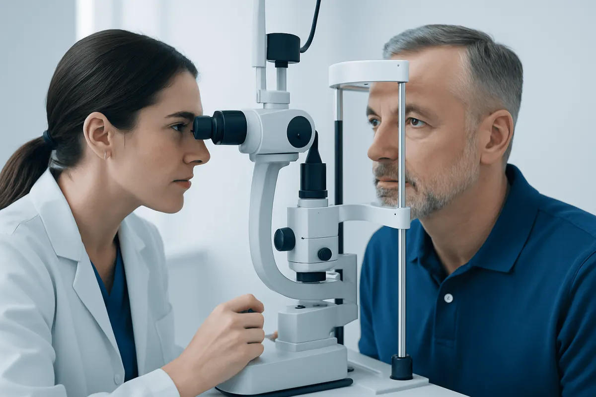
An eye stroke is a sudden interruption of blood flow in the eye, usually affecting the retina or optic nerve. It can cause painless, sudden loss or change in vision and should be assessed urgently, as it may also signal a risk of stroke elsewhere in the body.
Eye Stroke Overview
An eye stroke is a non-medical term for a sudden problem with blood flow to structures at the back of the eye. Most often, it refers to a blockage in a retinal artery or retinal vein. The retina is the light-sensitive layer at the back of the eye that sends visual information to the brain, so an interruption in its blood supply can affect sight quickly.
The eye stroke medical terminology depends on the vessel involved. A blockage in an artery is called retinal artery occlusion, while a blockage in a vein is called retinal vein occlusion. Another condition that may sometimes be described as an eye stroke is ischemic optic neuropathy, in which blood flow to the optic nerve is reduced.
These conditions are not the same as a brain stroke, but they may share similar causes and risk factors. Sudden vision loss should never be watched at home to see whether it passes. It needs urgent assessment by an eye specialist or emergency medical team.
How Serious Is a Stroke in Your Eye?
A stroke in the eye is serious because it can cause permanent visual loss and may point to an increased risk of stroke, heart disease, or other blood-vessel problems. In particular, a retinal artery occlusion is treated as a medical emergency because it can be linked with a clot or narrowing in the arteries supplying the brain and eye.
The severity varies. Some people notice a small missing area in their vision, blurred vision, or new distortion. Others experience a marked loss of vision in one eye. Eye stroke complications can include persistent visual field loss, difficulty reading or driving, swelling at the retina, abnormal blood-vessel growth, and, in some cases, painful glaucoma.
Timely assessment is valuable even when lost vision cannot be fully restored. It gives clinicians the opportunity to identify treatable causes, address cardiovascular risks, and help reduce the chance of future events affecting the eye, brain, or heart.
Symptoms That May Suggest an Eye Stroke

The most typical symptom is sudden, painless change in vision in one eye. The change may be complete or partial and may happen over seconds to minutes. Vision can become dim, blurry, shadowed, distorted, or reduced in one area of the visual field.
Some people describe a curtain, gray patch, dark spot, or blank area in their sight. With retinal vein occlusion, visual symptoms may occasionally develop more gradually over hours or days. Pain is not typical, although a painful red eye with visual loss can indicate other urgent eye conditions and also requires immediate care.
Not all transient visual symptoms are caused by an eye stroke. Migraine aura, retinal detachment, inflammation, bleeding, and neurological conditions can also affect vision. A clinician needs to examine the eye and assess the wider medical context to determine the cause.
- Sudden reduced or lost vision in one eye
- A new dark area, curtain-like shadow, or missing visual field
- Sudden distortion, blurred central vision, or difficulty seeing detail
- New flashes or floaters, especially when combined with reduced vision
- Vision symptoms with facial weakness, speech difficulty, imbalance, or numbness, which may indicate a brain stroke
Is an Eye Stroke Considered a Mini-Stroke?
An eye stroke is not simply another name for a mini-stroke. A mini-stroke, medically called a transient ischemic attack (TIA), is a temporary interruption of blood flow to part of the brain that causes symptoms which resolve. An eye stroke involves circulation in the eye or optic nerve and may leave lasting visual changes.
However, brief episodes of vision loss in one eye can sometimes be caused by temporary reduced blood flow, known as amaurosis fugax. This may be considered a TIA-like warning symptom and needs urgent evaluation. It can be associated with plaque or a clot in the carotid arteries, heart rhythm problems, or other vascular conditions.
Retinal artery occlusion is often approached with the same urgency as an ischemic stroke because it may reflect a vascular event affecting the central nervous system. Clinicians may recommend assessment of the heart, blood vessels, blood pressure, cholesterol, blood sugar, and other possible clotting risks.
Causes and Risk Factors for Eye Stroke
An eye stroke can occur when a clot, cholesterol-containing plaque, or other material blocks an artery supplying the retina. It can also happen when a retinal vein becomes blocked, causing blood and fluid to back up in the retina. In some cases, inflammation of blood vessels or disorders that increase blood clotting may contribute.
Eye stroke is more common with increasing age, but it can occur in younger adults as well. Important risk factors include high blood pressure, diabetes, high cholesterol, smoking, obesity, sleep apnea, kidney disease, and cardiovascular disease. Irregular heart rhythms such as Neurology for Stroke Prevention: Which Patients May Benefit?" class="ahp-ilk">Stroke Prevention in Atrial Fibrillation: How Doctors Choose Between Drugs and Procedures" class="ahp-ilk">atrial fibrillation can raise the risk of clots traveling through the bloodstream.
For some people, a specific cause is not immediately clear. The care team may consider carotid artery disease, heart conditions, inflammatory disorders, medications, and blood disorders where appropriate. This investigation is important because eye symptoms can provide an early opportunity to identify and manage a broader health concern.
Diagnosis and Tests After Sudden Vision Loss
Diagnosis begins with a prompt history and detailed eye examination. An ophthalmologist may check visual acuity, pupils, eye pressure, visual fields, and the retina after dilating the pupils. Photographs of the retina and optical coherence tomography, a noninvasive scan of retinal layers, can help show swelling, reduced blood flow, or damage.
Fluorescein angiography may be used in selected cases to study circulation in retinal blood vessels. Depending on the suspected type of eye stroke, urgent medical testing may include blood pressure measurement, blood tests, electrocardiography, heart imaging, carotid artery imaging, and brain imaging.
Diagnosis also involves excluding other causes of sudden visual loss. In adults over 50 with symptoms such as new headache, scalp tenderness, jaw pain while chewing, fever, or muscle aches, clinicians may urgently investigate giant cell arteritis. This inflammatory condition can threaten sight in both eyes if not treated promptly.
What Is the Best Treatment for Eye Stroke?
There is no single best treatment for every eye stroke. Treatment depends on whether an artery, vein, or optic nerve circulation is affected, how long symptoms have been present, the condition of the retina, and the person’s wider health. The first priority is rapid specialist assessment.
For retinal artery occlusion, emergency teams focus on urgent stroke-style evaluation and identifying possible causes such as a heart or carotid artery source of clots. Current treatment may include managing blood pressure, cholesterol, diabetes, and heart rhythm problems, as well as medicines recommended by the treating clinician to reduce future vascular risk. Time-sensitive interventions may be considered in carefully selected circumstances, but they are not suitable for everyone.
For retinal vein occlusion, treatment often focuses on complications such as macular edema, which is swelling in the central retina. Ophthalmologists may use medicines injected into the eye to reduce swelling or abnormal blood-vessel growth, and laser treatment may be appropriate in some situations. eye injection treatments can be used when a retinal specialist determines they are suitable.
Acıbadem Health Point’s multidisciplinary specialists and JCI-accredited hospitals support international patients needing assessment and treatment for urgent eye and vascular conditions. Ongoing care should be individualized, with regular monitoring by an ophthalmologist and coordination with primary care, neurology, or cardiology teams when needed.
Can You Recover Vision After an Eye Stroke?
Vision recovery after an eye stroke is possible for some people, but it is unpredictable. Recovery depends on the type of eye stroke, the extent of reduced blood flow, the area of the retina or optic nerve affected, and how quickly complications are identified and treated.
Some people regain part of their vision over days, weeks, or months, particularly if swelling in the retina improves. Others have permanent blind spots, reduced sharpness of vision, or major loss of sight in the affected eye. Even when central vision improves, changes in contrast sensitivity, color vision, depth perception, or peripheral vision may remain.
Visual rehabilitation can be helpful when visual loss persists. This may include low-vision aids, practical changes at home and work, mobility support, and advice about safe driving. Protecting the unaffected eye through regular eye care and cardiovascular risk management is also an important part of recovery.
When to Seek Medical Care
Anyone with sudden vision loss, a new visual field defect, or sudden major blurring in one eye should seek emergency medical care immediately. This is especially important when symptoms began within the last few hours, because some causes require time-sensitive evaluation. Do not drive yourself if vision is impaired.
Emergency services should also be contacted if visual symptoms occur with weakness, facial drooping, speech problems, severe dizziness, confusion, severe headache, or loss of coordination. These can be signs of a brain stroke and require urgent treatment.
People who have already been diagnosed with an eye stroke should attend follow-up appointments as advised. New worsening vision, eye pain, redness, flashes, floaters, or a curtain-like shadow should be reported promptly, as these symptoms may indicate a complication or another eye emergency.
Frequently asked questions
01What is the medical name for an eye stroke?
The eye stroke medical name depends on the affected structure. Common terms include central retinal artery occlusion, branch retinal artery occlusion, central retinal vein occlusion, branch retinal vein occlusion, and ischemic optic neuropathy. An ophthalmologist can determine the exact diagnosis after examining the eye.
02Is eye stroke common?
Eye stroke is less common than many everyday eye problems, but retinal vascular occlusions are important causes of sudden visual loss, particularly in older adults and people with vascular risk factors. The likelihood rises with conditions such as high blood pressure, diabetes, smoking, and high cholesterol. Sudden visual changes should always be assessed promptly regardless of age.
03Does an eye stroke hurt?
Most retinal artery and retinal vein occlusions cause painless visual loss or blurring. Eye pain does not rule out a serious problem, because other urgent eye conditions can cause pain and reduced vision. A painful red eye with vision change needs immediate medical assessment.
04Can stress cause an eye stroke?
Stress alone is not generally considered a direct cause of an eye stroke. However, long-term stress may contribute indirectly to high blood pressure, smoking, sleep disruption, and other factors that affect vascular health. Managing stress is helpful for overall health but does not replace medical treatment of risk factors.
05Can an eye stroke happen again?
A further vascular event can occur, especially when underlying risk factors are not well controlled. Follow-up with eye and medical specialists helps identify causes and reduce risk through individualized management of blood pressure, diabetes, cholesterol, heart conditions, and lifestyle factors. Regular monitoring is particularly important after retinal vein occlusion because eye complications can develop later.
06What should someone do while waiting for care?
A person with sudden vision loss should seek emergency help without delay and avoid driving. They should note when symptoms started, what changes occurred, and any accompanying symptoms or relevant medications. They should not use unprescribed eye drops or attempt home remedies, as these may delay appropriate care.
This article is for general information only and is not a substitute for professional medical advice. Please consult a qualified doctor about your individual situation.
Heart care in Turkey — expert evaluation and treatment
JCI-accredited · board-certified surgeons · reply within 24h
Add us as a Preferred Source to see more of our trusted health content across Google Search, AI Overviews and Discover.