Eye Melanoma: Early Signs, Risk Factors, and How It Is Treated
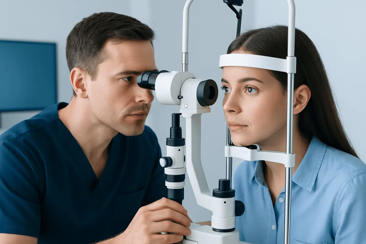
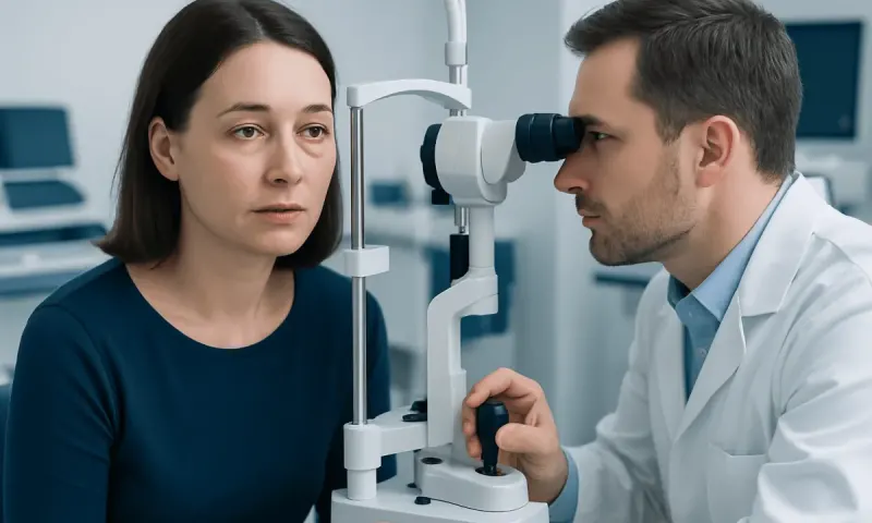
You go in for a routine eye check, your pupils get dilated, and the doctor pauses a little longer than usual over one spot at the back of your eye. That is how many eye melanomas are first noticed — quietly, before anything feels wrong.
Eye melanoma is a rare cancer that develops from pigment-producing cells in or around the eye, most often in the uvea. Early on, it may cause no symptoms at all. That is exactly why regular dilated eye examinations matter, and why any new change in your vision deserves prompt assessment.
Overview: what is eye melanoma?
Eye melanoma, also called ocular melanoma, is a cancer arising from melanocytes, the cells that make pigment. Most cases start in the uvea, the middle layer of the eye that includes the iris, ciliary body, and choroid. For this reason, the most common type is called uveal melanoma. It is different from melanoma of the eyelid skin and from other, less common cancers that can affect the eye.
Uveal melanoma can develop in the back of the eye, where it may not be visible without special examination. This means it may be found during a routine dilated eye examination before it causes noticeable changes in sight. Some tumors grow slowly, while others require more urgent treatment because of their size, location, or features seen on imaging.
A diagnosis like this can feel overwhelming, and that is understandable. Your care is planned around you. The main goals are to control the tumor, preserve the eye and useful vision whenever safely possible, and monitor for spread beyond the eye. A team may include an ophthalmologist specializing in ocular oncology, radiation specialists, medical oncologists, radiologists, and pathology experts.
Early signs and symptoms
Eye melanoma often has no symptoms in its earliest stages. This is particularly true when a tumor is small or located in the back of the eye. A clinician may notice a suspicious pigmented lesion or a change in the shape of the eye during a routine examination, then arrange further tests to determine whether it is a harmless mole-like spot or a melanoma.
When symptoms occur, they vary according to the tumor’s position. Possible changes include blurred or reduced vision, a shadow or missing area in the visual field, flashing lights, new floaters, or distortion of straight lines. A person may also notice that the pupil looks different, a dark spot on the iris appears to enlarge, or the eye becomes red or uncomfortable. These symptoms have many possible causes and do not by themselves mean cancer.
In more advanced cases, a tumor can cause retinal detachment, raised pressure inside the eye, or pain. Any sudden or persistent change in vision should be assessed promptly by an eye-care professional. Early evaluation is valuable because some eye conditions need timely treatment even when they are not melanoma.
Causes and risk factors
The precise cause of eye melanoma is not fully understood. It occurs when melanocytes develop DNA changes that allow them to grow and survive abnormally. These changes are usually acquired during a person’s lifetime rather than inherited directly from a parent. Researchers continue to study the genetic features of uveal melanoma and how these features influence prognosis and follow-up planning.
Risk factors associated with uveal melanoma include older age, fair skin, light-colored eyes, and a tendency to develop freckles or moles. Certain pre-existing pigmented eye lesions, such as an ocular nevus, may need periodic monitoring. Rare inherited conditions, including BAP1 tumor predisposition syndrome, can increase the likelihood of uveal melanoma and some other cancers, but most people with eye melanoma do not have a known inherited syndrome.
Ultraviolet exposure is an established risk factor for skin melanoma, but its role in uveal melanoma is less clear. Wearing sunglasses that block UVA and UVB rays and using a wide-brimmed hat in strong sunlight remain reasonable measures for general eye and skin protection. No lifestyle step can reliably prevent eye melanoma. If it develops, it is not your fault.
How eye melanoma is diagnosed and assessed
An ophthalmologist begins with a review of symptoms and medical history, followed by a comprehensive eye examination. Dilating drops allow the clinician to inspect the retina and choroid at the back of the eye. Photographs of the inside of the eye may be taken so that a lesion can be measured and compared accurately over time.
Eye ultrasound is a central test for evaluating a suspected uveal melanoma. It can show the lesion’s size, shape, thickness, and internal characteristics. Additional tests may include optical coherence tomography, fluorescein angiography, fundus autofluorescence imaging, or magnetic resonance imaging when more detail is needed. These tests also help distinguish melanoma from other conditions, such as a benign nevus, bleeding under the retina, or a metastatic tumor from another cancer.
A biopsy is not required in every case because experienced ocular specialists can often diagnose uveal melanoma from examination and imaging findings. When tissue or tumor cells are collected, genetic testing may provide information about the likelihood of spread and help guide surveillance. If melanoma is confirmed or strongly suspected, scans and blood tests may be used to check for disease elsewhere in the body, with particular attention to the liver.
Treatment options and follow-up care
Treatment depends on the tumor’s size, location, growth pattern, effects on vision, and whether there is evidence that it has spread. A very small lesion with uncertain features may be monitored closely with repeat imaging rather than treated immediately. If a lesion shows growth or high-risk features, the specialist will discuss active treatment.
For many uveal melanomas, focused radiation is used to control the tumor while keeping the eye in place. Options may include plaque brachytherapy, in which a small radioactive plaque is temporarily positioned on the outside of the eye, or external-beam approaches such as proton beam therapy. Selected small tumors may be treated with laser therapy, sometimes alongside radiation. The most suitable approach depends on where the tumor sits relative to structures needed for vision.
Surgery may be appropriate for selected tumors. In some situations, removing the eye, known as enucleation, provides the safest and most effective local control, especially for a very large tumor or an eye that has lost useful vision and is painful. An artificial eye can usually be fitted after healing to support appearance. If disease has spread, treatment is managed by oncology specialists and may involve systemic medicines, surgery, or liver-directed procedures based on the individual situation.
Follow-up is essential after any treatment. Eye visits monitor tumor control, pressure in the eye, retinal health, and treatment-related effects such as cataract, dry eye, retinal changes, or vision loss. Ongoing body imaging and laboratory assessment may also be advised because uveal melanoma can recur or spread after initial Lazy Eye Treatment in Adults: Can Amblyopia Still Improve?" class="ahp-ilk">eye treatment, sometimes years later.
Living with eye melanoma and supporting eye health
After treatment, visual recovery and daily adjustment vary widely. Some people retain good vision, while others need support for reduced sight, depth-perception changes, glare, or blind spots. Low-vision services can offer practical tools, lighting advice, magnifiers, screen adaptations, and strategies for work, reading, travel, and safe mobility.
Bring a family member or someone you trust to appointments. Write your questions down beforehand, and ask your team to explain scan results and follow-up schedules in plain language. Emotional support is also an important part of care. A counselor, support group, specialist nurse, or patient organization may help individuals and families manage uncertainty and the practical effects of treatment.
General health measures, including not smoking, staying physically active within personal ability, eating a balanced diet, and keeping regular medical appointments, support overall wellbeing. They do not replace follow-up for melanoma. Tell your team about any new visual symptoms, and keep every planned eye and body surveillance visit — even on the days you feel perfectly well.
When to seek medical care
Book an eye examination soon if you notice persistent blurred vision, flashes of light, a new shower of floaters, a curtain-like shadow across your vision, a growing dark area on the iris, or an unexplained change in the shape of your pupil. These signs are more often caused by conditions other than eye melanoma, but they should be assessed because some eye problems require prompt attention.
Urgent same-day eye care is appropriate for sudden loss of vision, a rapidly developing curtain or shadow, severe eye pain, marked redness, or symptoms after an eye injury. These symptoms may indicate retinal detachment, acute glaucoma, inflammation, or another urgent condition. Emergency services should be used when local urgent eye-care access is unavailable.
If you have been treated for eye melanoma before, call your clinical team about any new vision change, pain, visible change in the eye, or anything else that worries you. Acıbadem Health Point’s multidisciplinary specialists and JCI-accredited hospitals diagnose and treat eye melanoma for international patients, with care plans based on individual clinical findings.
Frequently asked questions
01Is eye melanoma the same as skin melanoma?
No. Both cancers begin in pigment-producing cells, but eye melanoma most often arises in the uvea inside the eye, whereas skin melanoma begins in the skin. Their risk factors, testing, treatment approaches, and patterns of spread can differ.
02Can eye melanoma be found during a routine eye test?
Yes. Many uveal melanomas cause no symptoms at first and are detected during a dilated eye examination or imaging performed for another reason. Regular eye checks are particularly useful for monitoring known pigmented lesions in the eye.
03Does a dark spot in the eye always mean melanoma?
No. A dark spot may be a benign nevus, normal pigment variation, or another non-cancerous change. An ophthalmologist can examine it and use photographs or ultrasound to determine whether monitoring or further investigation is needed.
04Can eye melanoma be cured?
Many tumors confined to the eye can be treated successfully with the aim of achieving local control. However, individual outlook depends on tumor features, location, size, genetic findings, and whether cancer has spread. Long-term follow-up remains important after treatment.
05What happens if the eye has to be removed?
Eye removal is considered when it is the safest option for controlling a tumor or when the eye is severely affected. After healing, most people can be fitted with a custom artificial eye that matches the appearance of the other eye. The care team can also arrange rehabilitation and emotional support.
06Can eye melanoma spread to other parts of the body?
Yes. Uveal melanoma can spread through the bloodstream, most commonly to the liver, and less often to other organs. This is why clinicians may recommend regular systemic monitoring after treatment, even if the tumor in the eye has been controlled.
This article is for general information only and is not a substitute for professional medical advice. Please consult a qualified doctor about your individual situation.
Oncology care in Turkey — second opinion and treatment plan
JCI-accredited · board-certified surgeons · reply within 24h
Add us as a Preferred Source to see more of our trusted health content across Google Search, AI Overviews and Discover.