

Epidural Hematoma: Symptoms, Causes and Treatment

Key Takeaways
- Epidural hematoma usually happens after a head injury and can become serious quickly.
- A person may seem alert at first, then develop worsening headache, confusion, vomiting, drowsiness, or weakness.
- CT imaging is the main test used to confirm the diagnosis and guide urgent treatment.
- Treatment may involve close hospital monitoring or emergency surgery to remove the blood and relieve pressure.
- Rapid evaluation after any significant head trauma is the safest approach, especially if symptoms change over time.
Epidural hematoma is a collection of blood between the skull and the outer covering of the brain, usually caused by head trauma. It needs urgent medical assessment because symptoms can worsen quickly, even after a brief period of seeming improvement.
Overview
An epidural hematoma is a build-up of blood between the inner surface of the skull and the dura, the tough outer layer that covers the brain. It most often follows a blow to the head, such as from a fall, road traffic accident, sports injury, or other impact that damages blood vessels inside the skull.
What makes this condition important is not only the bleeding itself, but the pressure it can create on the brain. In some people, the early phase may look deceptively mild. A person might speak normally, walk, or even feel well for a period of time before symptoms begin to change.
For that reason, epidural hematoma is treated as a neurosurgical emergency. Anyone with a significant head injury and new neurological symptoms should be evaluated promptly, whether they are at home, at a local emergency department, or traveling abroad and trying to decide how urgently to seek care.
Symptoms
Symptoms can develop within minutes or over several hours, and their pattern depends on the size of the bleed and how quickly pressure rises. A classic warning sign is a short period of recovery or clarity after the injury, followed by a noticeable decline. That improvement can be misleading, so it should never be used as proof that the injury is minor.
Common symptoms include a worsening headache, nausea or vomiting, confusion, increasing sleepiness, trouble speaking, unequal pupil size, dizziness, or difficulty walking. Some people may develop seizures, blurred vision, weakness on one side of the body, or loss of consciousness.
In children, older adults, and people taking blood-thinning medicines, symptoms may be less typical and the condition can still be serious. Any change in alertness, behavior, or coordination after a head injury deserves medical attention.
- Headache that keeps getting worse
- Repeated vomiting
- Sleepiness or hard-to-wake behavior
- Confusion, agitation, or unusual behavior
- Weakness, numbness, or speech changes
- Seizure or fainting
Causes & Risk Factors

The most common cause is trauma that tears a blood vessel, often the middle meningeal artery, leading to pressure from the accumulating blood. Because the skull is a fixed space, even a relatively small amount of bleeding can become dangerous if it expands rapidly.
Risk increases with high-impact injuries such as car crashes, falls from height, contact sports, assault, or industrial accidents. People with skull fractures are more likely to develop an epidural hematoma, although the bleed can also occur without an obvious fracture.
Certain factors can make bleeding more likely or more concerning. These include the use of anticoagulant or antiplatelet medicines, alcohol or drug intoxication, advanced age, and any condition that affects clotting. In international patients, it is especially important to share a full medication list and any prior brain injury history, even if records are from another country.
Diagnosis
Doctors start by assessing consciousness, pupil response, movement, speech, and other neurological signs. A careful history of the injury is important, including whether there was a moment of blackout, vomiting, confusion, or a symptom-free interval after the impact.
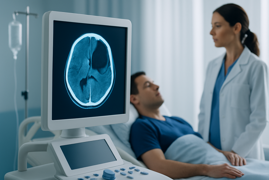
A head CT scan is the key test because it can quickly show the location, size, and shape of the bleed. Epidural hematomas often appear as a lens-shaped collection on CT, which helps specialists distinguish them from other types of intracranial bleeding.
Additional tests may be used to support safe treatment planning, such as blood tests to check clotting status, oxygen levels, and overall stability. When someone is being evaluated while away from home, doctors may also ask about previous imaging, past surgery, allergies, and regular medicines so the right treatment can be started without delay.
Treatment Options
Treatment depends on the size of the hematoma, the patient’s neurological status, and whether the bleeding is causing pressure on the brain. Small, stable bleeds in carefully selected patients may be monitored in the hospital with repeat examinations and follow-up imaging.
When the hematoma is large, expanding, or linked to neurological decline, urgent surgery is often needed. The aim is to remove the blood and relieve pressure before brain injury becomes more severe. Neurosurgeons may perform a craniotomy, and in some situations other urgent procedures may be considered based on the scan and the person’s condition.
Supportive care is part of treatment as well. This may include airway and breathing support, seizure management if needed, reversal of certain blood-thinning effects, pain control, and close monitoring in an intensive care or neurosurgical setting. After surgery or observation, follow-up care helps track recovery, symptoms, and return to normal activity.
Prevention & Self-care
Not every epidural hematoma can be prevented, but the risk of head injury can often be lowered. Wearing seat belts, using appropriate helmets for cycling or motorcycling, improving fall prevention at home, and following sports safety guidance all make a meaningful difference.
After any head injury, a cautious approach is better than a wait-and-see attitude if symptoms are evolving. Rest, avoid alcohol, and do not return to sports or demanding activity until a clinician says it is safe. If a person is advised to go home after evaluation, they should follow the discharge instructions closely and have someone observe them when possible.
It is also wise to keep a current list of medicines, especially blood thinners and aspirin-containing products, and to tell emergency staff about any medical condition that affects clotting. For travelers, carrying digital copies of scan reports, medication lists, and emergency contacts can make care smoother if urgent treatment is needed away from home.
- Use helmets and seat belts consistently
- Reduce fall hazards in living spaces
- Follow return-to-play advice after concussion or head injury
- Seek prompt review if symptoms worsen after discharge
- Keep medication and allergy information accessible
When to See a Doctor
Any significant head injury followed by worsening symptoms should be assessed urgently, even if the person seemed fine at first. Medical review is especially important after loss of consciousness, repeated vomiting, Neurology May Be Urgently Needed" class="ahp-ilk">severe headache, confusion, seizures, or any new weakness, numbness, or speech difficulty.
Emergency care is needed if the person becomes very drowsy, difficult to wake, or less responsive. Unequal pupils, abnormal walking, or a rapid decline in mental status are also reasons to seek immediate help.
If care is being arranged across borders, a neurology or neurosurgery team can help interpret scans, coordinate monitoring, and decide whether surgery is needed. Acibadem Health Point’s multidisciplinary specialists and JCI-accredited hospitals diagnose and treat this condition for international patients, with coordinated support from emergency assessment through follow-up.
Recovery and Outlook
Recovery depends on how quickly the bleed was treated, how much pressure it caused, and whether the brain was injured at the same time. Some people recover well after timely surgery or observation, while others may need rehabilitation for headaches, fatigue, memory changes, balance problems, or weakness.
Follow-up matters because symptoms can evolve after discharge. Doctors may recommend repeat imaging, activity restrictions, wound care if surgery was done, and a gradual return to work, travel, or exercise. For people returning home to another country, clear discharge paperwork and a follow-up plan can help local doctors continue care smoothly.
Although epidural hematoma can be alarming, prompt recognition and treatment often improve the outlook. The safest strategy is to take head injury seriously, especially when symptoms change after the initial impact.
Not applicable in body section.
Frequently asked questions
01How is an epidural hematoma different from a concussion?
A concussion is a functional brain injury, while an epidural hematoma is bleeding that creates pressure inside the skull. Both can happen after head trauma, but epidural hematoma is much more likely to need urgent imaging and sometimes surgery.
02Can someone have an epidural hematoma and still seem normal at first?
Yes. A person may look surprisingly well for a period after the injury, which is one reason this condition can be dangerous. Any later change in headache, alertness, behavior, or movement should be treated seriously.
03Is surgery always necessary?
No. Some small, stable epidural hematomas can be monitored closely in the hospital. Surgery is more likely when the bleed is large, growing, or causing neurological symptoms or pressure on the brain.
04What test confirms the diagnosis?
A head CT scan is the main test because it can quickly show the bleed and its size. Doctors may also use blood tests and neurological checks to guide treatment decisions.
05What should family members do after a head injury at home?
They should watch for worsening headache, vomiting, confusion, sleepiness, weakness, or seizure. If any of these appear, emergency medical care should be sought right away.
06Can epidural hematoma happen without a skull fracture?
Yes, although skull fracture is common, a fracture is not required. The key concern is bleeding inside the skull that can raise pressure on the brain.
This article is for general information only and is not a substitute for professional medical advice. Please consult a qualified doctor about your individual situation.
Persistent digestive symptoms? Get evaluated in Turkey
JCI-accredited · board-certified surgeons · reply within 24h
Add us as a Preferred Source to see more of our trusted health content across Google Search, AI Overviews and Discover.


