

ECG vs EKG Test: Why the Names Mean the Same
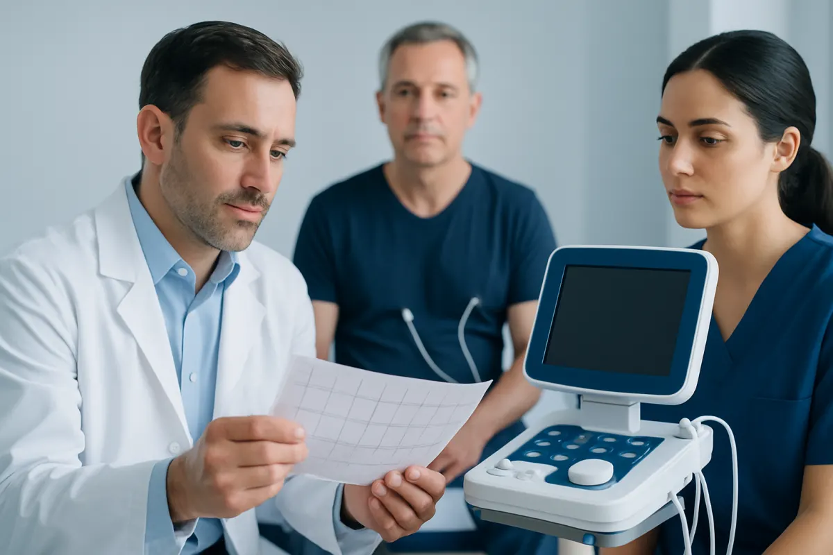
An ECG and an EKG are exactly the same test: both record the heart’s electrical activity through small skin electrodes. The different names reflect spelling conventions, not a difference in accuracy, procedure, or purpose.
ECG vs EKG test: side-by-side comparison
An ECG vs EKG test comparison has a simple answer: there is no medical difference. ECG stands for electrocardiogram, while EKG stands for elektrokardiogram, a spelling based on the German word Elektrokardiogramm. Both terms describe a test that measures the heart’s electrical signals and displays them as waves on a tracing.
| Feature | ECG | EKG |
|---|---|---|
| Full name | Electrocardiogram | Elektrokardiogram |
| What it measures | Electrical activity of the heart | Electrical activity of the heart |
| How it is performed | Electrodes placed on the chest and limbs | Electrodes placed on the chest and limbs |
| Accuracy and uses | The same | The same |
| Why the name differs | Common international and UK-style abbreviation | Commonly used in the United States |
During a standard resting test, adhesive electrodes are placed on the chest, arms, and legs while the person lies still for a few minutes. The machine records electrical impulses; it does not send electricity into the body. The test is painless, non-invasive, and does not involve radiation.
ECG vs EKG which is better is therefore not a choice between two different tests. A clinician may use either term depending on local practice, the medical record system, or personal preference. What matters is why the test is being requested and whether a resting ECG, exercise ECG, or longer-term rhythm monitor is most suitable.
How clinicians interpret ECG and EKG results
Clinicians do not tell an ECG apart from an EKG, because the recordings are the same. Instead, they assess the tracing in a structured way: heart rate, rhythm regularity, the timing of electrical conduction, and the shape and position of individual wave patterns. These features may provide clues about rhythm disorders, previous heart injury, strain on parts of the heart, or medication and electrolyte effects.
The ECG is interpreted together with the reason for testing. For example, palpitations may lead a clinician to look closely for an irregular rhythm, while chest discomfort may prompt assessment for changes that can occur with reduced blood flow to the heart. A single tracing is only one part of clinical decision-making and is not interpreted in isolation.
“Normal” means the tracing does not show a clear abnormality at the time it was recorded. “Abnormal” does not automatically mean a dangerous condition is present. Some findings are harmless variations, while others need prompt review, repeat testing, blood tests, monitoring, or imaging. The clinician who knows the person’s symptoms and health history is best placed to explain the significance of the result.
How do you tell if ECG results are normal or abnormal?
A formal report may describe sinus rhythm, meaning the heartbeat is being initiated by the heart’s natural pacemaker. It may also note heart rate and whether intervals or wave patterns are within expected ranges. However, patients should avoid trying to diagnose themselves from printed values or automated machine comments, as these can be incomplete or occasionally incorrect. A qualified clinician should confirm whether a result is normal for that individual and advise on any next steps.
What an ECG can and cannot show

An ECG can be especially helpful when symptoms are happening during the recording. It may detect abnormal rhythms such as atrial fibrillation, unusually slow or fast rhythms, conduction delays, and electrical patterns that can occur during or after reduced blood flow to heart muscle. It can also provide a useful baseline before certain treatments or procedures.
How accurate is an ECG or EKG?
An ECG is highly accurate at recording the electrical activity present at that moment, provided the electrodes are correctly placed and the recording quality is good. Its ability to identify a particular condition depends on the condition, whether it is active during the test, and the clinical context. An intermittent rhythm disturbance can be missed if the heartbeat is normal during a brief resting recording.
For this reason, a clinician may recommend a wearable monitor that records for longer, an exercise stress test, blood tests, or imaging. An ECG can support urgent assessment of symptoms suggestive of a heart attack, but a normal initial ECG alone cannot exclude every serious cause of chest symptoms. Ongoing or concerning symptoms should always be assessed promptly.
Does an ECG detect clogged arteries?
An ECG does not directly show clogged or narrowed coronary arteries. It may reveal changes suggesting that heart muscle is not receiving enough blood, especially if this is occurring during the recording or exercise, but it can also be normal when coronary artery narrowing is present. Depending on symptoms and risk factors, further assessment may include an exercise test, echocardiogram, coronary CT imaging, or invasive coronary angiography.
ECG vs EKG vs echocardiogram and ultrasound
ECG vs EKG vs echocardiogram can be confusing because all are used in heart care, but they answer different questions. An ECG or EKG records the heart’s electrical activity. An echocardiogram is a heart ultrasound that uses sound waves to create moving images of the heart muscle, chambers, valves, and blood flow.
In other words, the ECG helps assess the heart’s rhythm and electrical pathways, while an echocardiogram helps assess the heart’s structure and pumping function. An ultrasound performed on another body area, such as the abdomen or leg veins, is also different from an ECG because it uses sound waves to view tissues or blood flow rather than recording electrical signals.
A clinician may request one test or combine tests depending on the concern. Palpitations may first require an ECG and rhythm monitoring. A new heart murmur, suspected valve problem, unexplained breathlessness, or concern about heart pumping strength may lead to an echocardiogram. When coronary artery disease is a concern, testing is selected according to symptoms, risk factors, and the urgency of the situation.
These tests complement one another rather than compete. Choosing the right investigation is based on the clinical question, not on whether the test is called ECG or EKG.
What can throw off an ECG?
Technical issues can affect the quality of an ECG recording. Movement, talking, shivering, tense muscles, loose electrodes, sweat or oily skin, and poor electrode contact can create electrical “artifact” that makes the tracing harder to interpret. Staff may repeat the ECG if the recording is unclear.
Correct electrode placement matters because moving an electrode even slightly can alter parts of the tracing. Body position, breathing pattern, and individual anatomy may also influence the appearance of some waves. These factors do not usually harm the person, but they can make it important to verify an unexpected result.
Medical factors can also change an ECG. These include fever, dehydration, anemia, electrolyte imbalances, thyroid disorders, certain prescription or non-prescription medicines, stimulants, alcohol, and recreational drugs. Patients should tell the clinician about symptoms, medical conditions, all medicines and supplements, and recent substance use so the result can be interpreted safely.
There is usually no special preparation for a resting ECG. Wearing a two-piece outfit may make chest access easier, and remaining relaxed and still during the recording can improve quality. Patients should not stop prescribed medicines unless their clinician specifically tells them to do so.
What to do after ECG or EKG results
If the ECG is normal and symptoms have settled, a clinician may provide reassurance or arrange follow-up based on the person’s overall risk and history. If symptoms are recurrent, a normal brief recording may lead to longer rhythm monitoring, because episodes of palpitations or dizziness may not happen during the test.
If the ECG is abnormal, the next step depends on the finding and symptoms. It may involve repeating the test, comparing it with prior tracings, checking blood pressure and blood tests, reviewing medications, arranging an echocardiogram, or referring to a cardiologist. Some abnormalities are longstanding or benign; others need more timely investigation.
Healthy habits support heart health but do not replace medical evaluation of new symptoms. Avoiding tobacco, being physically active within personal limits, following a balanced eating pattern, managing blood pressure and diabetes, getting adequate sleep, and taking prescribed treatment as directed can all be helpful. People with known cardiovascular conditions should follow their individual care plan.
Acıbadem Health Point’s multidisciplinary specialists and JCI-accredited hospitals assess heart symptoms and arrange appropriate diagnostic testing and treatment for international patients.
When to seek medical care
Medical advice should be sought for new or recurring palpitations, unexplained dizziness, fainting, chest discomfort, shortness of breath, reduced exercise tolerance, or a family history of certain inherited heart conditions. A clinician can decide whether an ECG, EKG, monitoring, imaging, or another assessment is appropriate.
Emergency medical care is needed for sudden or persistent chest pressure, pain that spreads to the arm, jaw, back, or upper abdomen, severe shortness of breath, fainting, a sustained racing or irregular heartbeat with feeling unwell, or symptoms that may indicate stroke. Stroke warning signs can include sudden facial drooping, arm weakness, speech difficulty, confusion, or loss of balance.
People should not drive themselves if they have severe symptoms or feel faint. Calling local emergency services is the safest option. Prompt assessment is particularly important when symptoms are new, severe, worsening, or occur in someone with known heart disease.
Frequently asked questions
01Are ECG and EKG the same test?
Yes. ECG and EKG are different abbreviations for electrocardiogram, a test that records the heart’s electrical activity. The procedure, information obtained, and clinical value are the same.
02Why do some doctors say EKG instead of ECG?
EKG is derived from the German spelling, Elektrokardiogramm, and is widely used in the United States. ECG is more commonly used in many other countries. Neither term indicates a different type of heart test.
03Can an ECG detect a heart attack?
An ECG can show patterns that may suggest an ongoing or previous heart attack, particularly when interpreted with symptoms and blood tests. However, an early or normal ECG does not exclude every heart attack or other serious cause of chest symptoms. Urgent symptoms require immediate medical assessment.
04Is an ECG painful or does it use radiation?
A resting ECG is painless and non-invasive. Sticky electrodes are placed on the skin to record electrical signals from the heart. The test does not use radiation and does not send electricity into the body.
05Can anxiety affect an ECG result?
Anxiety can increase heart rate and may contribute to palpitations, shaking, or muscle tension that affects recording quality. It does not mean that symptoms should automatically be attributed to anxiety. A clinician should consider the ECG alongside symptoms and the person’s health history.
06What happens if my ECG is abnormal?
An abnormal result may lead to a repeat ECG, review of medicines, blood tests, longer rhythm monitoring, imaging, or specialist assessment. Some findings are harmless or temporary, while others need further evaluation. The appropriate next step depends on the exact tracing and the person’s symptoms.
This article is for general information only and is not a substitute for professional medical advice. Please consult a qualified doctor about your individual situation.
Heart care in Turkey — expert evaluation and treatment
JCI-accredited · board-certified surgeons · reply within 24h
Add us as a Preferred Source to see more of our trusted health content across Google Search, AI Overviews and Discover.


