What a Carotid Ultrasound Shows and How the Test Is Done
Your doctor listens to your neck with a stethoscope, pauses, and says they’d like to arrange a scan. That moment can be unsettling — but the test being suggested is a simple one.
Carotid ultrasound is a noninvasive imaging test that uses sound waves to examine the carotid arteries in your neck, measure blood flow, and look for plaque or narrowing. It is commonly used to help evaluate stroke risk, investigate certain symptoms, and guide decisions about monitoring or treatment.
What is a carotid ultrasound?
A carotid ultrasound is a medical imaging test that looks at the carotid arteries, the major blood vessels on each side of the neck that carry blood to the brain. The scan uses high-frequency sound waves rather than radiation, making it a safe and widely used way to assess blood flow and look for plaque buildup or narrowing.
Most carotid ultrasound exams include Doppler ultrasound, which shows how blood is moving through the artery. This allows the clinician to evaluate not only the artery’s structure but also whether blood flow has become faster or more turbulent, which can suggest narrowing. For this reason, the test is often called a carotid Doppler ultrasound or carotid artery ultrasound.
Carotid artery disease tends to build up quietly, which is why this scan is so useful. A person may have no symptoms until reduced blood flow or a blood clot leads to a transient ischemic attack or stroke. Carotid ultrasound helps identify changes early enough for monitoring, medication, or further treatment planning.
Why doctors order this test
Doctors may request a carotid ultrasound for different reasons. One common reason is to investigate possible carotid artery disease in someone who has had stroke-like symptoms, such as sudden weakness, trouble speaking, temporary vision loss, or numbness on one side of the body. The test can help determine whether narrowing in the neck arteries may be contributing.
It may also be used in people who have a carotid bruit, which is an unusual sound a doctor hears over the artery with a stethoscope. In other cases, carotid ultrasound is ordered because a person has risk factors for vascular disease, such as high blood pressure, high cholesterol, diabetes, smoking, older age, or a history of heart disease.
Carotid ultrasound can also be used to follow known disease over time or to assess arteries before and after treatment. If narrowing is significant, a doctor may discuss medical therapy or procedures such as carotid endarterectomy or, in selected situations, carotid angioplasty and stenting. It may be part of the broader evaluation of carotid artery disease and its relationship to stroke.
How the test is done and how to prepare

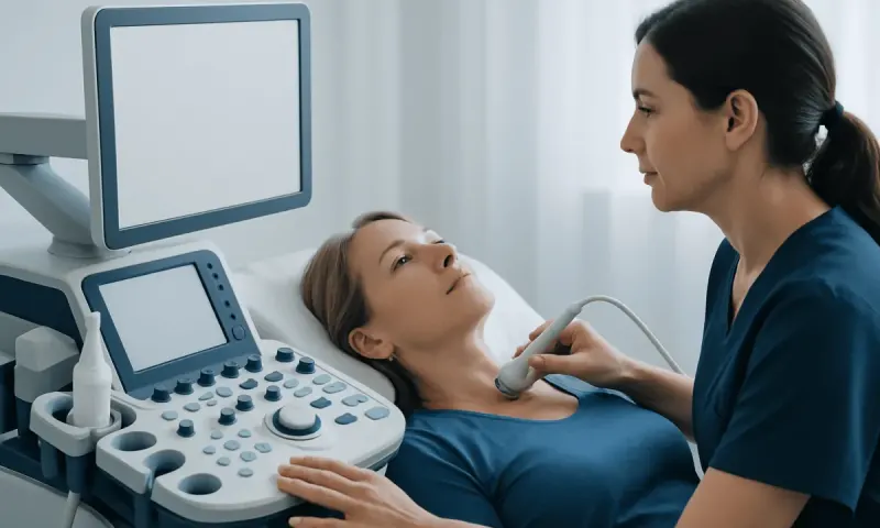
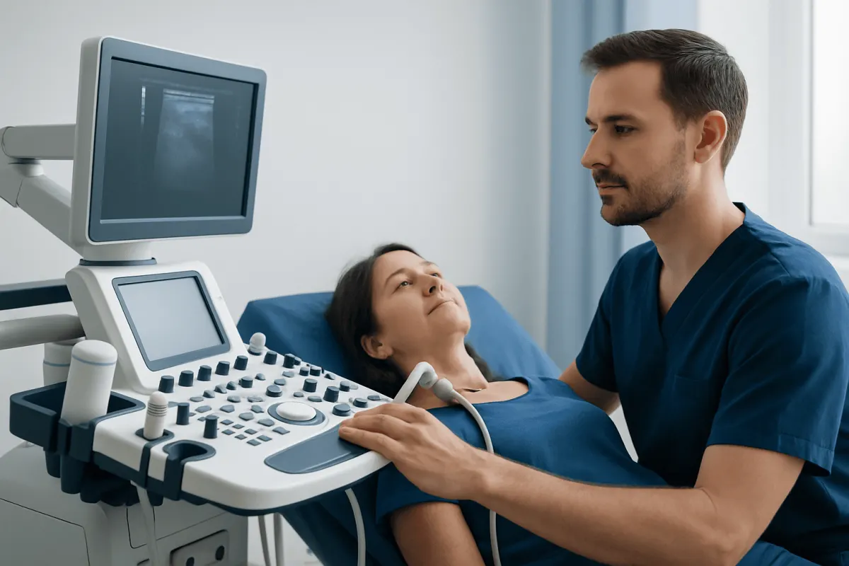
Carotid ultrasound is usually performed in a hospital imaging department, vascular laboratory, or outpatient clinic. The person lies on an examination table with the neck slightly extended. A clear gel is applied to the skin, and a small handheld device called a transducer is moved over the neck to create images of the arteries.
The test is painless, although light pressure from the transducer may be felt. The sonographer may ask the person to turn the head slightly or hold still for a few moments while images and Doppler measurements are recorded. In many cases, the exam takes about 15 to 30 minutes, though timing can vary.
There is not much to do beforehand. People may be asked to avoid necklaces, turtlenecks, or clothing that makes the neck difficult to access. Sometimes a doctor may suggest avoiding smoking or caffeine shortly before the test because these can affect blood vessels, but local instructions should always be followed. Most people can return to normal activities immediately afterward.
What carotid ultrasound can show
The main purpose of carotid ultrasound is to evaluate the carotid arteries for plaque, narrowing, and blood flow changes. Plaque is made of fatty deposits, cholesterol, inflammatory material, and calcium that build up in the artery wall. Over time, this process can make the artery stiffer and narrower, reducing blood flow to the brain or increasing the risk of clot formation.
Here is what the scan may show:
- Normal artery structure and blood flow
- Mild plaque without significant blockage
- Moderate or severe narrowing, also called stenosis
- Complete blockage in rare cases
- Changes after surgery or stent placement
In some situations, ultrasound can also provide information about plaque appearance. Certain plaque features may suggest a higher risk of causing symptoms, although ultrasound is only one part of the clinical picture. Doctors interpret the scan together with symptoms, physical examination, medical history, and sometimes other tests such as CT angiography, MR angiography, or additional interventional radiology procedures when needed.
Understanding the results
A normal carotid ultrasound means blood flow looks appropriate and there is no meaningful narrowing in the arteries examined. This can be reassuring, especially when the test was done to evaluate risk or investigate a symptom. Still, a normal result does not rule out every possible cause of neurological symptoms, so doctors may recommend further evaluation if symptoms remain unexplained.
If plaque or narrowing is present, the report often describes the degree of stenosis as mild, moderate, or severe. This estimate is based on both the appearance of the artery and Doppler blood flow measurements. In general, the more narrowed the artery is, the more closely it may need to be monitored or treated, particularly if the person has had symptoms such as a transient ischemic attack or stroke.
Results do not always mean that a procedure is necessary. Many people with mild or moderate disease are managed with lifestyle changes and medication to reduce the risk of progression and stroke. A specialist may recommend repeat carotid ultrasound after a period of time to see whether the narrowing is stable or changing.
Treatment and follow-up after an abnormal scan
Treatment depends on how much narrowing is present, whether the person has symptoms, and their overall health. For many patients, the foundation of care is medical management. This often includes controlling blood pressure, lowering cholesterol, treating diabetes, stopping smoking, maintaining a healthy weight, and using antiplatelet or other medications when appropriate under a doctor’s guidance.
When narrowing is more advanced, especially in people who have already had symptoms, doctors may consider a procedure to restore blood flow and lower future stroke risk. The two main approaches are surgery to remove plaque from the artery and stenting to open the narrowed area from inside the vessel. The most suitable option depends on anatomy, symptoms, age, other medical conditions, and expert assessment.
Narrowing in the neck arteries often points to changes elsewhere, so your follow-up may include checks on your heart and other blood vessels. Neurologists, vascular surgeons, cardiologists, and radiology specialists usually work together on this. Some international patients choose centers such as Acıbadem Health Point, where multidisciplinary specialists in JCI-accredited hospitals assess and treat vascular conditions using individualized plans.
Reducing risk and protecting vascular health
Carotid ultrasound can identify disease, but long-term health also depends on addressing the causes that lead to artery narrowing. A heart-healthy lifestyle supports both the carotid arteries and the rest of the circulation. Small changes, kept up over the years, add up more than most people expect.
Helpful steps often include:
- Not smoking and avoiding tobacco exposure
- Keeping blood pressure in the target range set by a doctor
- Managing cholesterol and blood sugar levels
- Following a balanced eating pattern rich in vegetables, fruits, whole grains, and healthy fats
- Being physically active most days of the week as advised by a clinician
- Taking prescribed medicines regularly and attending follow-up visits
People who already have plaque should not assume they will necessarily develop severe blockage. With good preventive care, many cases remain stable. Regular medical review helps ensure that any change in symptoms or ultrasound findings is addressed promptly and appropriately.
When to seek medical care
Medical advice should be sought promptly if a person experiences symptoms that could suggest reduced blood flow to the brain. These include sudden weakness or numbness of the face, arm, or leg; difficulty speaking or understanding speech; sudden loss of vision in one eye; dizziness with neurological symptoms; or a severe, unexplained change in balance or coordination. These symptoms can be temporary, but they still require urgent evaluation.
Even without symptoms, a person should speak with a doctor if they have multiple vascular risk factors, a known carotid bruit, or a history of stroke, transient ischemic attack, or heart disease. A clinician can decide whether carotid ultrasound is appropriate and whether other tests are needed.
Routine screening is not right for everyone, so booking a scan on your own may not help you. What works better is a plan built around your own risk, your symptoms, and what the examination shows. A qualified doctor can explain what the test may show, how to interpret the result, and what next steps, if any, are sensible.
Frequently asked questions
01Is a carotid ultrasound painful?
No. A carotid ultrasound is usually painless and noninvasive. A person may feel mild pressure from the probe on the neck, but the test does not involve needles or radiation.
02How long does a carotid ultrasound take?
Most exams are completed in about 15 to 30 minutes. The exact timing depends on how easily the arteries can be seen and whether additional Doppler measurements are needed.
03What does it mean if plaque is found?
Plaque means there is some buildup in the artery wall, often related to atherosclerosis. It does not always mean there is a dangerous blockage, but it does signal the need to review stroke risk factors and follow a doctor’s advice about monitoring and treatment.
04Can a carotid ultrasound prevent a stroke?
The test itself does not prevent a stroke, but it can help identify carotid narrowing or plaque that may increase stroke risk. That information can guide treatment and prevention strategies such as lifestyle changes, medication, or procedures in selected cases.
05Do normal results mean there is no stroke risk?
Not completely. A normal carotid ultrasound is reassuring for the arteries examined, but stroke can have other causes, including heart rhythm problems or disease in other blood vessels. Doctors consider the scan together with the person’s full medical history and symptoms.
06Who may need a carotid ultrasound?
It may be recommended for people with stroke-like symptoms, a carotid bruit, known vascular disease, or certain risk factors such as smoking, diabetes, high blood pressure, or high cholesterol. Whether the test is needed depends on individual circumstances and should be decided with a clinician.
This article is for general information only and is not a substitute for professional medical advice. Please consult a qualified doctor about your individual situation.
Explore treatments in Turkey — costs, top hospitals & a free quote
JCI-accredited · board-certified surgeons · reply within 24h
Add us as a Preferred Source to see more of our trusted health content across Google Search, AI Overviews and Discover.