Cancer Cells vs Normal Cells: How They Differ
Cancer cells differ from normal cells because they can grow without usual controls, avoid normal cell death, and sometimes invade nearby tissue or spread elsewhere. Doctors distinguish them through clinical assessment, imaging, laboratory tests and, most importantly, examination of tissue or cells under a microscope.
Cancer Cells vs Normal Cells: A Side-by-Side Comparison
Cancer cells and normal cells are both made from the body’s own cells, but they behave differently. Normal cells follow carefully regulated instructions for growth, repair and removal. Cancer cells have acquired changes that can allow them to grow or survive when they should not, although the exact behavior varies greatly between cancer types.
These differences are not usually visible to the naked eye. A clinician combines a person’s symptoms, medical history, examination findings, imaging and laboratory results to decide whether further testing is needed. When cancer is suspected, a pathologist examines sampled cells or tissue to identify the changes that matter most.
| Feature | Normal cells | Cancer cells |
|---|---|---|
| Growth and division | Divide in response to the body’s needs and stop when appropriate. | May continue dividing despite signals that normally limit growth. |
| Response to damage | Repair damage or undergo programmed cell death when damage cannot be repaired. | May survive despite important damage and continue multiplying. |
| Organization | Usually have a predictable shape and arrangement within a tissue. | May look irregular, vary in size and lose their usual tissue organization. |
| Function | Perform specialized jobs, such as carrying oxygen, producing hormones or forming protective barriers. | May lose some normal functions or use resources mainly to support continued growth. |
| Boundaries | Generally remain within their normal tissue and respect neighboring structures. | Some can invade nearby tissue and, in malignant cancers, spread to distant organs. |
| Communication | Respond to neighboring cells and chemical signals that coordinate tissue behavior. | May become less responsive to these signals and create signals that support their own growth. |
Not every abnormal-looking cell is cancerous. Inflammation, infection, healing, harmless growths and some precancerous changes can also alter how cells look or behave. This is why diagnosis depends on the complete clinical picture rather than one feature alone.
How Normal Cells Maintain Healthy Tissues
Healthy tissues are continuously renewed. For example, skin cells replace cells shed from the surface, intestinal cells renew the lining of the bowel, and blood-forming cells make new blood cells in the bone marrow. This renewal is controlled by genes, proteins and chemical signals that tell cells when to divide, mature, pause or die.
Normal cells also tend to remain specialized. A liver cell, lung cell and nerve cell have different structures and roles because they use different sets of genetic instructions. In many tissues, cells are arranged in orderly layers or patterns, with support from surrounding connective tissue, blood vessels and immune cells.
When a normal cell becomes damaged, the body may repair it. If repair is not possible, the cell can be removed through a regulated process called programmed cell death, or apoptosis. This protective process helps prevent damaged cells from building up. The immune system also helps recognize and remove some abnormal cells.
Normal growth can still produce lumps or enlarged areas. Cysts, fibroids, moles, enlarged lymph nodes and benign tumors are examples of changes that may require assessment but are not necessarily cancer. The key question is not simply whether there is a growth, but how it behaves and what testing shows.
What Makes Cancer Cells Behave Differently
Cancer develops when changes in a cell’s DNA affect genes involved in growth, repair and survival. These changes may arise over time as cells divide, from exposure to certain carcinogens, through inherited gene variants, chronic inflammation, some infections or other influences. In many people, there is no single identifiable cause.
A cancer cell may become less sensitive to signals that normally stop cell division. It may also evade apoptosis, continue multiplying after damage, and recruit new blood vessels to supply oxygen and nutrients. These are common biological features of cancer, but they are not identical in every tumor.
Some cancer cells can break through normal tissue boundaries. This is called invasion. If cells enter lymphatic vessels or blood vessels, they may travel to another part of the body and form a new tumor there, a process known as metastasis. Benign tumors can grow or cause symptoms by pressing on nearby structures, but they do not metastasize.
Cancers can change over time and can contain groups of cells with different genetic features. This helps explain why tumors from the same organ may behave differently and why testing the individual tumor can be important when planning treatment.
How Clinicians Tell Cancer Cells Apart From Normal Cells
Doctors begin with the person, not just the cells. They ask about symptoms, their duration and progression, family history, medications, medical conditions and possible risk factors. A physical examination may identify a lump, skin change, enlarged organ, swollen lymph node or other finding that needs clarification.
Blood tests and other laboratory studies can provide useful information about general health, organ function, inflammation or blood-cell changes. However, most blood tests cannot independently diagnose cancer. Tumor markers may be helpful in selected situations, but they can be elevated for non-cancer reasons and are interpreted alongside other findings.
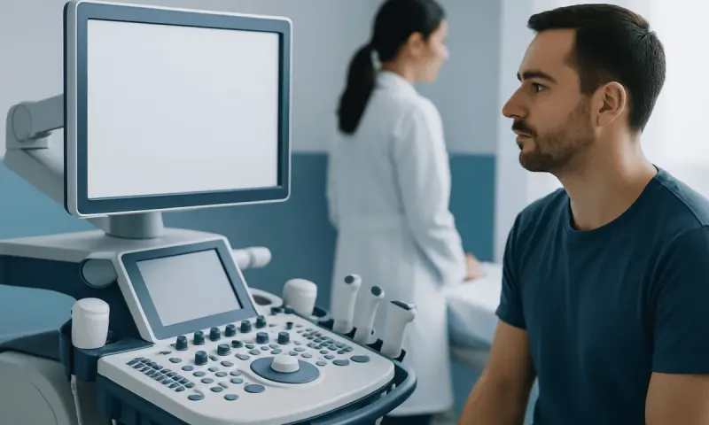
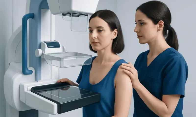
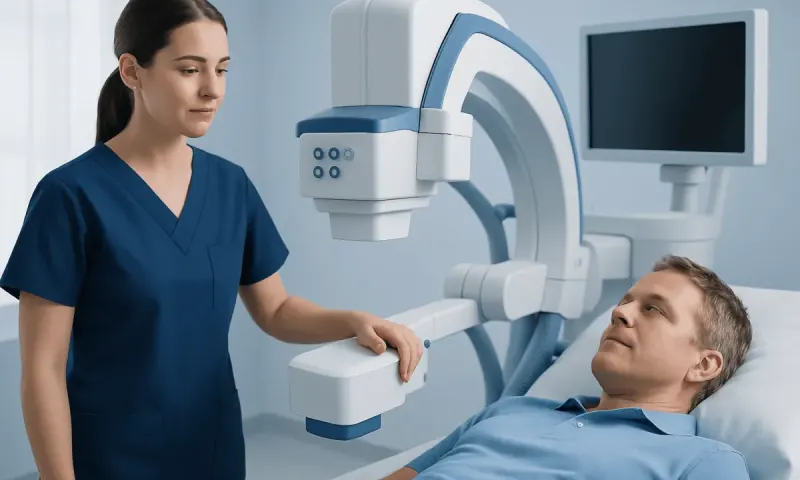
Imaging tests such as ultrasound, X-ray, mammography, computed tomography, magnetic resonance imaging and positron emission tomography can show the size, location and characteristics of an abnormal area. Imaging can help determine whether a finding appears more likely to be benign or concerning, but it often cannot establish a final diagnosis on its own.
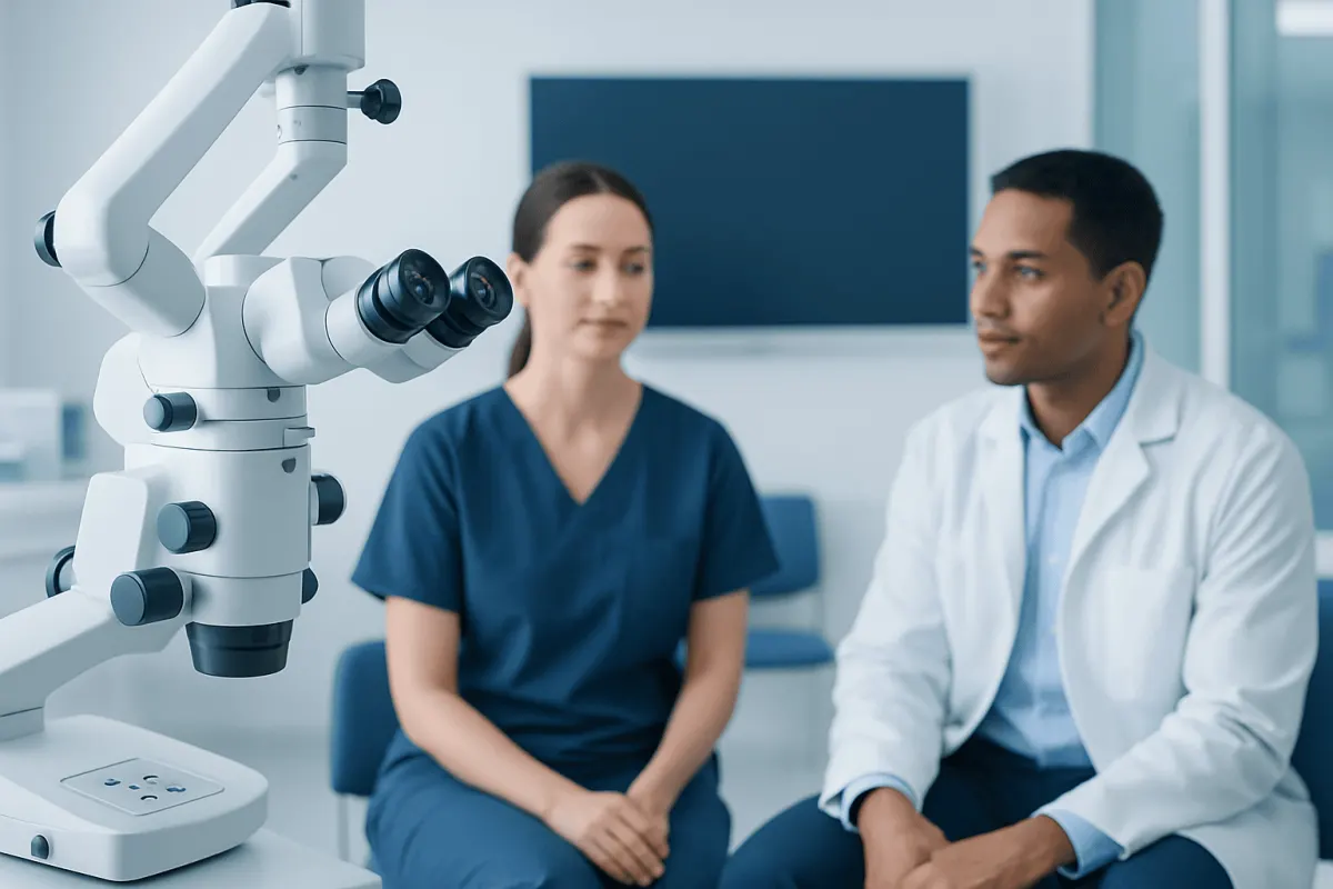
A biopsy is commonly the most reliable way to confirm cancer. A clinician removes a small sample of tissue or cells using a needle, endoscope, surgery or another technique. A pathologist examines the sample under a microscope and may use special stains, protein tests or molecular tests to identify the cell type, grade and genetic features relevant to diagnosis and treatment planning.
What Pathologists Look for in a Biopsy
Pathologists compare a tissue sample with the expected appearance of healthy tissue from the same area. They assess cell size, shape, internal structures, how often cells are dividing, and whether the normal tissue pattern has been disrupted. Cancer cells may have enlarged or irregular nuclei, variable shapes or a disorganized arrangement, but no one microscopic feature is sufficient in every case.
The pathologist also evaluates whether abnormal cells are confined to their place of origin or have invaded nearby tissue. In some cancers, the report describes grade, which reflects how closely the cells resemble normal cells. Lower-grade tumors often look more like the original tissue, while higher-grade tumors may look less differentiated; grade is not the same as stage.
Additional tests can help classify a cancer more precisely. Immunohistochemistry uses antibodies to detect proteins in cells. Molecular and genetic testing may identify gene changes, gene fusions or other biomarkers. These tests can sometimes clarify the diagnosis, estimate likely behavior or help determine whether certain treatments are appropriate.
Pathology results are interpreted with imaging and clinical information. Occasionally, more tissue, repeat testing or expert review is needed before a diagnosis is finalized. This careful process is designed to avoid making important treatment decisions based on incomplete information.
What to Do After an Abnormal Finding
An abnormal result does not automatically mean cancer. A scan may show a nodule, mass or enlarged lymph node that later proves to be benign. A screening test may need follow-up because it detects a possible change, not because it confirms a diagnosis. It is reasonable to ask what the result means, how urgent follow-up is and what test, if any, is recommended next.
If a biopsy confirms cancer, the next steps usually include determining the cancer’s type, location, grade and stage. A multidisciplinary team may include medical oncologists, surgeons, radiation oncologists, pathologists, radiologists, nurses and other specialists. Treatment may involve active surveillance, surgery, radiation therapy, systemic medicines or a combination, depending on the individual diagnosis.
For benign conditions, care may range from reassurance and observation to treatment if the finding causes pain, bleeding, pressure, functional problems or uncertainty about the diagnosis. Some precancerous changes can be monitored or treated to reduce the chance that they progress. Follow-up plans should be individualized by the treating clinician.
People can support their health by attending recommended screening, avoiding tobacco, limiting alcohol, maintaining a balanced diet and regular physical activity where possible, and discussing family history with a doctor. These measures can lower risk for some cancers, but they cannot eliminate all risk or replace appropriate medical evaluation.
When to Seek Medical Care
A person should arrange a medical review for a new lump that persists or grows, unexplained bleeding, a lasting change in bowel or bladder habits, persistent difficulty swallowing, a cough or hoarseness that does not improve, an unexplained change in a mole, or ongoing unexplained pain, fatigue or weight loss. These symptoms are common and often have non-cancer causes, but persistent changes deserve professional assessment.
More prompt care is appropriate for severe symptoms such as coughing up blood, significant bleeding, new weakness or numbness, severe headache with neurological changes, worsening shortness of breath, or intense pain. Urgent services may be needed when symptoms are sudden, severe or accompanied by confusion, fainting or inability to function safely.
People with a strong family history of cancer, a known inherited cancer-related gene variant, prior cancer treatment or a previous precancerous condition may benefit from individualized screening advice. A doctor or genetic counselor can explain which tests may be useful and when they should begin.
Acıbadem Health Point’s multidisciplinary specialists and JCI-accredited hospitals support international patients with diagnostic assessment and cancer care plans when further evaluation is needed. A qualified clinician can explain test results in the context of the person’s overall health and preferences.
Frequently asked questions
01What is the main difference between cancer cells and normal cells?
Normal cells generally grow, divide and die according to the body’s regulatory signals. Cancer cells may acquire changes that let them keep growing, resist normal cell death, invade surrounding tissue or spread to other parts of the body.
02Can doctors identify cancer from a blood test alone?
Most cancers cannot be diagnosed from a blood test alone. Blood tests may show changes that warrant further evaluation or help monitor known cancer, but imaging and tissue testing are often needed to establish a diagnosis.
03Is every abnormal cell cancerous?
No. Cells can look or behave abnormally because of inflammation, infection, injury, healing or benign growths. A pathologist and treating clinician assess the complete pattern of findings before making a diagnosis.
04Why is a biopsy important for cancer diagnosis?
A biopsy provides cells or tissue that can be examined directly by a pathologist. It can confirm whether cancer is present and often identifies the specific cancer type and features that help guide treatment.
05Can a benign tumor become cancerous?
Most benign tumors do not become cancerous. However, some conditions involve precancerous cells or lesions that may progress over time, which is why recommended follow-up and treatment should not be delayed.
06Do cancer cells always spread?
No. Some cancers are detected before they have spread, and some remain localized. The likelihood of spread depends on the cancer type, biological features, location and how early it is diagnosed.
This article is for general information only and is not a substitute for professional medical advice. Please consult a qualified doctor about your individual situation.
Oncology care in Turkey — second opinion and treatment plan
JCI-accredited · board-certified surgeons · reply within 24h
Add us as a Preferred Source to see more of our trusted health content across Google Search, AI Overviews and Discover.