Can You See Arthritis on an X-Ray?
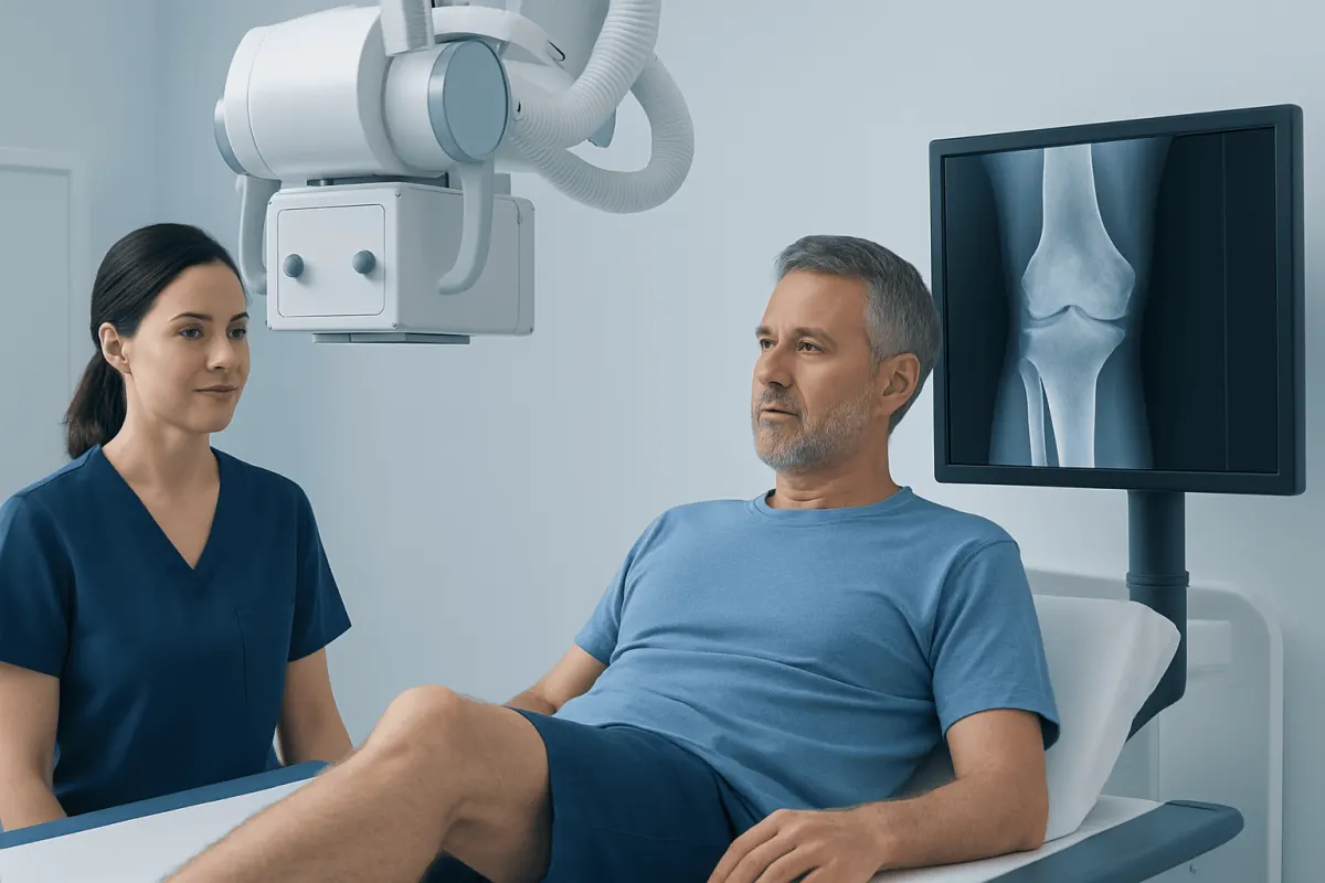
Can you see arthritis in an X-ray? X-rays can often show established arthritis through changes in bone shape, joint spacing, and alignment, but they may appear normal in early disease or in conditions affecting mainly soft tissues. A clinician combines imaging with symptoms, examination findings, and sometimes blood tests or other scans to reach an accurate diagnosis.
Overview: can you see arthritis in an X-ray?
Yes, arthritis can often be seen on an X-ray, particularly when it has caused changes to the joint structure over time. An X-ray may show narrowing of the space between bones, extra bone growth, changes in bone contour, altered alignment, or damage around the joint. These findings are especially common in osteoarthritis, the most frequent form of arthritis.
However, an X-ray is not a complete test for every type or stage of arthritis. Early arthritis may cause pain, stiffness, and swelling before visible structural changes have occurred. X-rays also do not directly show cartilage, tendons, ligaments, or most inflammation in the joint lining. For this reason, clinicians consider the person’s symptoms, physical examination, and medical history alongside imaging.
Arthritis is a broad term for conditions that affect joints. Osteoarthritis involves gradual wear and changes within a joint, while inflammatory types of arthritis result from immune-related inflammation. The pattern of symptoms and imaging findings helps guide the next steps in assessment and care.
What an arthritis X-ray can and cannot show
X-rays use a small amount of radiation to create images of dense structures, particularly bones. In a joint affected by osteoarthritis, a radiologist or clinician may look for joint-space narrowing, which can indirectly suggest cartilage loss. Other possible findings include osteophytes, often called bone spurs, increased density of bone beneath cartilage, small fluid-related bone cavities, and changes in the joint’s alignment.
In inflammatory arthritis, X-rays may eventually show erosions at bone edges, joint-space loss, or deformity. Yet early inflammatory changes are often subtle or absent on a plain X-ray. Ultrasound can identify fluid and inflammation in some joints, while MRI can show cartilage, bone marrow changes, joint lining inflammation, and other soft-tissue structures in greater detail.
An X-ray finding does not always match the amount of pain a person feels. Some people have notable X-ray changes with limited discomfort, whereas others have significant symptoms before changes are visible. Imaging is useful evidence, but it is one part of a person-centered assessment rather than a stand-alone measure of severity.
- Can show: bone shape, alignment, joint-space changes, bone spurs, fractures, and some later-stage damage.
- Cannot show well: cartilage directly, early inflammation, tendons, ligaments, nerves, and many causes of soft-tissue pain.
Can you confirm arthritis with an X-ray?

An X-ray can strongly support a diagnosis of arthritis when the image shows a characteristic pattern and the findings fit the person’s symptoms and examination. For example, joint-space narrowing and bone spurs in a painful knee, hip, hand, or spine joint may support osteoarthritis. X-rays can also help rule out or identify other concerns, such as a fracture, certain bone disorders, or joint misalignment.
Still, an X-ray alone usually cannot confirm every form of arthritis. A person may have rheumatoid arthritis, psoriatic arthritis, gout, or another inflammatory joint condition with a normal or nearly normal X-ray early on. Blood tests, joint fluid analysis, ultrasound, MRI, or referral to a rheumatologist may be appropriate depending on the symptoms.
Diagnosis is therefore based on the full picture: where symptoms occur, how long they last, whether stiffness is worse after rest, whether joints are swollen or warm, family and medical history, and the results of relevant tests. A clinician can explain what a report means in the context of the individual rather than relying only on the wording of an imaging result.
The X-ray procedure: how it works, candidacy, steps, benefits and risks
Joint X-rays are commonly used when a person has persistent pain, stiffness, reduced movement, suspected injury, swelling, or a change in joint shape. They may be taken of the hands, wrists, knees, hips, feet, shoulders, spine, or other areas. A clinician decides whether X-ray is the right first test based on the affected joint and the likely cause of symptoms.
During the procedure, the person is positioned by a radiology professional while the body part being examined is placed near an imaging detector. Several views may be needed, sometimes including standing or weight-bearing images for the knees, hips, or feet. The person needs to remain still briefly for each image. The scan itself is painless and usually takes only a short time; no needles, sedation, or recovery period are typically required.
The main benefits are that X-rays are widely available, fast, and helpful for assessing bony joint changes and alignment. The radiation exposure is low, but it is still important to tell the imaging team about a possible or confirmed pregnancy so the examination can be planned appropriately. There is no meaningful aftercare for most people: normal activities can usually resume immediately, and a clinician reviews the images and report with the patient.
When symptoms and standard imaging suggest a joint problem needing specialist assessment, orthopedic evaluation can help determine whether exercise-based care, medication, injections, rehabilitation, or another approach is appropriate.
What are the first signs of arthritis in the legs?
Early signs of arthritis in the legs can vary according to the joint involved. In the hips, knees, ankles, or feet, people may notice pain during or after walking, climbing stairs, standing for a long time, or getting up after sitting. Stiffness after rest that improves with gentle movement is also common, particularly with osteoarthritis.
Other possible early signs include reduced ability to bend or straighten a joint, clicking or grinding sensations, mild swelling, tenderness, and a feeling that the joint is less stable. Hip arthritis may be felt in the groin, buttock, thigh, or even the knee. Knee arthritis may make stairs, squatting, or getting up from a chair more difficult.
Symptoms that involve several joints, prolonged morning stiffness, visible warmth or swelling, fatigue, skin changes, or episodes of sudden intense pain may point to a condition other than typical osteoarthritis. A medical assessment is important when symptoms persist or affect daily activities, as early guidance can help preserve movement and function.
Does barometric pressure affect arthritis?
Many people report that joint pain or stiffness changes with weather, including shifts in barometric pressure, temperature, or humidity. Research has not established a consistent effect for every person, but some studies suggest that weather conditions may be associated with small changes in symptoms for some people with arthritis. Individual experiences can be genuine even when the exact biological mechanism remains uncertain.
Weather does not appear to cause arthritis or permanently damage a joint. On days when symptoms seem worse, gentle movement, warm clothing, heat or cold therapy when appropriate, regular sleep, and following an agreed care plan may help maintain comfort. Keeping a simple symptom diary can help identify personal patterns and support a useful conversation with a clinician.
If pain is increasing over time, occurring at rest, or limiting walking and self-care, it should not simply be attributed to the weather. New or changing symptoms deserve medical review to check for treatable causes.
What foods cause osteoarthritis flare-ups?
No specific food has been proven to directly cause osteoarthritis flare-ups in everyone. Osteoarthritis is primarily related to changes in joint tissues and can be influenced by age, previous injury, joint mechanics, muscle strength, genetics, and body weight. However, some people feel that highly processed foods, excess alcohol, or meals high in added sugars are associated with worse overall symptoms or energy levels.
Rather than focusing only on foods to avoid, many clinicians recommend a balanced eating pattern that supports general health and a weight that is comfortable for the individual. This may include vegetables, fruits, whole grains, beans, nuts, fish or other protein sources, and unsaturated fats. For people with weight-bearing joint osteoarthritis, gradual weight management where needed can reduce load on the hips, knees, and feet.
Food triggers are more relevant in certain other conditions, such as gout, where particular dietary choices may contribute to attacks in susceptible individuals. Before making restrictive diet changes or using supplements, a person should discuss their plans with a qualified clinician or registered dietitian, especially if they have diabetes, kidney disease, digestive conditions, or take regular medication.
When to seek medical care
Medical assessment is advisable for joint pain, stiffness, or swelling that lasts more than a few weeks, keeps returning, limits work or daily activities, or does not improve with reasonable self-care. A clinician can assess whether symptoms are likely due to arthritis, an injury, a tendon problem, a nerve-related condition, or another cause. Treatment often begins with education, activity modification, strengthening, physical therapy, and symptom-relief strategies tailored to the person.
Prompt medical care is important for a joint that becomes suddenly very painful, hot, red, or markedly swollen, particularly if fever or feeling unwell is present. Urgent assessment is also appropriate after a significant injury, inability to bear weight, a visibly deformed joint, new weakness, or loss of sensation.
For people who need coordinated evaluation, Acıbadem Health Point’s multidisciplinary specialists and JCI-accredited hospitals diagnose and treat joint conditions for international patients. Depending on the diagnosis, a plan may include physical therapy and rehabilitation to improve strength, mobility, and confidence with everyday movement.
Frequently asked questions
01Can an X-ray show early arthritis?
An X-ray may be normal in early arthritis, especially before cartilage loss, bone changes, or erosions develop. A clinician may use symptoms, examination, blood tests, ultrasound, or MRI when early inflammatory arthritis or soft-tissue disease is suspected.
02What does osteoarthritis look like on an X-ray?
Common X-ray features include narrowing of the joint space, bone spurs, changes in the bone beneath the cartilage, and altered joint alignment. The findings may occur in knees, hips, hands, feet, and the spine, among other joints.
03Why do I have joint pain if my X-ray is normal?
Pain can come from early arthritis, inflammation, tendons, ligaments, muscles, bursae, nerves, or other structures that are not well seen on a plain X-ray. A normal image is useful information, but it does not exclude all causes of joint pain.
04Do I need an MRI after an arthritis X-ray?
Not always. Many people can be assessed and managed using symptoms, examination, and X-ray findings, while MRI is generally reserved for unanswered questions or suspected soft-tissue, cartilage, or early inflammatory changes.
05Can arthritis be prevented from worsening?
Although existing joint changes cannot always be reversed, regular appropriate exercise, muscle strengthening, injury prevention, weight management when relevant, and timely treatment of symptoms can support joint function. A clinician or physiotherapist can tailor advice to the affected joint and overall health.
06How long does it take to get X-ray results for arthritis?
The images are available immediately after the examination, but the formal interpretation may take longer depending on the imaging service. The requesting clinician explains the report alongside the person’s symptoms and examination findings.
This article is for general information only and is not a substitute for professional medical advice. Please consult a qualified doctor about your individual situation.
Joint and spine care in Turkey — expert assessment & treatment
JCI-accredited · board-certified surgeons · reply within 24h
Add us as a Preferred Source to see more of our trusted health content across Google Search, AI Overviews and Discover.