Calcium Oxalate Kidney Stones: Causes and Prevention

Kidney stones and calcium oxalate are closely connected because calcium oxalate is the most common material found in urinary stones. Many small stones pass naturally, while larger, painful or obstructing stones may need a procedure; long-term prevention focuses on urine volume, food choices and identifying individual risk factors.
Overview: kidney stones and calcium oxalate
Kidney stones and calcium oxalate are linked because calcium oxalate is the most common type of kidney stone. These crystals can form when certain substances in urine become concentrated and join together. A stone may stay in the kidney without causing symptoms, or it may move into the ureter, the narrow tube that carries urine from the kidney to the bladder.
Management depends on the stone’s size, location, symptoms, whether urine flow is blocked and whether infection is present. Small stones often pass without an operation. Larger stones, persistent pain, infection, reduced kidney function or ongoing obstruction may require a procedure to break up or remove the stone.
Having one stone does not mean another is inevitable. Stone analysis, urine testing and practical prevention measures can help a clinician identify why a calcium oxalate stone formed and lower the chance of recurrence.
Symptoms and why blockage matters
A kidney stone may cause no symptoms until it moves. When a stone irritates or blocks the ureter, it can cause sudden, severe pain in the side or back that may move toward the lower abdomen or groin. The pain often comes in waves. Nausea, vomiting, urinary urgency, burning during urination and blood in the urine can also occur.
The severity of pain does not reliably show the stone’s size. A small stone can be very painful if it blocks a narrow part of the ureter, while a larger stone in the kidney may be quiet. Doctors also look for signs that urine is not draining properly, as prolonged obstruction can affect kidney function.
Fever, chills, feeling unwell or confusion together with urinary symptoms can indicate infection behind an obstruction. This situation needs urgent medical care because drainage of the kidney and antibiotics may be necessary.
How calcium oxalate stones form and who is at risk

Calcium oxalate stones develop when calcium and oxalate in urine become concentrated enough to form crystals. Low urine volume, usually from not drinking enough fluid or losing fluid through sweating, vomiting or diarrhea, is one of the most important contributors. Urine also contains natural inhibitors of crystallization, including citrate; low citrate can increase stone risk.
Risk can be influenced by high sodium intake, high amounts of animal protein, a diet very high in oxalate-rich foods, obesity, certain bowel conditions, repeated urinary infections and some medicines. Family history and medical conditions that affect calcium, uric acid or intestinal absorption may also matter. A clinician can review personal risks rather than assuming that one food caused a stone.
Dietary calcium should not usually be eliminated. Normal calcium intake with meals can bind oxalate in the gut and may reduce the amount absorbed into the bloodstream. Calcium supplements may need individualized advice, especially for people who have had stones or have another medical reason to take them.
- High-oxalate foods can include spinach, rhubarb, beets, nuts, wheat bran and some chocolate products.
- High sodium intake can increase calcium excretion in urine.
- Very high doses of vitamin C may raise urinary oxalate in some people.
Diagnosis and stone-removal procedures
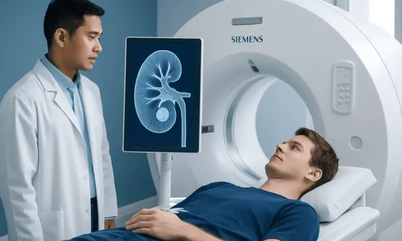
Evaluation usually begins with a medical history, examination, urine testing and blood tests when needed. Imaging helps confirm the stone’s location, size and any blockage. Non-contrast computed tomography is highly detailed in many situations, while ultrasound may be preferred in selected patients, including some pregnant patients, to avoid radiation.
If a stone is passed or removed, laboratory analysis can identify its composition. For recurrent stones, young age at onset, bilateral stones or higher-risk medical conditions, clinicians may recommend 24-hour urine collections. These tests measure factors such as urine volume, calcium, oxalate, citrate, sodium and uric acid, helping to guide prevention.
Procedures are selected according to the stone and the person’s health. Shock wave lithotripsy uses sound waves from outside the body to fragment selected stones. Ureteroscopy involves passing a small instrument through the urinary tract to remove or break up a ureter or kidney stone, often with laser energy. For very large or complex kidney stones, percutaneous nephrolithotomy removes stones through a small incision in the back. In urgent obstruction with infection, a ureteric stent or a drainage tube through the skin may first be used to drain urine safely.
These approaches are part of individualized kidney stone treatment. The treating urologist explains the expected benefit, alternatives, anesthesia, possible need for a temporary stent and follow-up imaging.
Recovery and results after a kidney stone
Recovery depends on whether the stone passed naturally or was treated with a procedure. After a small stone passes, soreness and urinary discomfort may settle within days, although fatigue can last longer after severe pain, vomiting or poor fluid intake. After ureteroscopy or shock wave lithotripsy, mild blood in the urine, burning and passing small fragments can occur temporarily. A stent can cause urinary frequency, urgency or flank discomfort until it is removed.
After percutaneous nephrolithotomy, recovery usually takes longer because it is a more invasive procedure. Follow-up is important to confirm that the stone has cleared or that remaining fragments are clinically insignificant. The care team may also repeat imaging to make sure urine drainage has returned to normal.
How long does it take for your kidneys to heal after a kidney stone?
If a stone did not cause significant obstruction, infection or underlying kidney disease, the kidneys often return to usual function after the stone passes or is removed. Symptoms can improve over days, while recovery from a period of obstruction may take days to weeks. The timeline is less predictable when obstruction was prolonged, both kidneys were affected, there was infection or there is pre-existing kidney disease; blood and urine tests can help monitor recovery.
People should follow their clinician’s instructions on fluid intake, activity, pain medicines and stent removal. New fever, worsening pain, inability to urinate or persistent vomiting should not be treated as routine recovery symptoms.
Can anything dissolve calcium oxalate kidney stones?
Calcium oxalate kidney stones generally cannot be dissolved with medicines or home remedies. This differs from some uric acid stones, which may sometimes dissolve when urine is made less acidic under medical supervision. Drinking fluids is useful for preventing concentration of urine and may support passage of a small stone, but it does not dissolve a formed calcium oxalate stone.
For a small ureteric stone, a clinician may recommend pain control, anti-nausea medicines and, in selected cases, a medicine that relaxes the ureter to support passage. If the stone is too large to pass, is causing ongoing obstruction or symptoms are difficult to control, a procedure may be the safer option. Herbal products and unproven “stone dissolving” treatments should not replace assessment, particularly if symptoms are severe.
What is the prognosis for people with calcium oxalate stones?
The outlook is usually good when stones are diagnosed and managed promptly. Many people recover fully from an individual episode, and kidney function is often preserved when blockage and infection are treated without delay. However, calcium oxalate stones can recur, so identifying modifiable risks and attending follow-up are important parts of long-term care.
People with recurrent stones may benefit from a tailored prevention plan that includes laboratory evaluation, nutrition advice and, where appropriate, medicines prescribed for their specific urine abnormalities. Regular review is especially valuable for people with a single kidney, chronic kidney disease or frequent stone episodes.
Prevention, daily habits and the “worst” stone type
Prevention is most effective when it is based on stone analysis and urine testing, but several measures are helpful for many people. Drinking enough fluid to produce pale urine through most of the day is a common goal, unless a clinician has advised fluid restriction for another condition. Reducing excess salt, eating a balanced diet with normal food-based calcium intake and avoiding very high amounts of oxalate-rich foods can be useful.
Rather than broadly restricting foods, it may help to pair oxalate-containing foods with calcium-containing foods during meals. Limiting excessive animal protein and avoiding unnecessary high-dose vitamin C supplements may also be advised for some people. A dietitian or clinician can adapt recommendations for diabetes, bowel disease, osteoporosis, kidney disease or other individual needs.
What’s the worst type of kidney stone to have?
There is no single “worst” kidney stone for every person. Any stone can become serious if it blocks urine flow or is associated with infection. Infection-related struvite stones may grow quickly and can form large branching stones, while cystine stones can recur and may be difficult to manage. Calcium oxalate stones are common and can be very painful, but they are often manageable with timely treatment and prevention.
At Acıbadem Health Point, multidisciplinary specialists in JCI-accredited hospitals can assess kidney stones, coordinate imaging and laboratory evaluation, and discuss appropriate treatment and prevention plans for international patients.
When to seek medical care
Medical assessment is appropriate for suspected kidney stone symptoms, especially first-time severe flank pain, visible blood in the urine, recurring symptoms or pain that is not improving. A clinician can confirm whether a stone is present and exclude other causes of abdominal or back pain that may need different care.
Urgent care is needed for fever or chills with possible stone symptoms, severe or worsening pain, repeated vomiting or inability to keep fluids down, inability to pass urine, marked weakness or confusion. Prompt assessment is also important for pregnant people, people with one functioning kidney, known kidney disease or a weakened immune system.
After a stone has passed or been treated, follow-up helps confirm recovery and supports prevention. Bringing in a collected stone, if possible, can allow analysis; it should be handled according to the healthcare team’s instructions.
Frequently asked questions
01Are calcium oxalate kidney stones common?
Yes. Calcium oxalate is the most common material found in kidney stones. It forms when urine becomes concentrated with calcium and oxalate and conditions favor crystal growth.
02Can drinking water make a calcium oxalate stone pass?
Adequate fluid intake may help a small stone pass and helps prevent future stones by diluting urine. It does not guarantee passage, and it cannot dissolve an established calcium oxalate stone. A clinician should guide care when pain is severe or there are signs of blockage or infection.
03Should people with calcium oxalate stones avoid calcium?
Usually, no. Normal dietary calcium consumed with meals can reduce oxalate absorption from the gut. People should ask their clinician before making major changes to calcium intake or using calcium supplements.
04Is blood in the urine always caused by a kidney stone?
No. A kidney stone can cause blood in the urine, but infections, kidney disease and other urinary conditions can also do so. Visible blood in urine should be assessed by a healthcare professional.
05Can calcium oxalate stones come back after removal?
Yes, recurrence is possible after a stone passes or is removed. Stone analysis, 24-hour urine testing when appropriate and long-term fluid and dietary measures can reduce risk. Some people also need prescription prevention treatment based on their test results.
06Do all kidney stones need surgery?
No. Many small stones pass without a procedure, supported by symptom management and medical follow-up. Procedures are considered when a stone is unlikely to pass, causes persistent obstruction, is associated with infection, or produces difficult-to-control symptoms.
This article is for general information only and is not a substitute for professional medical advice. Please consult a qualified doctor about your individual situation.
Persistent digestive symptoms? Get evaluated in Turkey
JCI-accredited · board-certified surgeons · reply within 24h
Add us as a Preferred Source to see more of our trusted health content across Google Search, AI Overviews and Discover.