Brain Tumor Stages and Grades: How They Differ
Brain tumor stages are often described as grades rather than the stage I–IV system used for many other cancers. The grade, tumor type, molecular features, location and whether it can be safely removed all help the care team plan treatment and discuss outlook.
Overview: What Do Brain Tumor Stages Mean?
When people ask about brain tumor stages, they are usually seeking to understand how serious a tumor is and what may happen next. Unlike many cancers that are staged from I to IV according to tumor size and spread, most tumors that begin in the brain or spinal cord are described primarily by their grade. Grade reflects how the tumor cells appear under a microscope and how likely the tumor is to grow or return.
The World Health Organization (WHO) classification generally uses grades 1 through 4. Grade 1 tumors are usually slower growing and may sometimes be cured with complete surgery. Grade 4 tumors are more aggressive, but treatment can still help control disease, relieve symptoms and support quality of life. A grade is important, yet it is only one part of the picture.
Brain tumors can be primary, meaning they started in the brain, or secondary (metastatic), meaning cancer spread to the brain from another part of the body. Metastatic brain tumors are assessed in relation to the original cancer as well as their number, size, location and response to treatment. This article focuses mainly on how primary brain tumors are graded.
Brain Tumor Grades 1 to 4
Grade 1 tumors are usually slow growing and have cells that look relatively close to normal. Examples include some meningiomas and pilocytic astrocytomas. If a grade 1 tumor can be safely removed completely, further treatment may not be needed, although follow-up scans remain important.
Grade 2 tumors tend to grow more slowly than higher-grade tumors but can extend into nearby brain tissue. They may recur after treatment and, in some cases, change over time. Some diffuse gliomas fall into this group. Their behavior is influenced strongly by molecular findings, not only their microscopic appearance.
Grade 3 tumors have more abnormal cells and are generally faster growing. They commonly require a combination of treatments after diagnosis, such as surgery followed by radiotherapy and drug treatment. Grade 4 tumors are the highest grade. They are typically fast growing and need prompt, coordinated specialist care. Glioblastoma is a well-known example of a grade 4 diffuse glioma.
Grades should not be interpreted as a fixed timetable or a personal prediction. People with tumors of the same grade can have different experiences because tumor biology, age, overall health, location, treatment response and access to specialist care all matter.
Why Modern Classification Is More Than a Grade
Today, specialists do not rely on grade alone. The WHO classification combines what the tumor looks like under the microscope with molecular and genetic features found in tumor cells. For certain gliomas, tests for changes such as IDH mutation, 1p/19q codeletion and MGMT promoter methylation can help clarify the diagnosis, estimate likely behavior and inform treatment discussions.
Location is also crucial. A small tumor in an area controlling speech, movement, vision or breathing may cause significant symptoms or be difficult to remove safely. In contrast, another tumor with the same grade in a more accessible area may have different treatment options. MRI findings, the amount of tumor that can be removed and the person’s neurological function are considered together.
For this reason, terms such as “early stage” and “late stage” can be misleading for primary brain tumors. Brain tumors do not usually spread through the body in the same pattern as many other cancers. A multidisciplinary neuro-oncology team can explain the pathology report in practical terms and relate it to the individual treatment plan.
How Brain Tumors Are Diagnosed and Classified
Assessment often starts with a medical history and neurological examination. Symptoms may include persistent or changing headaches, seizures, weakness, balance difficulties, personality or memory changes, speech problems, visual changes, nausea or vomiting. These symptoms can also have many non-tumor causes, so evaluation is needed rather than self-diagnosis.
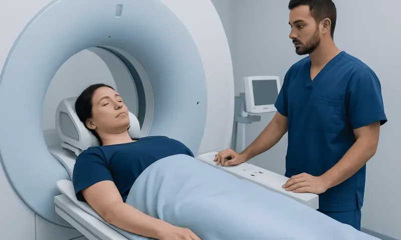
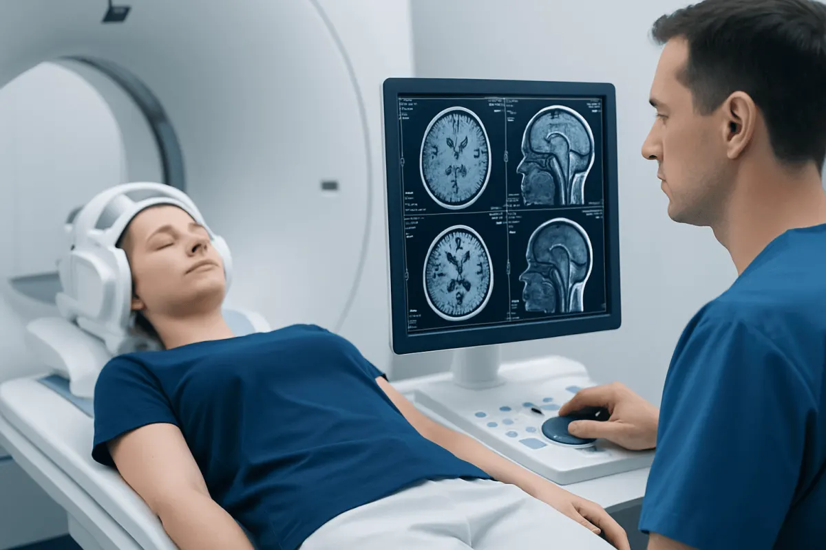
MRI with contrast is the main imaging test used to examine a suspected brain tumor. CT may be used in urgent situations or when MRI is not suitable. Imaging can show the tumor’s size, position and effect on nearby structures, but it cannot always identify the precise tumor type or grade.
In many cases, diagnosis is confirmed through surgery to remove tissue or through a stereotactic biopsy. A neuropathologist examines the sample and may arrange molecular testing. The team then brings together the scan results, pathology findings and the person’s overall health to make a diagnosis and recommend next steps. More detailed information about assessment and care is available through brain tumor treatment.
Treatment Options: How Care Is Planned
Treatment is individualized rather than determined by grade alone. Some small, slow-growing tumors that are not causing symptoms may be monitored with scheduled MRI scans. This active surveillance approach can be appropriate when immediate treatment is unlikely to offer more benefit than careful observation.
When treatment is needed, surgery is often considered first if the tumor can be reached with an acceptable level of risk. The aim may be complete removal, removal of as much tumor as safely possible, or obtaining tissue for diagnosis. Eligibility depends on tumor location, size, relationship to important brain functions and the patient’s general health. Advanced surgical planning, mapping techniques and image guidance may help protect neurological function.
Radiotherapy uses focused radiation to damage tumor cells and may be recommended after surgery, as a primary treatment when surgery is not suitable, or for tumor recurrence. Drug treatments may include chemotherapy, targeted therapy or other medicines selected for the tumor type and molecular profile. Some patients may also be eligible for clinical trials. Steroids, anti-seizure medicines, rehabilitation and psychological support can be important parts of care.
After treatment begins, regular MRI scans and clinical visits monitor the response and identify recurrence or treatment effects. A treatment plan can change over time as new scan results, symptoms or pathology information become available. The goal is to balance tumor control with safety, independence and quality of life.
Surgery: What to Expect, Benefits and Risks
Brain tumor surgery is usually performed by a neurosurgical team under general anesthesia, although an awake procedure may be considered for selected tumors close to areas responsible for language or movement. Before surgery, the team reviews scans, explains expected goals and discusses whether removal, biopsy or both are planned. Not every tumor is suitable for surgery, particularly if it is located in a highly sensitive area or if another treatment offers a safer option.
During a craniotomy, the surgeon creates an opening in the skull to reach the tumor and removes tissue while protecting normal brain structures as much as possible. In a stereotactic biopsy, a small sample is taken through a precisely planned pathway. The tissue is then examined to establish the diagnosis and grade. The procedure approach depends on the tumor’s position and the information needed.
Potential benefits include reducing pressure on the brain, improving symptoms, providing an accurate diagnosis and decreasing the amount of tumor that remains for further treatment. Risks vary by procedure and tumor location and can include bleeding, infection, seizures, blood clots, stroke-like deficits, swelling, temporary or lasting changes in speech, movement, sensation, memory or vision, and anesthesia-related complications.
Recovery differs widely. Hospital stay may range from a short period after a biopsy to several days or longer after major surgery. Fatigue, headaches and scalp discomfort are common early on, while neurological recovery can take weeks or months. Follow-up appointments review wound healing, pathology results and plans for rehabilitation, scans or additional treatment.
Living With a Brain Tumor and Ongoing Follow-Up
A brain tumor diagnosis can affect physical abilities, emotions, work, family life and daily routines. Supportive care should begin early and continue alongside tumor-directed treatment. Depending on symptoms, helpful services may include physiotherapy, occupational therapy, speech and language therapy, neuropsychology, nutrition advice, social work support and palliative care.
People with seizures should follow their clinician’s guidance about medicines, driving, swimming alone, working at heights and other safety-sensitive activities. Family members or caregivers can be helpful in noticing subtle changes in speech, behavior, balance or memory that may need to be reported to the healthcare team.
It is reasonable to ask for a clear explanation of the tumor’s integrated diagnosis, grade, treatment aim, likely benefits and possible side effects. Keeping a list of questions and bringing a trusted support person to appointments may make discussions easier. Acıbadem Health Point’s multidisciplinary specialists and JCI-accredited hospitals diagnose and treat brain tumors for international patients.
When to Seek Medical Care
Anyone with new, persistent or worsening neurological symptoms should arrange medical assessment. Symptoms that deserve attention include a new seizure, a persistent headache that is changing in pattern, progressive weakness or numbness, new difficulty speaking, changes in vision, unexplained balance problems or notable changes in memory or personality.
Emergency care is needed for a first seizure, sudden severe headache unlike usual headaches, loss of consciousness, sudden weakness on one side of the body, new confusion, persistent vomiting with severe headache, or rapidly worsening neurological symptoms. These signs are not always caused by a brain tumor, but urgent evaluation is important.
For people already diagnosed with a brain tumor, the treating team should be contacted promptly about new symptoms, seizures, medication side effects, fever after surgery, wound redness or drainage, or a meaningful change in function. Timely communication can help clinicians identify treatable complications and adjust supportive care.
Frequently asked questions
01Are brain tumor stages the same as brain tumor grades?
For most primary brain tumors, doctors use grades rather than traditional cancer stages. Grade describes the tumor’s microscopic features and expected behavior, while a full assessment also includes molecular findings, location and imaging results.
02Does a grade 4 brain tumor mean there are no treatment options?
No. Grade 4 indicates a high-grade, faster-growing tumor, but treatment can still be used to control the tumor, manage symptoms and maintain quality of life. The plan may include surgery, radiotherapy, drug treatment and supportive therapies, depending on the individual situation.
03Can a brain tumor grade change over time?
Some tumors can recur or develop features of a higher grade over time. This is one reason ongoing MRI monitoring and specialist follow-up are important, even after initial treatment.
04Can an MRI scan determine the grade of a brain tumor?
MRI can suggest whether a tumor may be more or less aggressive and helps guide treatment planning. However, a definite grade often requires tissue obtained through surgery or biopsy and examined by a neuropathologist.
05What factors affect outlook besides brain tumor grade?
Outlook is influenced by the exact tumor type, molecular profile, location, age, general health, neurological function, amount safely removed and response to treatment. A specialist team can explain what these factors mean for a particular person.
06Is it possible to live well with a brain tumor?
Many people continue meaningful daily activities during and after treatment, although needs and abilities vary. Symptom control, rehabilitation, emotional support and regular follow-up can all help support function and quality of life.
This article is for general information only and is not a substitute for professional medical advice. Please consult a qualified doctor about your individual situation.
Joint and spine care in Turkey — expert assessment & treatment
JCI-accredited · board-certified surgeons · reply within 24h
Add us as a Preferred Source to see more of our trusted health content across Google Search, AI Overviews and Discover.