Bone Demineralization: Causes, Testing, and Care
Bone demineralization means that bones have lost some of the minerals, mainly calcium and phosphate, that help make them strong. It may be found before symptoms develop, and early assessment can identify reversible contributors and lower the risk of fractures.
What bone demineralization means
Bone demineralization is the gradual reduction of minerals within bone, especially calcium and phosphate. These minerals are part of the hard structure that gives bones their strength. Bone is living tissue that is continuously broken down and rebuilt; demineralization can occur when breakdown happens faster than rebuilding over time.
The term may appear on an imaging report, laboratory discussion or clinical note. It does not always mean a person has osteoporosis, and it does not by itself predict whether a fracture will occur. Its significance depends on the degree of mineral loss, a person’s age, medical history, medicines and overall risk of falling or fracturing a bone.
Reduced bone mineral density is often described as osteopenia when it is mild to moderate, or osteoporosis when it is more pronounced. Osteoporosis can make fractures more likely, particularly at the hip, spine and wrist. Osteoporosis is a specific condition that requires individualized assessment and management.
How bone strength changes over time
Bone health reflects more than mineral density alone. Bone size, internal structure, the rate of remodeling, muscle strength, balance and fall risk all influence how likely a bone is to break. A bone density result is therefore an important piece of information, but clinicians interpret it alongside the wider clinical picture.
Most people build bone during childhood, adolescence and early adulthood. After peak bone mass is reached, gradual loss can occur as part of normal ageing. In women, the decline in estrogen around menopause can accelerate bone loss. Men may also develop low bone density, particularly with advancing age or when other risk factors are present.
Bone loss is usually silent. Many people feel well and do not know their bone density is low until a screening test, an X-ray taken for another reason or a fracture brings it to attention. Vertebral compression fractures can sometimes cause back pain, height loss or a more rounded upper back, but these signs are not present in everyone.
Causes and factors that can raise risk
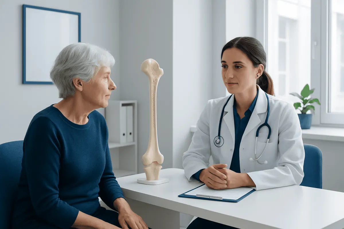
Age, family history and a previous low-trauma fracture are important risk factors. A low-trauma fracture is one that happens from a fall at standing height or less, rather than from a major injury. Smaller body frame, early menopause, removal of the ovaries before natural menopause and prolonged absence of menstrual periods may also be relevant for some people.
Nutrition and lifestyle can affect bone remodeling. Low intake or poor absorption of calcium and vitamin D, limited physical activity, smoking and heavy alcohol use may contribute to lower bone density. Long periods of immobility, including recovery from illness or injury, can also lead to bone loss because bones respond to regular loading and movement.
Some medical conditions can affect bones, including overactive thyroid or parathyroid disorders, chronic kidney or liver disease, inflammatory bowel disease, celiac disease, rheumatoid arthritis and conditions that reduce sex hormone levels. Long-term use of certain medicines, particularly corticosteroids, may increase risk. Other medicines may be relevant depending on the individual, so it is important not to stop prescribed treatment without medical advice.
A clinician may look for secondary causes when bone loss occurs at a younger age, is unexpectedly severe, progresses quickly or is associated with fractures. Identifying and treating a contributing condition can be an important part of protecting bone health.
Assessment and diagnosis
A healthcare professional will usually begin with a medical history, including fractures, menstrual or menopause history where relevant, family history, diet, physical activity, smoking, alcohol use and current medicines. They may also ask about digestive symptoms, endocrine conditions and other illnesses that can influence calcium balance or bone remodeling.
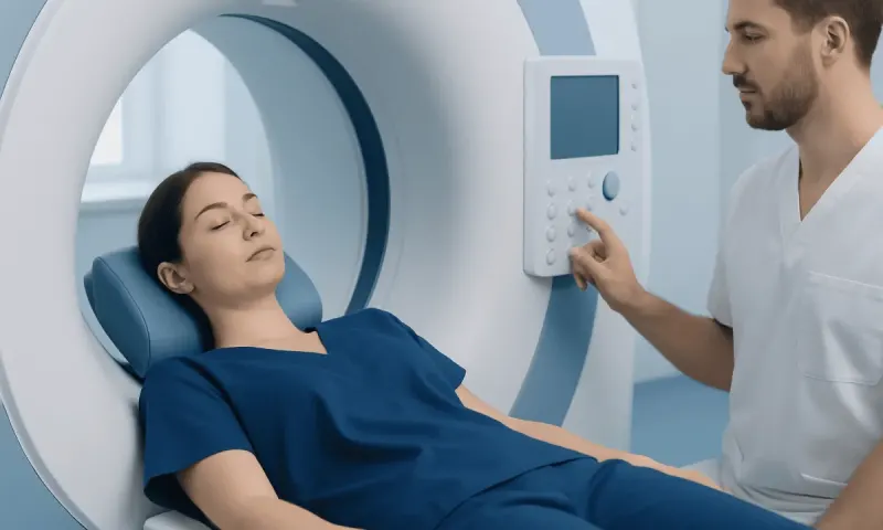
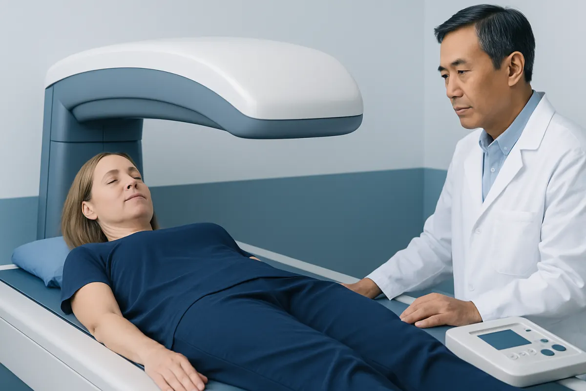
The standard test for measuring bone mineral density is a dual-energy X-ray absorptiometry scan, commonly called a DXA or DEXA scan. It is quick, painless and uses a very low dose of radiation. The scan commonly measures the hip and lower spine and can help classify bone density and estimate future fracture risk in the appropriate clinical context.
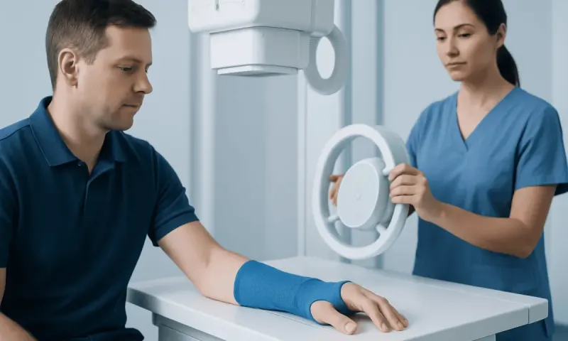
Plain X-rays can sometimes suggest demineralization, but they are not as precise as a DXA scan for measuring bone density. A clinician may recommend blood or urine tests to investigate possible contributors, such as vitamin D status, calcium levels, kidney function, thyroid function or parathyroid hormone, depending on the person’s history.
Not everyone needs routine bone density testing at the same age. Screening recommendations vary according to sex, age, menopausal status and risk factors. A doctor can advise whether testing is appropriate and how often it should be repeated.
Treatment options and monitoring
Care for bone demineralization is tailored to the cause and level of fracture risk. For some people, the main approach is improving nutrition, increasing suitable physical activity, reducing fall risk and addressing factors such as smoking or excess alcohol. If a medical condition or medicine is contributing, the clinician may treat the underlying issue or review whether alternatives are appropriate.
Calcium and vitamin D are important for bone health, but supplements are not automatically necessary for everyone. Healthcare professionals commonly encourage meeting nutritional needs through food where possible and may recommend supplements when intake is insufficient, absorption is impaired or a deficiency is identified. The safest amount depends on diet, health conditions and other medicines.
Prescription medicines that slow bone breakdown or support bone formation may be considered for people with osteoporosis, fragility fractures or a sufficiently high calculated fracture risk. The choice depends on bone density, fracture history, kidney function, other health conditions and treatment preferences. Osteoporosis treatment may include medication alongside lifestyle and fall-prevention planning.
Follow-up may involve repeat DXA testing after an interval chosen by the clinician, rather than frequent scans. Monitoring also includes checking for new fractures, reviewing medicines and reassessing risks as health circumstances change.
Everyday steps that support bone health
Regular movement helps maintain bone and muscle strength. Weight-bearing activities, such as walking, stair climbing, dancing or jogging when appropriate, place healthy load on bones. Resistance training can strengthen muscles and support balance. The right activity depends on current fitness, joint health, fracture history and mobility, especially for people who already have osteoporosis or spinal fractures.
A balanced eating pattern should provide enough protein as well as calcium-containing foods and vitamin D from safe sun exposure, diet or supplements when advised. Dairy foods, fortified alternatives, leafy green vegetables, beans, nuts, seeds and certain fish can contribute calcium, although the best choices vary with dietary needs. People with kidney disease or conditions affecting calcium should seek individualized advice before changing supplements.
Fall prevention is a practical part of fracture prevention. Helpful measures include having vision checked, reviewing medicines that may cause dizziness, using stable footwear, improving lighting, removing loose rugs and maintaining strength and balance. A physiotherapist can recommend exercises and mobility strategies when falls or unsteadiness are concerns.
- Avoid smoking and seek support to stop if needed.
- Limit alcohol according to local health guidance.
- Discuss persistent digestive symptoms or unexplained weight loss with a clinician.
- Continue prescribed medicines unless the prescribing clinician advises otherwise.
When to seek medical care
Medical advice is appropriate after a fracture caused by a minor fall or everyday movement, especially after midlife. Prompt assessment is also sensible for new, persistent back pain after a minor strain or fall, unexplained loss of height, a noticeable change in posture or repeated falls. These symptoms have many possible causes, but they deserve evaluation.
People taking long-term corticosteroids, those with early menopause or prolonged loss of periods, and those with conditions that affect nutrient absorption or hormones should ask whether bone health assessment is needed. A person with a parent or sibling who had a hip fracture, or with a personal history of fragility fracture, may also benefit from a risk review.
Urgent medical care is needed after a significant fall, severe pain, inability to bear weight, sudden weakness or symptoms suggesting a serious injury. Acıbadem Health Point’s multidisciplinary specialists and JCI-accredited hospitals can assess bone health concerns and coordinate care for international patients when needed.
Frequently asked questions
01Is bone demineralization the same as osteoporosis?
Not exactly. Bone demineralization is a descriptive term for loss of mineral from bone, while osteoporosis is a defined condition involving low bone density and increased fracture risk. A DXA scan and clinical risk assessment help determine whether osteoporosis is present.
02Can bone demineralization be reversed?
Whether bone density can improve depends on the cause, age, hormone status, medicines and overall fracture risk. Treating an underlying problem, improving nutrition and exercise, and using bone-specific medicines when indicated may stabilize bone loss and sometimes improve density. A clinician can set realistic goals based on the individual situation.
03What test checks for bone demineralization?
A DXA, also called a DEXA scan, is the standard test for measuring bone mineral density. It is painless and usually scans the hip and lower spine. Blood tests may also be used to look for conditions that contribute to bone loss.
04Does low vitamin D cause bone demineralization?
Vitamin D helps the body absorb calcium and supports normal bone mineralization. Low vitamin D can contribute to weaker bones, particularly when it is prolonged or combined with low calcium intake or another health condition. Testing and treatment should be guided by a healthcare professional.
05Can exercise help low bone density?
Yes, appropriately chosen weight-bearing and resistance exercises can support bone and muscle strength. Balance training can also reduce fall risk. People with fractures, severe osteoporosis, pain or major mobility limitations should seek tailored guidance before beginning a new program.
06Who should discuss bone density screening with a doctor?
Older adults, postmenopausal women, people with a previous fragility fracture and those using long-term corticosteroids should discuss screening. It may also be appropriate for people with early menopause, low body weight, certain chronic conditions or a strong family history of fractures. The decision is based on individual risk factors.
This article is for general information only and is not a substitute for professional medical advice. Please consult a qualified doctor about your individual situation.
Joint and spine care in Turkey — expert assessment & treatment
JCI-accredited · board-certified surgeons · reply within 24h
Add us as a Preferred Source to see more of our trusted health content across Google Search, AI Overviews and Discover.