Aortic Aneurysm Diagnosis: Imaging and Testing
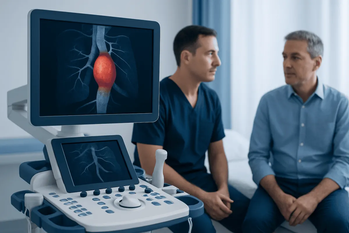
Aortic aneurysm diagnosis is usually confirmed with imaging that shows the aorta’s size, location and shape. Ultrasound, CT angiography, MRI and echocardiography help clinicians determine whether monitoring, further assessment or treatment is appropriate.
Overview: How Aortic Aneurysms Are Diagnosed
Aortic aneurysm diagnosis involves imaging tests that identify an enlarged area of the aorta, measure it accurately and show whether nearby branches or heart structures are involved. The right test depends on where the suspected aneurysm is located, whether symptoms are present and whether the situation needs urgent assessment.
The aorta is the body’s largest artery. It carries blood from the heart through the chest and abdomen. An aneurysm occurs when part of its wall weakens and widens. Aneurysms may affect the thoracic aorta in the chest, the abdominal aorta, or both. They can remain stable for years, so diagnosis also includes establishing a reliable baseline for future comparison.
Imaging does more than confirm an aneurysm. It helps the clinical team evaluate its diameter, length, shape, growth over time and relationship to important arteries. These findings guide decisions about surveillance, risk-factor management and, when appropriate, referral for aortic aneurysm treatment.
Why an Aortic Aneurysm May Be Suspected
Most aortic aneurysms do not cause noticeable symptoms, particularly when they are small. They are often discovered incidentally during an ultrasound, chest X-ray, CT scan or MRI performed for another health concern. Some are found through screening in people with risk factors.
A clinician may consider testing when a person has a family history of aortic aneurysm or dissection, a known connective-tissue condition, high blood pressure, a history of smoking, atherosclerosis, or certain heart valve differences such as a bicuspid aortic valve. Age and sex can also influence screening recommendations, especially for abdominal aortic aneurysm.
When symptoms occur, they may include persistent chest, back or abdominal discomfort, a pulsating feeling in the abdomen, breathlessness, hoarseness, coughing or difficulty swallowing. These symptoms are not specific to aneurysms and often have other causes, but they should be assessed by a qualified clinician, particularly in someone with known risk factors.
Initial Assessment and Clinical Evaluation

The diagnostic process usually begins with a medical history and physical examination. The clinician asks about symptoms, cardiovascular health, previous imaging, tobacco exposure, family history and any conditions that may affect the aortic wall. They may also review medicines and assess blood pressure.
During the examination, the clinician listens to the heart and checks pulses and circulation. In some people with an abdominal aneurysm, an enlarged pulsating area may be felt in the abdomen. However, a normal examination does not exclude an aneurysm, particularly in the chest or in people with more abdominal tissue.
Blood tests may be used to check kidney function, blood count and other aspects of general health before contrast-enhanced imaging or a procedure. They cannot diagnose an aneurysm on their own. Their role is to help the team select and prepare for imaging safely.
- Medical history: identifies inherited and cardiovascular risk factors.
- Physical examination: may detect signs but cannot reliably rule out an aneurysm.
- Blood pressure assessment: is important because hypertension can contribute to aortic disease.
- Laboratory tests: support safe use of contrast agents and treatment planning.
Imaging Tests Used for Aortic Aneurysm Diagnosis
Ultrasound uses sound waves to create images without radiation or injections. It is often the first test for suspected abdominal aortic aneurysm and is commonly used for follow-up because it is quick, painless and effective for measuring the abdominal aorta. A transesophageal echocardiogram, performed with an ultrasound probe passed into the esophagus after sedation, may provide detailed views of parts of the thoracic aorta and heart.
CT angiography (CTA) combines CT imaging with contrast dye administered through a vein. It produces detailed cross-sectional and three-dimensional images of the aorta, including its branches. CTA is frequently used when symptoms need urgent evaluation, when the chest aorta must be assessed, or when detailed measurements are needed for procedural planning.
MRI or MR angiography (MRA) uses magnetic fields rather than ionizing radiation. It can give highly detailed images of the aorta and may be particularly useful for long-term monitoring in selected patients or when repeated radiation exposure is a concern. MRI may take longer than CT and may not be suitable for everyone, such as people with some implanted devices or severe claustrophobia.
A standard echocardiogram through the chest wall may assess the aortic root, ascending aorta and aortic valve. Chest X-rays can occasionally suggest an enlarged aorta but cannot confirm or accurately measure an aneurysm. The imaging team selects the test that provides the necessary information as efficiently and safely as possible.
What Happens During CT, MRI and Ultrasound Imaging
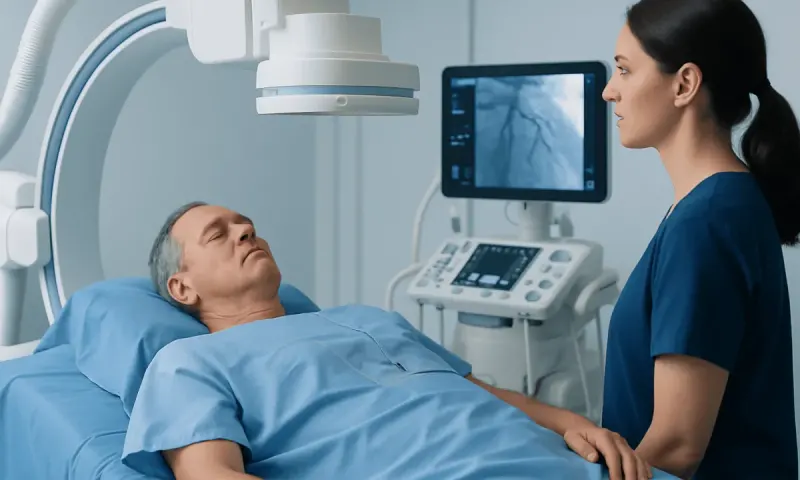
For an ultrasound, the person usually lies on an examination table while a small handheld probe is moved over the abdomen or chest. Gel is applied to improve contact with the skin. The examination is generally comfortable and often takes less than an hour, depending on the area being assessed.
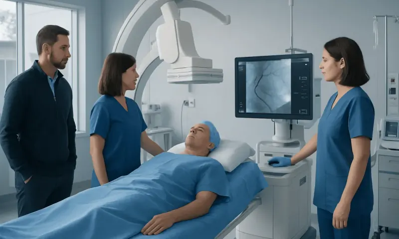
For CT angiography, a cannula is placed in a vein so contrast dye can be injected. The person lies still on a table that moves through the scanner. The scan itself is brief. Some people experience a temporary warm sensation or metallic taste when contrast is given. The radiology team checks for relevant allergies, kidney concerns and previous contrast reactions beforehand.
For MRI, the person lies on a table that moves into the scanner. The machine makes loud tapping sounds, and ear protection is provided. Contrast may or may not be needed. The examination commonly takes longer than CT, and the team can communicate with the person throughout the scan.
In urgent situations, rapid CT access may be used because it can quickly show the size and extent of an aortic problem. Imaging is interpreted by appropriately trained radiologists and reviewed alongside the person’s symptoms, examination findings and prior scans.
Understanding Results and Planning the Next Steps
After an aneurysm is identified, the report describes its location and maximum diameter. It may also describe the shape of the enlargement, whether there is thrombus within the aneurysm, involvement of branch arteries and signs of complications. Comparing current imaging with older scans is important because growth rate can affect management.
A diagnosis does not automatically mean surgery or an endovascular procedure is needed. Many aneurysms are monitored with scheduled imaging and managed by controlling blood pressure, avoiding tobacco and addressing cardiovascular risk factors. Follow-up intervals are individualized according to aneurysm type, size, growth and the person’s overall health.
If imaging suggests that treatment should be considered, the team discusses the available approaches, including open surgery and endovascular repair where suitable. Planning may involve detailed CTA or MRA measurements to assess whether a minimally invasive device can be safely positioned. This is part of the broader care pathway for aortic aneurysm.
Potential benefits of accurate imaging include timely recognition of a changing aneurysm, personalized surveillance and well-informed treatment planning. Imaging also has limitations and risks: CT involves radiation, contrast can occasionally affect kidney function or cause a reaction, and MRI may not be appropriate in all circumstances. These factors are weighed carefully against the clinical need.
Recovery, Follow-Up and Everyday Self-Care
Most diagnostic imaging does not require recovery time. After ultrasound, people can usually return to normal activities immediately. Following CT or MRI with contrast, the care team may advise drinking fluids if appropriate and may provide specific instructions for people with kidney disease or diabetes.
If a transesophageal echocardiogram is performed, recovery includes observation until sedation wears off. The throat can feel mildly sore for a short time, and the person should arrange transportation home because driving is not recommended after sedation.
Once an aneurysm has been diagnosed, follow-up imaging is one of the most important parts of care. Attending scheduled scans enables clinicians to identify changes before they cause problems. People should also work with their healthcare team on blood pressure control, cholesterol management when indicated, smoking cessation, regular activity suited to their condition and a heart-healthy eating pattern.
Acıbadem Health Point’s multidisciplinary specialists and JCI-accredited hospitals support international patients with diagnostic imaging, cardiovascular assessment and individualized aortic care planning.
When to Seek Medical Care
Anyone with a known aortic aneurysm should keep scheduled appointments and contact their clinician if new or persistent symptoms develop. It is also sensible to request medical advice about screening when there is a close family history of aortic aneurysm, dissection or certain inherited connective-tissue conditions.
Emergency medical care is needed for sudden, severe or tearing chest, upper back or abdominal pain; fainting; sudden weakness; marked shortness of breath; or signs of shock such as cold, clammy skin and confusion. These symptoms can have several serious causes, including an acute aortic problem, and should not be managed at home or by waiting for a routine appointment.
For non-urgent concerns, a primary care clinician, cardiologist or vascular specialist can determine whether imaging is needed. Prompt assessment is especially important if symptoms are new, worsening or associated with a known aneurysm.
Frequently asked questions
01What is the best test for aortic aneurysm diagnosis?
The best test depends on the location of the suspected aneurysm and the clinical situation. Ultrasound is commonly used for abdominal aortic aneurysms, while CT angiography, MRI and echocardiography can provide more detailed views of the chest aorta and surrounding structures.
02Can an aortic aneurysm be diagnosed with a blood test?
No blood test can confirm an aortic aneurysm. Blood tests may be performed before contrast imaging or treatment to assess kidney function and general health, but imaging is needed to see and measure the aorta.
03Does a CT scan always require contrast dye?
Not always, but contrast-enhanced CT angiography is often preferred when detailed images of the aorta and its branches are needed. The clinical team considers kidney function, previous contrast reactions and the purpose of the scan before deciding.
04How long does an aortic aneurysm scan take?
An abdominal ultrasound may take around 20 to 60 minutes, depending on the examination. A CT scan is usually completed quickly once preparation is finished, while an MRI often takes longer because it collects detailed images over multiple sequences.
05If an aneurysm is found, will treatment be needed immediately?
Not necessarily. Many aneurysms are safely monitored with repeat imaging and risk-factor management. The need for treatment depends on factors including the aneurysm’s location, size, growth rate, symptoms and the person’s overall health.
06How often is follow-up imaging needed after diagnosis?
Follow-up timing varies considerably. A clinician uses the type and measurements of the aneurysm, changes seen on previous scans and individual health factors to create an imaging schedule.
This article is for general information only and is not a substitute for professional medical advice. Please consult a qualified doctor about your individual situation.
Explore treatments in Turkey — costs, top hospitals & a free quote
JCI-accredited · board-certified surgeons · reply within 24h
Add us as a Preferred Source to see more of our trusted health content across Google Search, AI Overviews and Discover.