X-Rays vs CT Scans: Which Image Shows More?
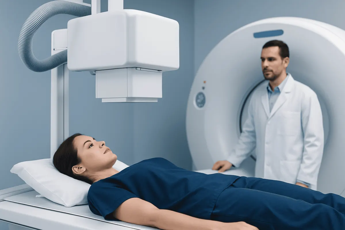
X-rays and CT scans both use radiation to create medical images, but they serve different purposes. An X-ray is usually a quick first test for bones or the chest, whereas a CT scan creates detailed cross-sectional views that can better assess internal organs, complex injuries, bleeding, infection, and some cancers.
X Rays vs CT Scans: A Side-by-Side Comparison
In the comparison of x rays vs CT scans, neither test is universally better. An X-ray is a fast, widely available imaging test that uses a small amount of ionizing radiation to create a flat image. It is often the first choice for checking a suspected broken bone, joint alignment, or certain chest problems.
A computed tomography (CT) scan also uses X-rays, but takes many images from different angles. A computer combines these images into thin cross-sections, sometimes called slices, that can be viewed individually or reconstructed in three dimensions. This gives clinicians a more detailed view of bones, organs, blood vessels, and many soft tissues.
- Image detail: CT offers much more detail and reduces overlap between structures; X-rays provide a simpler two-dimensional view.
- Typical uses: X-rays are common for fractures, lung assessment, and dental or joint imaging. CT is useful for complex trauma, internal bleeding, stroke assessment, abdominal pain, lung disease, and surgical planning.
- Time: X-rays are usually completed within minutes. CT scanning is also fast, especially in emergencies, but preparation and contrast use may add time.
- Radiation: Both use ionizing radiation, but a CT scan generally delivers more radiation than a single standard X-ray.
- Contrast material: CT may involve an iodine-based contrast agent to make blood vessels, organs, inflammation, or tumors easier to see. Most routine X-rays do not require injected contrast.
How Clinicians Tell the Tests Apart and Choose One
Clinicians do not choose imaging based on a general preference for one machine. They begin with the medical question: what body part needs to be examined, what condition is suspected, how urgent the situation is, and whether a result is likely to change care. Medical history, examination findings, previous imaging, pregnancy status, age, and kidney function may also guide the decision.
An X-ray may be appropriate when the main concern is a straightforward bone injury or a first assessment of the lungs and chest. For example, an X-ray can often identify a displaced fracture, a dislocated joint, or signs that may suggest pneumonia. However, a normal X-ray does not always exclude a small fracture, early infection, or a condition involving soft tissue.
A CT scan may be selected when clinicians need a more complete internal picture quickly. It is particularly valuable after significant trauma, when symptoms may indicate bleeding in the brain, when abdominal symptoms require assessment of several organs, or when a complex fracture needs definition. The scan protocol is tailored to the concern, so a head CT, chest CT, and abdominal CT are not interchangeable tests.
In x rays vs CT scan vs MRI comparisons, MRI uses magnetic fields and radio waves rather than ionizing radiation. MRI is often better for the brain, spinal cord, ligaments, tendons, and some soft-tissue conditions, though it usually takes longer. In an X-ray vs CT scan vs ultrasound comparison, ultrasound uses sound waves and is especially useful for pregnancy, the gallbladder, kidneys, blood flow, and many pelvic or soft-tissue concerns.
What Is More Accurate, an X-ray or CT Scan?

CT is generally more accurate than an X-ray when the clinical question requires detailed images of internal structures. Because CT creates cross-sectional views, it can detect findings that may be hidden by overlapping bone, air, or soft tissue on a standard X-ray. It can also define the location, size, and extent of many abnormalities more precisely.
However, accuracy depends on what is being investigated. A well-positioned X-ray can be highly useful and sufficiently accurate for many common problems, including obvious fractures or initial chest assessment. Choosing CT when an X-ray already provides the information needed may not improve care and would expose the person to additional radiation.
CT is particularly helpful when an X-ray is normal but symptoms and examination findings still raise concern. For example, a clinician may consider CT for a suspected subtle fracture, a complicated joint injury, or a potentially serious chest, head, or abdominal condition. Results should always be interpreted alongside symptoms, physical examination, and other tests.
Imaging also has limits. A scan can reveal an abnormality that is unrelated to a person’s symptoms, and some findings need follow-up rather than immediate treatment. A radiologist interprets the images, and the treating clinician explains what the result means in the person’s overall clinical context.
What Can a CT Scan Show That an X-ray Cannot?
A CT scan can show much finer detail within the body than a conventional X-ray. Rather than viewing structures as overlapping shadows, clinicians can assess thin slices through the head, chest, abdomen, pelvis, spine, or limbs. This makes it easier to distinguish bone, fat, fluid, air, organs, and many types of soft tissue.
For the head, CT can rapidly identify many cases of acute bleeding, skull fractures, and swelling after injury. For the chest, it can show small lung nodules, areas of infection, clots in lung blood vessels when contrast is used, and complex injuries. For the abdomen and pelvis, CT can help evaluate appendicitis, kidney stones, bowel obstruction, internal injury, and inflammation around organs.
CT also provides more detailed assessment of complicated fractures. It can show the number and direction of fracture lines, involvement of a joint surface, and small bone fragments that may not be clear on an X-ray. This information may help with treatment planning, including decisions about immobilization, monitoring, or surgery.
Still, CT is not the ideal test for every issue. MRI may better show certain brain, nerve, muscle, tendon, ligament, and spinal conditions. Ultrasound may be preferred for real-time assessment of blood flow, pregnancy-related imaging, and some abdominal or pelvic conditions. The most informative scan is the one matched to the suspected diagnosis.
Why Don't Doctors Like to Do CT Scans?
Doctors do use CT scans when the expected benefit outweighs the potential risks. The reason they avoid unnecessary CT imaging is not that CT is inherently unsafe, but that it uses more ionizing radiation than most single X-ray examinations. Radiation exposure is cumulative over a lifetime, so clinicians aim to use the lowest appropriate dose and avoid tests that are unlikely to affect care.
CT may also require contrast material. Iodine-based contrast is generally well tolerated, but some people can have reactions. Clinicians may take additional precautions for people with a previous contrast reaction, certain kidney problems, thyroid conditions, or other relevant health concerns. Patients should tell the imaging team about allergies, medications, kidney disease, pregnancy, and prior imaging experiences.
Another consideration is incidental findings. CT is sensitive and may detect changes that are harmless but require further imaging or review to clarify. This can lead to understandable worry and sometimes additional tests. Careful ordering helps ensure that the information gained is likely to be useful.
Children and pregnant people require particular consideration because radiation decisions should be individualized. Imaging teams can often adapt protocols to reduce dose, use ultrasound or MRI when suitable, or postpone non-urgent imaging. In emergencies, a CT scan may still be the safest and most informative option.
How Can a CT Scan Prove More Effective Than X-rays?
A CT scan can prove more effective than X-rays when rapid, detailed information is needed to identify or rule out a serious problem. In emergency care, it can evaluate several internal structures during a single examination and help clinicians make time-sensitive decisions. This is especially relevant after major trauma or when symptoms suggest internal bleeding, a serious infection, obstruction, or a complication affecting an organ.
CT can also be more effective when the diagnosis is uncertain after an X-ray. A person may have persistent pain despite a normal initial X-ray, or the affected area may be difficult to assess with a two-dimensional image. CT can clarify subtle fractures, complex anatomy, and the extent of injury in ways that support more targeted care.
For some conditions, contrast-enhanced CT provides information about blood vessels and organ blood supply that a standard X-ray cannot provide. This may help evaluate certain vascular emergencies or inflammatory conditions. The decision to use contrast is based on the suspected problem and the person’s health history.
Effectiveness is therefore not simply about producing the clearest picture. It means getting the right information at the right time while limiting unnecessary exposure and procedures. A clinician may start with an X-ray, proceed directly to CT, or select MRI or ultrasound depending on the circumstances.
Preparing for Imaging and Understanding Results
Most X-rays require little preparation. Patients may be asked to remove jewelry, glasses, piercings, or clothing with metal near the area being imaged. They may need to hold still briefly or change position so the radiographer can obtain clear views. It is important to mention possible pregnancy before any examination involving radiation.
Preparation for CT depends on the body area and whether contrast is planned. Some people may be asked not to eat for a period beforehand. If contrast is used, the patient may feel a brief warm sensation or a metallic taste; these feelings usually pass quickly. The imaging team will provide specific instructions and check relevant medical information before the scan.
A radiologist reviews the images and prepares a report for the referring clinician. Results may be available quickly in urgent situations, while non-urgent reporting times vary. The clinician who requested the test can explain whether the findings account for the symptoms and whether follow-up, treatment, or additional imaging is needed.
Keeping copies of previous imaging reports can be helpful, particularly for recurrent symptoms or long-term monitoring. Comparing new images with earlier studies may reduce unnecessary repeat testing and help clinicians recognize whether a finding is new, stable, or changing.
When to Seek Medical Care
Urgent medical assessment is important after a serious injury, especially if there is loss of consciousness, confusion, worsening headache, repeated vomiting, weakness, severe neck pain, difficulty breathing, chest pain, or severe abdominal pain. Emergency evaluation is also appropriate for a visibly deformed limb, uncontrolled bleeding, or an inability to use an injured arm or leg.
A person should arrange medical review for persistent or worsening pain after an injury, swelling that does not improve, fever with breathing symptoms, unexplained weight loss, ongoing severe headaches, or symptoms that are interfering with daily life. A clinician can determine whether imaging is needed and which method is most appropriate.
Patients should not delay urgent care because they are uncertain whether they need an X-ray or CT scan. The care team will assess the symptoms and choose imaging based on medical need. Acıbadem Health Point’s multidisciplinary specialists and JCI-accredited hospitals evaluate and treat patients who may need diagnostic imaging, including international patients.
Frequently asked questions
01Is a CT scan more radiation than an X-ray?
Yes. A CT scan generally uses more ionizing radiation than a single standard X-ray because it collects images from many angles. The actual dose varies by the body area, scan protocol, equipment, and whether more than one scan phase is needed.
02Can a CT scan detect fractures missed on an X-ray?
Yes, CT can identify some small, hidden, or complex fractures that are not clear on an initial X-ray. It is often considered when pain and examination findings suggest a fracture despite normal or uncertain X-ray results.
03Should a person have an X-ray before a CT scan?
Not always. An X-ray may be a sensible first test for many uncomplicated bone or chest concerns, but CT may be the first choice in trauma or when detailed internal imaging is urgently needed. The sequence depends on the suspected condition and clinical urgency.
04Is MRI safer than CT because it has no radiation?
MRI does not use ionizing radiation, which can make it preferable for certain situations. However, MRI is not automatically the best test because it takes longer, may not suit people with some implanted devices, and answers different clinical questions than CT.
05Can CT contrast cause side effects?
Most people tolerate iodine-based CT contrast well. Some experience brief warmth or a metallic taste, while allergic-like reactions are less common. People should tell the medical team about prior contrast reactions, kidney disease, pregnancy, and relevant medications.
06Can a person request a different scan instead of CT?
A person can discuss concerns and ask why a CT scan is recommended. The clinician can explain whether an X-ray, MRI, or ultrasound could provide the necessary information, but the final choice should be based on what will best answer the medical question safely.
This article is for general information only and is not a substitute for professional medical advice. Please consult a qualified doctor about your individual situation.
Joint and spine care in Turkey — expert assessment & treatment
JCI-accredited · board-certified surgeons · reply within 24h
Add us as a Preferred Source to see more of our trusted health content across Google Search, AI Overviews and Discover.