What Is Pilon Bone Fracture? A Complete Medical Guide

Key Takeaways
- A pilon bone fracture affects the tibia at the ankle joint and can involve the joint surface.
- High-energy falls and vehicle accidents are common causes, but lower-energy twisting injuries can also occur.
- Swelling, severe pain, inability to bear weight, and deformity are common warning signs.
- Diagnosis usually relies on X-rays and often CT scans to map the fracture pattern.
- Treatment may involve temporary stabilization, surgery, immobilization, and rehabilitation.
- Prompt orthopedic assessment helps protect joint function and reduce long-term stiffness or arthritis.
A pilon bone fracture is a serious break in the lower end of the tibia, where the bone forms part of the ankle joint. It usually happens after high-energy trauma and often needs careful imaging, staged treatment, and a structured rehabilitation plan.
Overview
A pilon bone fracture is a break in the lower end of the tibia, the larger bone in the lower leg, where it meets the ankle joint. Because this area helps carry body weight and guides ankle motion, a pilon fracture is more than a simple broken bone; it is often a joint-involving injury that needs precise treatment.
The term pilon refers to the “plafond,” the flat weight-bearing surface of the tibia at the ankle. When this surface is damaged, the injury can affect not only bone healing but also how smoothly the ankle moves later. For that reason, doctors usually evaluate both the fracture and the surrounding soft tissues before deciding on treatment.
These fractures are often linked to significant force, such as a fall from height or a road traffic accident. Some cases are less dramatic in appearance yet still complex inside the joint, which is why people should not assume the injury is minor if they can still stand or if swelling develops later.
Symptoms

The first signs of a pilon bone fracture usually appear right away after the injury. Pain is typically severe and concentrated around the ankle or lower shin, and the person may not be able to put weight on the leg. Swelling often develops quickly, and the area may look bruised, misshapen, or tense.
Other symptoms can include tenderness to touch, reduced ankle movement, numbness, or a feeling that the foot is cold or weak. If the skin is broken, the fracture may be open, which needs urgent medical attention because the risk of infection is higher.
Some people notice that the discomfort changes over the first several hours as swelling increases. Even if the ankle still seems aligned, persistent pain after a major twist, fall, or impact should be checked by a clinician, especially when walking is difficult.
Causes & Risk Factors
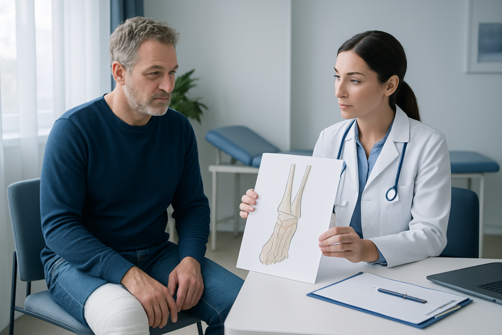
Pilon fractures usually happen when the tibia is driven into the ankle joint with enough force to split or crush the bone. Common causes include falls from ladders or heights, vehicle collisions, sports accidents, and direct impacts to the lower leg. Because the injury mechanism can compress the joint surface, the fracture pattern is often complex.
Some people are more vulnerable to worse injury patterns. Risk can be higher in those with osteoporosis, previous ankle injuries, or jobs and activities that expose them to falls or heavy impact. In older adults, even a lower-energy fall can cause a significant fracture if the bone is fragile.
Soft-tissue condition also matters. The ankle has little padding, so swelling, skin injury, or blistering can accompany the fracture and affect when surgery can safely be done. Doctors often consider the condition of the skin and circulation as carefully as the bone itself.
Diagnosis
Diagnosis starts with a physical examination and a detailed description of how the injury happened. A doctor will check the ankle’s alignment, swelling, skin condition, circulation, and nerve function before moving to imaging studies. This helps identify urgent problems such as open injury or compromised blood flow.
X-rays are usually the first imaging test and can show the position of the fracture and whether the ankle joint is involved. A CT scan is commonly used after X-rays because it gives a more detailed view of the number of fracture lines, joint surface fragments, and whether the bone is shifted. That information guides treatment planning.
In some cases, additional tests may be needed if there are concerns about blood vessels, nerves, or nearby injuries. For people traveling from another country for treatment, bringing any initial X-rays, emergency notes, or scan reports can save time and help the orthopedic team build a clearer treatment plan.
Treatment Options
Treatment depends on the fracture pattern, the amount of swelling, whether the skin is open, and the person’s overall health. Many pilon fractures are not treated with a single immediate operation because the surrounding tissues need time to settle. Temporary splinting or an external fixator may be used first to keep the ankle stable while swelling improves.
Once it is safe, surgery is often needed for fractures that are displaced or involve the joint surface. The goal is to restore alignment as accurately as possible and support the bone while it heals. Methods may include plates, screws, or other fixation devices, chosen according to the fracture shape and soft-tissue condition.
Not every fracture follows the same path. Some stable injuries may be treated without surgery using casting or protected immobilization, particularly when the bone pieces remain well aligned. After the initial phase, pain control, elevation, and careful skin care are important parts of treatment regardless of whether surgery is performed.
Recovery is usually gradual. A period of limited weight bearing is common, followed by a step-by-step increase in activity under orthopedic guidance. Physiotherapy often plays an important role in restoring ankle motion, strength, balance, and confidence with walking.
Prevention & Self-care
Not every pilon fracture can be prevented, especially when trauma is severe. Still, general injury prevention can lower risk in daily life: use fall-prevention measures at work and home, wear appropriate footwear, and follow safety practices for driving, cycling, and sports.
For people with weaker bones, improving bone health can make a meaningful difference. Adequate calcium and vitamin D intake, regular weight-bearing exercise when appropriate, and assessment for osteoporosis may all be part of a broader prevention plan recommended by a doctor.
After the injury, self-care supports healing. Elevating the leg, keeping dressings clean and dry if instructed, attending follow-up visits, and avoiding weight bearing until cleared are all important. It is also helpful to watch for new redness, fever, increasing pain, or drainage, since these can signal a complication that should be reviewed promptly.
When to See a Doctor
Anyone who has a severe ankle injury with an inability to bear weight, obvious deformity, or major swelling should seek urgent medical evaluation. Open wounds, numbness, a pale or cold foot, or worsening pain also deserve immediate attention because they may indicate a more serious injury pattern.
People who have been told they may have a pilon fracture should follow up with an orthopedic specialist even if the pain seems manageable after the first day. These fractures can look straightforward from the outside while still involving the joint surface in a way that affects long-term ankle function.
If treatment is being planned across borders, it is reasonable to ask for a clear explanation of the fracture pattern, expected recovery steps, and the timeline for imaging, surgery, or rehabilitation. Acibadem Health Point’s multidisciplinary specialists and JCI-accredited hospitals diagnose and treat pilon bone fractures for international patients, with coordinated care from initial assessment through follow-up.
Frequently asked questions
01What is a pilon bone fracture in simple terms?
A pilon bone fracture is a break in the lower part of the tibia near the ankle joint. It often affects the joint surface, which makes it more complex than a typical ankle fracture.
02Is a pilon fracture the same as an ankle fracture?
Not exactly. A pilon fracture involves the tibia at the top of the ankle joint, while many ankle fractures involve the smaller bones around the ankle. The two can overlap, but pilon fractures usually need more detailed imaging and planning.
03Do all pilon fractures need surgery?
No, but many displaced or joint-involving fractures do. Stable fractures may sometimes be treated without surgery, depending on alignment, soft-tissue condition, and the doctor’s assessment.
04How long does recovery take?
Recovery can be slow because the bone and joint surface need time to heal. The exact timeline varies, but many people need weeks of protection followed by rehabilitation to regain motion and strength.
05Can a pilon fracture cause arthritis later?
Yes, joint surface injury can increase the chance of post-traumatic arthritis. Good alignment, appropriate treatment, and rehabilitation may help reduce that risk, but they cannot remove it completely.
06What should someone do right after this injury?
They should avoid putting weight on the leg, keep it elevated if possible, and seek urgent medical assessment. If the skin is broken, the foot feels numb or cold, or pain is severe, emergency care is especially important.
This article is for general information only and is not a substitute for professional medical advice. Please consult a qualified doctor about your individual situation.
Joint and spine care in Turkey — expert assessment & treatment
JCI-accredited · board-certified surgeons · reply within 24h
Add us as a Preferred Source to see more of our trusted health content across Google Search, AI Overviews and Discover.