Varus Knee Stress Test: Purpose, Procedure and Results

Key Takeaways
- The varus knee stress test checks the outer support structures of the knee for laxity or pain.
- It is commonly used after a twist, direct impact, sports injury, or when chronic knee instability is suspected.
- A positive result does not confirm one single diagnosis; it is interpreted together with symptoms, other physical tests, and imaging when needed.
- The test is usually quick and does not require special preparation.
- Persistent swelling, instability, or difficulty bearing weight should be evaluated by a qualified clinician.
A varus knee stress test is a hands-on orthopedic exam used to assess the stability of the outer side of the knee, especially the lateral collateral ligament and nearby structures. It helps a clinician understand whether pain or looseness may be related to injury, degeneration, or other knee conditions.
Overview
The varus knee stress test is a clinical examination used to see how well the outer, or lateral, side of the knee resists inward pressure. In practical terms, a clinician gently applies force to the lower leg while the knee is positioned in a specific way to look for pain, opening of the joint line, or unusual looseness.
This test is most often used when a person reports a twisting injury, a sports collision, unexplained knee instability, or pain along the outer knee. It helps assess the lateral collateral ligament (LCL) and other lateral structures that help keep the knee aligned and steady.
For patients deciding whether to seek care at home or travel for assessment, the value of the test is simple: it gives an orthopedic specialist real-time information during the exam. That hands-on detail can shape the next step, whether that is rest and rehabilitation, imaging, bracing, or a broader evaluation for more complex injury.
What the test is designed to show
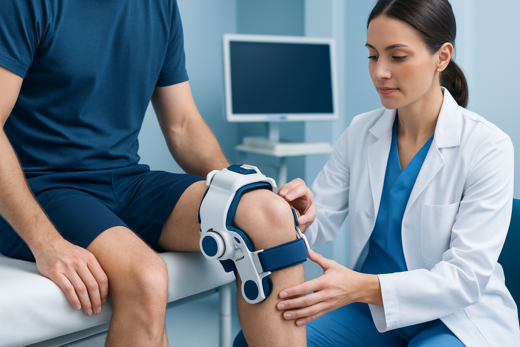
The varus stress test is intended to check whether the lateral side of the knee is stable under stress. If the outer structures are intact, the joint should feel firm with only a small, expected amount of movement. If there is injury or weakening, the clinician may notice excessive opening of the joint space or a clear difference compared with the other knee.
The test is especially useful for evaluating the LCL, but it may also provide clues about nearby tissues, including the posterolateral corner of the knee. Because the knee works as a coordinated unit, a positive finding may point to more than one structure being involved.
It is important to know that the test is not a stand-alone diagnosis. A result is interpreted together with the story of the injury, where the pain is felt, swelling, range of motion, walking ability, and whether other ligaments may also be affected.
Symptoms that may lead to a varus stress exam

A clinician may choose this test when a person describes pain on the outer side of the knee, a sense that the knee is giving way, or symptoms that began after a side impact or twisting injury. Some people notice that stairs, walking on uneven ground, or changing direction makes the knee feel unstable.
Swelling, bruising, stiffness, or tenderness along the lateral knee can also prompt the exam. In more significant injuries, a person may have trouble fully straightening the knee or may avoid putting weight on the leg because it feels insecure.
The test can also be part of a broader orthopedic assessment for people with chronic knee problems. In those cases, the clinician may be looking for subtle looseness that suggests a ligament imbalance, prior injury, or joint wear that has altered how the knee tracks and carries load.
Causes and risk factors linked to abnormal findings
Abnormal varus stress findings are often associated with trauma. Common causes include sports injuries, falls, direct blows to the inside of the knee, or sudden changes in direction that place stress on the outer support structures.
Some people are at higher risk because of their activity level or health history. Contact sports, high-speed pivoting sports, occupational demands that involve kneeling or twisting, and a previous knee injury can all raise the chance of lateral knee strain. Muscle weakness, poor movement mechanics, and balance problems may also contribute to repeated stress over time.
Not every positive exam means a major tear. Pain, swelling, or muscle guarding can make the knee feel less stable during testing. That is one reason clinicians consider the full picture before drawing conclusions.
How the procedure is performed
The exam is usually done in a clinic, emergency department, sports medicine setting, or orthopedic office. The person lies down or sits while the clinician positions the knee, commonly with slight bending, and applies gentle outward or inward pressure to assess the lateral structures.
At one angle, the test looks more closely at the LCL; at another, it helps assess additional structures that support the posterolateral knee. The clinician compares how far the joint opens on the injured side with the opposite knee and notes whether pain is present during the maneuver.
The procedure is brief and does not require fasting, sedation, or special preparation. Some tenderness may occur if the knee is already injured, but the test should be performed carefully by a trained professional to avoid unnecessary discomfort and to ensure an accurate interpretation.
- It is often combined with other knee stability tests.
- The unaffected knee may be checked for comparison.
- Imaging is not part of the test itself, but may be ordered later if needed.
How results are interpreted
A normal result generally means the knee feels stable and does not open excessively under stress. Mild discomfort without looseness may suggest irritation, soft-tissue strain, or inflammation rather than a major ligament disruption.
A positive result usually means there is more sideways opening than expected, pain with the maneuver, or a clear difference from the other knee. This can suggest injury to the LCL or nearby structures, but the pattern must be matched with other exam findings and, when appropriate, imaging such as X-rays or MRI.
Results are especially meaningful when the clinician compares findings at different knee angles. The amount of looseness, the quality of the endpoint, and whether other tests are also abnormal help determine whether the injury is isolated or part of a larger ligament problem.
Treatment options after an abnormal exam
Treatment depends on the cause and severity of the findings. Mild sprains may improve with activity modification, temporary bracing, pain-relief strategies recommended by a clinician, and a structured rehabilitation plan focused on strength, balance, and movement control.
More significant ligament injuries may need closer orthopedic follow-up. Some cases are managed non-surgically, while others—especially when more than one ligament is involved or the knee is markedly unstable—may require surgical evaluation. The choice depends on stability, symptoms, activity goals, and the overall injury pattern.
For international patients, this is often the stage where clear planning matters: understanding the diagnosis, expected recovery timeline, and the need for follow-up once back home. In well-coordinated centers, multidisciplinary orthopedic teams can align imaging, consultation, and rehabilitation so that care feels connected rather than fragmented.
Prevention, recovery, and self-care
People can reduce knee strain by keeping the muscles around the hip and thigh strong, using good landing and cutting techniques in sport, and paying attention to footwear and terrain. Gradual conditioning is often safer than returning quickly to intense activity after time off.
If a clinician suspects a lateral knee injury, early self-care usually includes resting from painful activity, using support if advised, and following a rehabilitation plan. It is generally wise to avoid forcing the knee through painful twisting or pivoting movements until it has been assessed.
Recovery is often smoother when people follow the plan consistently and report changes in symptoms. Swelling that increases, a knee that repeatedly gives way, or a loss of motion can signal that reassessment is needed.
When to see a doctor
A knee should be evaluated promptly if pain follows an injury and there is visible swelling, instability, difficulty walking, or a feeling that the joint is slipping outward. Urgent assessment is also important if the knee cannot bear weight, looks deformed, or the person cannot fully straighten or bend it.
Even if symptoms are mild, a persistent sense of looseness deserves medical review. Early examination can help identify ligament injury before compensatory walking patterns or ongoing instability lead to further strain.
For patients traveling for care, timely review with an orthopedic specialist can be especially helpful when the diagnosis is uncertain or when coordinated imaging and follow-up are needed. Acibadem Health Point’s multidisciplinary specialists and JCI-accredited hospitals diagnose and treat knee conditions for international patients with coordinated, evidence-based care.
Frequently asked questions
01What does a varus knee stress test check for?
It checks how stable the outer side of the knee is when gentle sideways force is applied. Clinicians use it mainly to assess the lateral collateral ligament and nearby support structures.
02Is the varus stress test painful?
It should not be painful when the knee is healthy, although it may feel uncomfortable if the area is already injured or inflamed. The clinician performs it carefully and stops if the discomfort is too great.
03Does a positive result mean the ligament is torn?
Not necessarily. A positive finding suggests looseness or pain on the lateral side of the knee, but swelling, muscle guarding, or other injuries can also affect the result. The test is interpreted with the rest of the exam and sometimes imaging.
04Do patients need to prepare for the test?
Usually, no special preparation is needed. The clinician may ask about the injury, symptoms, prior surgery, or any previous knee problems before beginning the exam.
05What happens after an abnormal result?
The next step may include more physical tests, X-rays, MRI, bracing, rehabilitation, or referral to an orthopedic specialist. The plan depends on how unstable the knee feels and whether other structures may also be injured.
06Can the test be done on both knees?
Yes. Clinicians often compare the injured knee with the other side because small differences can help them judge whether the movement is normal or abnormal.
This article is for general information only and is not a substitute for professional medical advice. Please consult a qualified doctor about your individual situation.
Joint and spine care in Turkey — expert assessment & treatment
JCI-accredited · board-certified surgeons · reply within 24h
Add us as a Preferred Source to see more of our trusted health content across Google Search, AI Overviews and Discover.