Ultrasound of Testis: Purpose, Procedure and Results

Key Takeaways
- An ultrasound of testis uses sound waves, not radiation, to create detailed images of the testicles and scrotum.
- It is commonly ordered for pain, swelling, a lump, injury, suspected infection, torsion, or fertility evaluation.
- The test is usually painless, brief, and performed while the person lies on an examination bed.
- Results may show normal anatomy or signs of cysts, fluid collection, inflammation, reduced blood flow, or a mass that needs follow-up.
- A prompt medical review is important if testicular pain is sudden, severe, or linked with swelling, fever, or nausea.
An ultrasound of testis is a quick, noninvasive imaging test used to examine the testicles and surrounding scrotal structures. It helps doctors evaluate pain, swelling, lumps, trauma, infertility concerns, and possible infections or torsion.
Overview
An ultrasound of testis is a standard imaging exam that helps doctors look closely at the testicles, epididymis, spermatic cord, and nearby scrotal tissues. It is often the first test chosen when a person develops pain, swelling, a lump, or other changes in the scrotum, because it is fast, painless, and does not use radiation.
The test is sometimes called a testicular ultrasound or scrotal ultrasound. In practical terms, it gives the clinician a clear view of structures that cannot be fully assessed by a physical examination alone. For many patients, the exam provides reassurance; for others, it helps guide the next step, whether that is treatment, follow-up imaging, or referral to a specialist.
In international-patient settings, this scan is often part of a broader workup arranged after an initial consultation. That may be especially helpful when someone is traveling for care and wants a concise, efficient way to clarify a symptom before returning home or moving ahead with a treatment plan.
Symptoms and reasons it is ordered
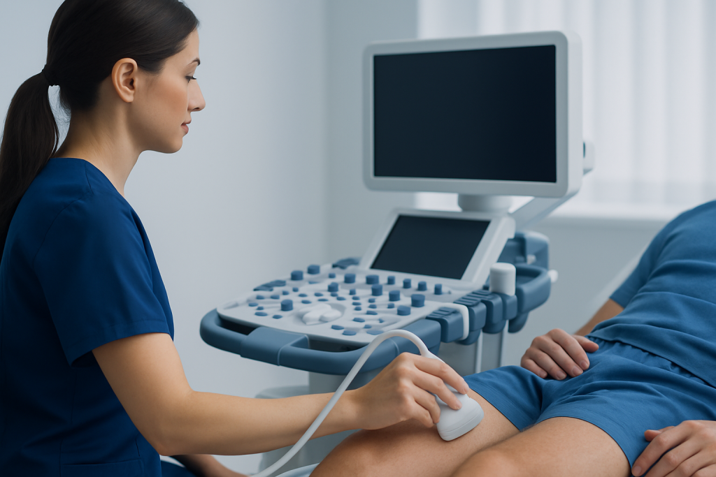
Doctors may request an ultrasound of testis for several common reasons. One of the most frequent is testicular pain, whether it started suddenly or developed gradually. The scan can help determine whether pain is related to torsion, infection, trauma, a hernia, fluid around the testicle, or another cause.
It is also used when there is a lump, enlargement, heaviness, or a change in the shape of the scrotum. Some patients are referred after a clinician feels a nodule during an examination. In other cases, the ultrasound is part of an infertility evaluation, especially if there are concerns about varicocele, testicular size, or prior injury.
- Sudden or persistent testicular pain
- Swelling or redness of the scrotum
- A lump or firm area in the testicle
- After a sports injury or direct trauma
- Fever with scrotal discomfort, suggesting infection
- Evaluation of fertility-related concerns
Although the scan is commonly ordered for symptoms, it can also be used when a clinician wants to monitor a known condition over time. That makes it useful not only for diagnosis, but also for confirming whether a problem is stable, improving, or in need of closer attention.
Causes and risk factors behind abnormal findings

An ultrasound does not cause the underlying condition; it reveals what may already be present. Abnormal findings can be linked to infection, inflammation, a twisted spermatic cord, a hydrocele, a spermatocele, a varicocele, a cyst, prior trauma, or, less commonly, a tumor. The meaning of any image always depends on the symptom history and physical examination.
Some risk factors make scrotal conditions more likely. Recent injury, heavy physical strain, untreated urinary or sexually transmitted infection, a history of undescended testicle, and prior episodes of testicular pain can all influence the reason a scan is ordered. In fertility workups, doctors may also look for enlarged veins or differences in testicular size that suggest a potentially correctable issue.
It is worth noting that not every unusual finding is dangerous. Fluid collections and small cysts are often benign, and some changes simply need observation. A careful interpretation by a qualified clinician is what turns the image into a meaningful diagnosis.
How the test is done
The procedure is usually straightforward. The patient lies on an examination table, and a technician or doctor applies a clear gel to the scrotal skin. A handheld probe, called a transducer, is then moved gently over the area to create images on a screen. The exam typically takes only a short time.
Most people do not need fasting or special preparation. Comfortable clothing is helpful, since the lower garments will need to be removed. If the scan is being performed because of significant pain, the clinician may be especially careful to minimize pressure from the probe.
The exam may include color Doppler imaging, which shows blood flow. This can be important when doctors need to check for torsion or inflammation. At the end, the images are reviewed by a radiologist or treating clinician, who then explains whether the findings are normal or suggest a specific condition.
What results may show
Normal results usually mean the testicles are well formed, the tissue appears uniform, and blood flow is appropriate. In that case, the doctor may look for other explanations for symptoms outside the scrotum or recommend simple follow-up if symptoms are improving.
Abnormal results vary widely. The scan may show inflammation of the epididymis or testicle, a collection of fluid around the testicle, a varicocele, signs of trauma, or reduced blood flow that raises concern for torsion. If a solid mass is seen, the finding requires careful medical assessment, though imaging alone cannot confirm the exact diagnosis in every case.
- Infection or inflammation: swelling, increased blood flow, tenderness
- Hydrocele: fluid around the testicle
- Varicocele: enlarged veins in the scrotum
- Cyst or spermatocele: usually benign fluid-filled structure
- Torsion: reduced or absent blood flow, urgent concern
- Mass: needs timely specialist review
Patients sometimes worry when they hear the word “abnormal,” but the result simply means the clinician has found something that needs interpretation. Many findings are treatable, and some only require observation or repeat imaging.
Treatment options after the scan
Treatment depends entirely on what the ultrasound shows. If the scan suggests infection, a clinician may prescribe treatment based on the likely cause and examine whether the urinary tract or sexually transmitted infections are involved. If the scan points to torsion, surgery is typically urgent because blood flow to the testicle can be threatened.
For hydrocele, varicocele, or benign cysts, treatment may range from observation to procedural management, depending on symptoms and size. A traumatic injury may simply need rest and pain control, but more significant damage can require specialist care. When a mass is identified, the next step may include blood tests, repeat imaging, or referral to a urologist for further evaluation.
For men seeking care while traveling, a clear diagnosis can be especially helpful because it allows the next appointment to be planned with purpose rather than guesswork. In this setting, multidisciplinary teams can coordinate imaging review, specialist consultation, and follow-up recommendations so the patient knows what happens next before leaving the hospital.
Prevention and self-care
Not every scrotal condition can be prevented, but sensible habits can reduce avoidable strain and support timely care. Wearing athletic protection during contact sports, seeking prompt treatment for urinary or genital infections, and avoiding delays when pain appears suddenly are all practical steps.
Self-care after an ultrasound is usually minimal because the test itself does not have a recovery period. If the clinician advises it, supportive underwear, rest, ice wrapped in cloth for short periods, and avoiding heavy lifting may help with discomfort while awaiting results or treatment.
Men who notice a new lump, persistent ache, heaviness, or swelling should not assume the symptom will settle on its own. A careful exam and imaging study are often the fastest route to clarity, especially when the concern is difficult to judge by touch alone.
When to see a doctor
A doctor should be contacted promptly for sudden testicular pain, especially if it is severe, associated with nausea, or comes with swelling. These symptoms can sometimes signal torsion, which needs immediate assessment. Even when pain is less intense, persistent discomfort should be evaluated rather than watched indefinitely.
Medical review is also important for a lump, scrotal redness, fever, a recent injury, or any change in the size or position of a testicle. If symptoms interfere with daily life, sexual function, or fertility concerns, a clinician can decide whether an ultrasound of testis is the right next step.
For patients arranging care from another country, it is reasonable to ask what the scan can answer, how results will be reported, and whether a urologist should review the images the same day. Acibadem Health Point’s multidisciplinary specialists and JCI-accredited hospitals diagnose and treat this condition for international patients, with coordinated imaging and specialist follow-up when needed.
Frequently asked questions
01Is an ultrasound of testis painful?
The exam is usually not painful. The probe is moved gently over the scrotum with gel, and most people feel only light pressure or mild discomfort if the area is already tender.
02Do I need to prepare before the scan?
In most cases, no special preparation is needed. Wearing loose clothing can make the visit easier, and the clinic may ask for a brief medical history before the exam begins.
03How long does a testicular ultrasound take?
It is typically a short procedure, often completed within a single visit. The exact time can vary depending on whether Doppler imaging is used or whether both testicles need close review.
04Can ultrasound tell if a testicle lump is cancer?
Ultrasound can identify whether a lump looks solid or fluid-filled and whether it needs further evaluation. It cannot always determine the exact cause on its own, so a doctor may recommend additional tests or specialist review.
05What does reduced blood flow on ultrasound mean?
Reduced blood flow can be an important finding, especially if torsion is suspected. It does not confirm every diagnosis by itself, but it usually prompts urgent medical assessment.
06Is ultrasound safe?
Yes, ultrasound uses sound waves rather than radiation. It is widely used in medicine because it is noninvasive and generally safe when performed by trained professionals.
This article is for general information only and is not a substitute for professional medical advice. Please consult a qualified doctor about your individual situation.
Explore treatments in Turkey — costs, top hospitals & a free quote
JCI-accredited · board-certified surgeons · reply within 24h
Add us as a Preferred Source to see more of our trusted health content across Google Search, AI Overviews and Discover.