TEE Procedure: How Heart Images Are Taken
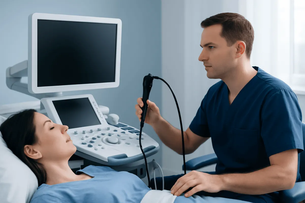
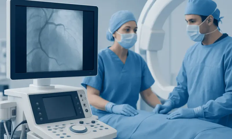
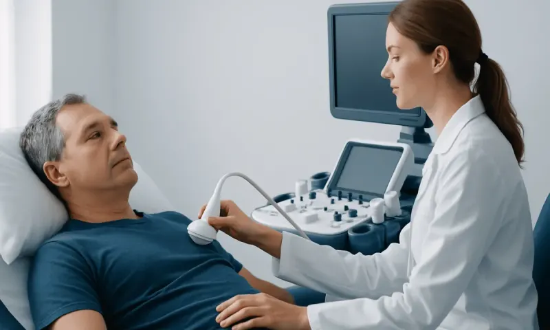
A TEE procedure, or transesophageal echocardiogram, is a heart ultrasound performed with a slim probe passed through the mouth into the esophagus. It gives clinicians highly detailed images of heart valves, chambers, blood flow, and nearby major blood vessels when standard imaging from the chest is not enough.
Overview: What Is a TEE Procedure?
A tee procedure is short for a transesophageal echocardiogram (TEE). It is a specialized ultrasound test that creates detailed pictures of the heart by placing a flexible ultrasound probe in the esophagus, the swallowing tube behind the heart. Because the probe is close to the heart and is not blocked by the ribs or lungs, it can provide clearer views than a standard echocardiogram performed through the chest wall.
The test uses high-frequency sound waves and does not involve X-rays or radiation. A cardiologist may use TEE to examine heart valves, pumping chambers, blood clots, infection involving a valve, congenital heart differences, blood flow, or the aorta. It may also guide certain heart procedures or help assess the heart around the time of surgery.
TEE is often recommended when a transthoracic echocardiogram, the usual chest ultrasound of the heart, does not answer an important clinical question. It is a diagnostic procedure rather than a treatment, and the findings are considered alongside symptoms, examination results, laboratory tests, and other scans.
How TEE Works and Why It May Be Recommended
The ultrasound probe at the end of the TEE tube sends sound waves toward the heart and receives returning echoes. A computer converts these echoes into moving images. The clinician can gently adjust the probe position to view structures from several angles, including the left atrium, mitral valve, aortic valve, and thoracic aorta.
A TEE procedure may be considered when clinicians need to look for a possible blood clot before a rhythm-restoring procedure for atrial fibrillation, assess a suspected valve infection, evaluate a prosthetic heart valve, investigate an unexplained stroke, or clarify a possible problem with the aorta. It can also help evaluate leaks or narrowing in heart valves and certain structural heart conditions.
For patients, tee procedure patient education should include the reason the test is being proposed and how the result may influence care. The clinician will explain whether TEE is the most useful next step, what alternatives may be appropriate, and whether the scan is being performed in an outpatient setting, hospital, or during another procedure.
Who Can Have a TEE Procedure?

Many adults can safely have a transesophageal echocardiogram, including people with complex heart disease who need more detailed imaging. Before scheduling the test, the care team reviews medical history, current symptoms, allergies, medications, prior anesthesia or sedation reactions, and any difficulty swallowing.
TEE may not be suitable, or may require special precautions, for people with an esophageal narrowing, tumor, tear, diverticulum, severe swallowing disorder, active bleeding in the digestive tract, or recent surgery involving the esophagus. A history of esophageal varices, prior chest radiation, significant reflux-related injury, or neck problems may also affect planning. Pregnancy, lung disease, obstructive sleep apnea, and kidney or liver disease should be discussed because they can influence sedation decisions.
Patients should tell the team about blood-thinning medicines, diabetes medicines, insulin, weight-loss injections, dentures, loose teeth, and any previous esophageal conditions. The clinician may coordinate with a cardiologist, gastroenterologist, anesthesiologist, or other specialist when additional evaluation is needed before the procedure.
What Should I Do to Prepare for a Transesophageal Echocardiogram (TEE)?
Preparation instructions vary somewhat by hospital and by the type of sedation planned, so patients should follow the instructions from their own care team. In general, people are asked not to eat or drink for several hours before the test. An empty stomach lowers the chance of vomiting and inhaling stomach contents into the lungs during sedation.
Medication instructions are individualized. Some medicines may be taken with a small sip of water, while diabetes medicines, insulin, blood thinners, or medicines affecting stomach emptying may need adjustment. Patients should not stop prescribed medication on their own; they should ask the clinician managing the TEE for clear instructions in advance.
It is usually necessary to arrange for a responsible adult to drive the patient home and stay available afterward, as sedatives can impair alertness and judgment for the rest of the day. Patients should bring an up-to-date medication list, wear comfortable clothing, and report a cold, fever, chest infection, new swallowing difficulty, or other illness before arriving.
- Confirm fasting times with the care team.
- Ask how to take regular medicines on the morning of the test.
- Tell staff about swallowing problems, allergies, sleep apnea, and dentures.
- Arrange transport home; driving after sedation is not safe.
What Happens During the TEE Procedure?
On arrival, staff confirm the patient’s identity, medical history, fasting status, and consent. An intravenous line is commonly placed, and monitors track heart rhythm, blood pressure, oxygen level, and breathing. The patient usually lies on the left side, and a mouth guard helps protect the teeth and the probe.
Most patients receive a numbing spray or gel for the throat and sedating medicine through the intravenous line. They may feel relaxed or sleepy and may remember little of the examination. In some circumstances, such as TEE performed during surgery or a complex intervention, deeper anesthesia may be used.
Once the throat is numb and sedation has taken effect, the clinician guides the lubricated probe through the mouth and into the esophagus. The patient does not need to swallow continuously, and the probe does not enter the windpipe. Image collection often takes less than an hour, although the total visit is longer because of preparation and recovery monitoring.
Afterward, the cardiologist reviews the images and creates a tee procedure report. A clinical tee procedure note documents relevant details such as the reason for the test, sedation, probe passage, key findings, and whether any complications occurred. A tee procedure template or tee procedure note template is a documentation tool used by clinicians; it is not something patients need to complete.
What Is the Recovery Like After a Transesophageal Echocardiogram (TEE)?
Recovery usually begins in a monitored area while the sedative wears off. The throat may remain numb for a short time, so patients should not eat or drink until staff confirm that swallowing has returned safely. Once swallowing is normal, small sips of water are often introduced first, followed by light food if tolerated.
For the first day, mild sore throat, hoarseness, dry mouth, tiredness, bloating, or a brief sensation of throat irritation can occur. These effects are generally temporary. Rest, fluids once permitted, and soft foods can be more comfortable for some people. The care team may provide specific advice about managing throat discomfort.
Because sedation can slow reactions and affect memory, patients should not drive, operate machinery, drink alcohol, sign important documents, or make major decisions until the following day or as instructed. Most people return to usual activities the next day, although timing can differ if TEE was performed alongside another procedure or if an underlying heart condition requires further care.
How Risky Is a Transesophageal Echocardiogram?
A transesophageal echocardiogram is widely used and is generally considered safe when performed by trained teams with appropriate monitoring. The most common effects are temporary throat discomfort and sedation-related drowsiness. The benefit is the ability to obtain detailed information that may guide important decisions about heart rhythm, valve disease, infection, blood clots, or surgery.
Less common risks include low oxygen levels, changes in blood pressure or heart rhythm, nausea, vomiting, aspiration, damage to a tooth, bleeding, or a reaction to sedation. Rarely, the probe can injure the throat or esophagus, including a tear or perforation. The risk may be higher in people with known esophageal disease, significant swallowing difficulties, or serious medical conditions.
The team monitors the patient throughout the examination and is prepared to manage complications. Before agreeing to the procedure, patients can ask why TEE is needed, what information it is expected to provide, whether another test could be suitable, and what specific risks apply to their health history.
What Not to Do After TEE and When to Seek Medical Care
After TEE, patients should not eat or drink until the numbness in the throat has resolved and staff say it is safe to swallow. They should also avoid driving, alcohol, strenuous activity, and activities requiring close concentration for the rest of the day if sedation was used. It is important not to stay alone immediately after outpatient sedation unless the care team confirms that this is appropriate.
Patients should contact the treating team promptly for worsening throat pain, trouble swallowing, persistent vomiting, fever, coughing up blood, or a new problem with breathing. Emergency medical care is needed for severe chest pain, severe shortness of breath, fainting, heavy bleeding, or symptoms that feel sudden or serious. These symptoms are uncommon, but timely assessment is important.
The result may be available soon after the test, while a full interpretation may take longer depending on the setting and urgency. Patients should ask when and how they will receive results and what follow-up is recommended. Acıbadem Health Point’s multidisciplinary specialists and JCI-accredited hospitals provide diagnostic assessment and treatment planning for international patients with heart and vascular conditions.
Frequently asked questions
01Is a TEE procedure painful?
TEE is not usually painful, but it can feel uncomfortable when the probe is first passed through the throat. Numbing medicine and sedation are commonly used to improve comfort. Some people have a mild sore throat afterward, which usually settles within a day or two.
02How long does a TEE procedure take?
The imaging portion often takes less than an hour. However, preparation, sedation, monitoring, and recovery mean the complete appointment may take several hours. The exact timing depends on the reason for the test and the patient’s recovery from sedation.
03Can I eat after a transesophageal echocardiogram?
Patients should wait until the numbness in the throat has worn off and staff confirm that swallowing is safe. They may then begin with small amounts of water and follow the discharge instructions provided. Soft foods may feel more comfortable if the throat is mildly sore.
04Will I be awake during a TEE?
Many people are awake but sleepy and relaxed because of moderate sedation. They may have little memory of the test afterward. Some situations require deeper sedation or anesthesia, particularly when TEE is performed during surgery or another intervention.
05Why would a doctor order TEE instead of a regular echocardiogram?
TEE places the ultrasound probe closer to the heart, allowing clearer images of some structures than a standard chest-wall echocardiogram can provide. It is particularly useful for examining valves, detecting certain clots or infections, and evaluating parts of the aorta. The choice depends on the clinical question and the quality of previous imaging.
06Can I go back to work after TEE?
If sedation is used, most patients should plan to rest for the remainder of the day and return to work the next day if they feel well. Driving and work that requires alertness should be avoided until the sedative effects have fully worn off. The treating team may recommend a different timeline after a combined procedure or if there are other medical concerns.
This article is for general information only and is not a substitute for professional medical advice. Please consult a qualified doctor about your individual situation.
Heart care in Turkey — expert evaluation and treatment
JCI-accredited · board-certified surgeons · reply within 24h
Add us as a Preferred Source to see more of our trusted health content across Google Search, AI Overviews and Discover.