Sonography: Uses, Types and What to Expect

Key Takeaways
- Sonography uses high-frequency sound waves to produce live images inside the body.
- It is often used to evaluate abdominal organs, the thyroid, blood vessels, pregnancy, and many soft tissues.
- The test is generally painless and safe, and most scans require little or no special preparation.
- Results help clinicians decide whether further imaging, monitoring, or treatment is needed.
- A doctor may recommend sonography when symptoms, exam findings, or follow-up needs call for a closer look.
Sonography, also called ultrasound imaging, uses sound waves to create real-time pictures of organs, blood vessels, and soft tissues. It is widely used because it is non-invasive, does not use radiation, and can help guide many everyday medical decisions.
Overview
Sonography is a form of medical imaging that helps clinicians look inside the body without making an incision or using ionizing radiation. It is sometimes called ultrasound imaging, and in everyday language many people simply refer to it as “an ultrasound.”
The scan works by sending sound waves into the body and measuring how they bounce back from different tissues. Those echoes are turned into real-time pictures that can show the size, shape, movement, and texture of organs and structures. Because the images are created live, sonography can be especially helpful when a clinician wants to see motion, such as blood flow, heart activity, or fetal movement.
For many patients, sonography is one of the first imaging tests recommended when a symptom needs a closer look. It is often used in outpatient care, emergency settings, prenatal care, and follow-up visits. For international patients deciding whether to travel for medical evaluation, it is also a practical test because it is widely available, usually quick, and rarely requires a long recovery period afterward.
How Sonography Works
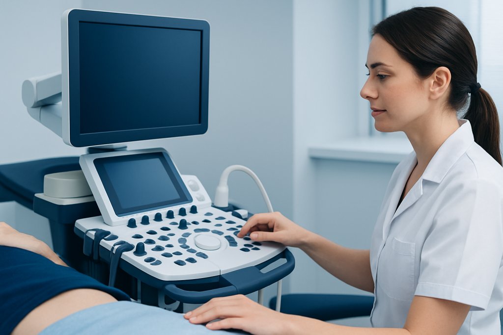
During a sonography exam, a handheld device called a transducer is placed on the skin or, in some cases, used internally. The transducer sends sound waves into the body and receives the echoes that return from tissues. A computer then converts those echoes into an image displayed on a monitor.
Different tissues reflect sound in different ways. Fluid-filled spaces often appear darker, while denser tissues may appear brighter. This contrast helps the clinician distinguish between normal structures and possible changes such as cysts, inflammation, swelling, or abnormal growths. Doppler ultrasound is a related technique that can also assess blood flow, which is useful for blood vessels, the heart, and certain pregnancy evaluations.
Because sonography is dynamic, the clinician may ask the patient to change position, hold a breath, or remain still for short periods. These small adjustments can improve image quality and help capture a more complete view of the area being examined.
Common Uses

Sonography is used in many areas of medicine because it can answer a broad range of practical questions. It is often ordered to evaluate abdominal pain, liver or gallbladder problems, kidney stones, thyroid nodules, breast lumps, pelvic discomfort, and suspected fluid collections. It is also frequently used to monitor pregnancy and to guide procedures such as biopsies or needle placement.
In some situations, sonography can provide enough information to support a diagnosis on its own. In others, it is one step in a wider evaluation that may include blood tests, physical examination, or other imaging studies. Clinicians often choose ultrasound first when they want a fast, bedside-friendly, and repeatable way to assess soft tissues.
- Abdominal organs such as the liver, gallbladder, pancreas, spleen, and kidneys
- Pelvic organs, including the uterus and ovaries
- Pregnancy and fetal development
- Thyroid and neck structures
- Breast lumps or focal areas of concern
- Arteries and veins using Doppler imaging
What to Expect During the Exam
Most sonography appointments are straightforward. The patient may be asked to change into a gown and lie on an exam table. A clear gel is usually applied to the skin to help the transducer move smoothly and transmit sound waves effectively. The gel may feel cool, but it is harmless and easily wiped away afterward.
The exam itself usually lasts from a few minutes to around half an hour, depending on the area being studied and how detailed the images need to be. Some scans require the patient to arrive with a full bladder, while others may require fasting for a short period beforehand. The imaging team gives clear instructions in advance so the appointment runs smoothly.
Sonography is generally painless. Patients may feel mild pressure when the transducer is pressed against the body, especially over tender areas, but the scan should not be painful. If internal sonography is needed, the clinician explains the process carefully beforehand so the patient knows what to expect and can ask questions in advance.
Benefits and Limitations
One of sonography’s main advantages is safety. It does not use radiation, which makes it especially useful for pregnancy and for repeat imaging when monitoring over time is important. It is also portable, relatively affordable compared with some other imaging tests, and often available quickly.
Sonography is particularly good at showing soft tissues, fluid, and blood flow. It can help distinguish whether a lump is solid or fluid-filled, whether an organ looks enlarged, or whether there is evidence of inflammation or blockage. In many cases, it gives clinicians enough detail to decide on next steps without delay.
At the same time, ultrasound does have limits. Image quality can be affected by body habitus, bowel gas, the depth of the target area, or difficult-to-reach structures. Some findings may still require follow-up with CT, MRI, laboratory testing, or specialist review. A useful scan is not always the final answer, but it often provides an important and efficient starting point.
Preparation and Safety
Preparation depends on the type of sonography being performed. An abdominal scan may require fasting for several hours so the gallbladder and upper abdominal organs are easier to assess. A pelvic scan may require a full bladder, while a vascular or thyroid scan may need little preparation at all. The appointment instructions should be reviewed carefully before arrival, especially when traveling from another country and coordinating tests around clinic visits.
Sonography is considered very safe. The sound waves used are not the same as radiation, and the test has a long record of routine clinical use. Side effects are uncommon. Most people can return to normal activities immediately after the exam, unless a procedure or additional test has been performed at the same visit.
Patients who are pregnant, have chronic medical conditions, or are undergoing repeated imaging can usually feel reassured that ultrasound is a standard, well-established tool. Still, it should be used under the guidance of a qualified clinician, who can determine whether it is the right test for the specific clinical question.
After the Scan: Understanding Results
After the images are reviewed, the clinician explains what they show in the context of the patient’s symptoms, examination, and other test results. A sonography report may describe normal findings, an area that needs monitoring, or a change that should be evaluated further. The wording can sound technical, so patients should feel comfortable asking for a plain-language explanation.
Sometimes the next step is simply reassurance and routine follow-up. In other cases, the result may lead to another imaging test, a specialist appointment, or treatment for the underlying condition. When care is being planned from abroad, it is useful to ask for copies of the report and images, along with written guidance about follow-up timing after returning home.
It is important to remember that an imaging result is only one part of the whole picture. A small finding may be harmless, while a normal-appearing scan does not always rule out every possible cause of symptoms. Clinicians interpret ultrasound results together with the full clinical story.
When to See a Doctor
Sonography is usually recommended when a clinician needs to investigate a symptom, confirm a suspected diagnosis, or monitor a known condition. It is also common when a physical exam suggests that imaging could provide more detail. Patients should seek medical advice if they have persistent abdominal pain, a new lump, unexplained swelling, pelvic symptoms, urinary changes, or concerns during pregnancy.
Prompt evaluation is especially helpful if symptoms are getting worse, if there is fever, significant pain, visible blood, or sudden shortness of breath, or if a clinician has already recommended follow-up imaging and the timeline is unclear. In international care pathways, it is often wise to complete both the scan and the review with enough time to discuss the result before travel home.
Acibadem Health Point’s multidisciplinary specialists and JCI-accredited hospitals diagnose and treat conditions evaluated with sonography for international patients, helping coordinate imaging and follow-up in a clear, organized way. The aim is to make the process understandable from the first appointment through the next step in care.
Frequently asked questions
01Is sonography the same as an ultrasound scan?
Yes. Sonography and ultrasound are commonly used to describe the same imaging method. Both refer to using sound waves to create pictures of structures inside the body.
02Does sonography use radiation?
No, sonography does not use ionizing radiation. That is one reason it is often chosen for pregnancy and for repeat examinations.
03Is the test painful?
Most sonography exams are not painful. Patients may feel some pressure from the transducer, especially if the area being examined is tender.
04Do all ultrasound scans require preparation?
No. Preparation depends on the body part being examined and the reason for the test. Some scans require fasting or a full bladder, while others need no special preparation.
05Can sonography diagnose everything on its own?
Not always. It is a valuable tool, but some conditions need blood tests, another imaging study, or specialist assessment to reach a complete diagnosis.
06How soon are results available?
That depends on the setting and the urgency of the case. In many clinics, the images are reviewed soon after the scan, and the patient is told whether any further tests or follow-up are needed.
This article is for general information only and is not a substitute for professional medical advice. Please consult a qualified doctor about your individual situation.
Explore treatments in Turkey — costs, top hospitals & a free quote
JCI-accredited · board-certified surgeons · reply within 24h
Add us as a Preferred Source to see more of our trusted health content across Google Search, AI Overviews and Discover.