Small White Spots on Skin Pictures: Possible Causes
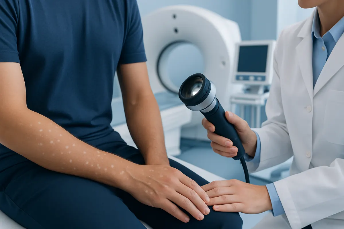
Small white spots on the skin may be caused by common and often harmless changes in skin pigment, sun exposure, dryness, inflammation, or a superficial fungal infection. Pictures can help describe a pattern, but they cannot reliably identify the cause because several conditions can look similar.
What Small White Spots on Skin Pictures Can—and Cannot—Show
Small white spots on skin pictures often show areas with less visible pigment than the surrounding skin. They may be tiny round marks, larger irregular patches, dry scaly areas, or small raised bumps. This difference matters: a flat light patch usually reflects a change in pigment or surface scale, while a raised white bump may have a different cause altogether.
Photographs can be useful for tracking whether spots are changing over time, but they cannot confirm a diagnosis. Lighting, camera settings, skin tone, tanning, and image quality can all make pale areas appear more or less noticeable. A clinician considers the location, texture, border, symptoms, timing, medical history, and examination findings rather than relying on an image alone.
In many cases, white spots are not dangerous. However, they can occasionally signal a treatable skin condition or an autoimmune pigment disorder. A calm, stepwise assessment is the most reliable way to understand what the spots represent and whether treatment is needed.
How White Spots May Look in Different Conditions
Images of white spots online may look alike even when the underlying causes differ. Acne Treatment Options: Topical, Oral, and Procedure-Based Care" class="ahp-ilk">Treatment Options" class="ahp-ilk">Idiopathic guttate hypomelanosis commonly causes multiple small, smooth, flat, porcelain-white spots on sun-exposed areas, especially the forearms, shins, and lower legs. These spots tend to be a few millimetres across, are not usually itchy or scaly, and become more noticeable when surrounding skin tans.
Tinea versicolor is a common superficial yeast-related skin condition. It may cause lighter, darker, pink, or tan patches, most often on the chest, upper back, neck, or upper arms. In pictures, the patches can look finely scaly and may join together. Mild itch can occur, particularly in warm or humid conditions, but some people have no symptoms.
Vitiligo usually appears as more clearly defined, milk-white patches caused by loss of pigment-producing cells. It may affect the hands, face, body folds, genital area, or skin around the eyes and mouth. Hair within an area may also become lighter. The pattern can be localized or more widespread, and it is important not to assume that every pale spot is vitiligo.
- Flat and smooth: may suggest pigment variation or sun-related spots.
- Fine scale or dryness: may occur with tinea versicolor, eczema-related changes, or pityriasis alba.
- Sharp borders: can occur in vitiligo and some other pigment conditions.
- Raised, firm white bumps: may be milia rather than pigment loss.
Common Causes of Small White Spots

Idiopathic guttate hypomelanosis is one of the most frequent explanations for small white spots in adults. It is associated with cumulative sun exposure and normal skin aging, although its exact cause is not fully understood. It is harmless and does not spread from person to person. The spots are often most visible on skin that has become tanned.
Post-inflammatory hypopigmentation occurs after the skin has healed from inflammation or injury. It may follow eczema, acne, a rash, a burn, a scratch, an insect bite, or a skin procedure. The affected area can temporarily make less pigment as it recovers. In many cases, color gradually returns, though the timeline varies according to the cause, skin type, and depth of inflammation.
Pityriasis alba is another common cause of pale, slightly dry patches, especially in children and teenagers with dry or eczema-prone skin. It often affects the face, upper arms, or upper body. The patches are typically not truly white; instead, they are lighter than the surrounding skin and may be more visible after sun exposure.
Less commonly, nutritional problems, certain medications, congenital pigment conditions, or inflammatory and autoimmune disorders may contribute to light areas of skin. A medical assessment is particularly helpful if spots are widespread, new in adulthood, or accompanied by other changes such as hair color loss, rash, fatigue, or joint symptoms.
Why Some Spots Are More Noticeable After Sun Exposure
Sunlight does not always cause white spots, but it can make them easier to see. When surrounding skin darkens after ultraviolet exposure, areas with less pigment do not tan in the same way. This contrast may make previously subtle spots seem new or more extensive.
For people with tinea versicolor, the yeast-related process can interfere with normal tanning and leave pale areas behind after the active infection is treated. The surface yeast can be cleared, but the skin color may take weeks or sometimes months to become even again. This does not necessarily mean treatment has failed.
Regular broad-spectrum sun protection is helpful for everyone, including people with pigment changes. It can reduce tanning contrast, protect vulnerable skin from sunburn, and support long-term skin health. Protective clothing, shade, and sunscreen used as directed are practical options, especially on frequently exposed areas such as the face, arms, chest, and legs.
How Doctors Diagnose the Cause
A clinician will usually begin by asking when the spots appeared, whether they are changing, and whether there is itch, scaling, pain, recent illness, new skin products, sun exposure, or a history of eczema. They may also ask about family history of vitiligo or other autoimmune conditions. Examining the complete skin surface can show patterns that are not obvious in a close-up photograph.
A Wood’s lamp examination, which uses ultraviolet light in a darkened room, can sometimes help highlight pigment differences and support assessment for vitiligo or certain infections. If tinea versicolor is suspected, a clinician may gently scrape a small amount of scale from the skin for microscopic examination. These tests are generally quick and straightforward.
Blood tests are not routinely needed for every person with pale spots. They may be considered when symptoms, examination findings, or confirmed vitiligo suggest an associated condition should be investigated. A skin biopsy is uncommon but may be used if the diagnosis remains uncertain after examination and initial tests.
Treatment and Skin Care Options
Treatment depends on the underlying cause. Harmless sun-related white spots often do not require medical treatment. Some people choose to focus on sun protection or cosmetic camouflage if the contrast concerns them. A dermatologist can discuss whether any procedure or topical approach is appropriate, but results vary and no single option is right for everyone.
If tinea versicolor is diagnosed, treatment commonly involves antifungal washes, creams, or, in more extensive cases, prescription oral medicine. A healthcare professional can recommend the right approach based on the location and extent of the rash, other health conditions, and current medicines. Even after the yeast is treated, normal pigment can return gradually rather than immediately.
For pityriasis alba or post-inflammatory hypopigmentation, gentle skin care is often important. Fragrance-free moisturizers, avoiding harsh exfoliants, and treating active eczema or irritation with clinician-guided care may help the skin recover. Vitiligo management is individualized and can include topical medicines, light-based treatment, camouflage, and support for the emotional effects of visible skin changes.
It is best to avoid self-treating unexplained white spots with strong steroid creams, bleaching agents, acids, or unverified home remedies. These products can irritate the skin, worsen color differences, or mask signs that would otherwise help establish an accurate diagnosis.
When to Seek Medical Care
Medical care is advisable when white spots are new, spreading, sharply defined, or causing concern. It is also sensible to arrange an appointment if they are itchy, scaly, painful, inflamed, or present on the face, genitals, hands, or around the eyes. A clinician can distinguish between conditions that have similar appearances but different management.
Prompt assessment is particularly important if pigment loss develops quickly, hair within the patch turns white, or there are other symptoms such as persistent rash, unexplained fatigue, weight changes, or joint discomfort. These findings do not always indicate a serious problem, but they deserve professional evaluation in context.
At an appointment, bringing a timeline and clear photographs taken in similar lighting can be helpful. Acıbadem Health Point’s multidisciplinary specialists and JCI-accredited hospitals diagnose and treat skin conditions for international patients, with care tailored to the individual findings and needs.
Frequently asked questions
01Are small white spots on the skin always vitiligo?
No. Many conditions can cause lighter skin areas, including sun-related pigment changes, tinea versicolor, dry skin conditions, and marks left after inflammation. Vitiligo is one possible cause, but it is usually diagnosed based on the pattern of pigment loss and a clinical examination.
02Can a picture diagnose white spots on the skin?
A picture can help show the size, distribution, and progression of spots, but it cannot provide a definite diagnosis. Texture, scaling, symptoms, and examination under appropriate lighting may be important. A qualified clinician should assess persistent or changing spots.
03Do white spots from tinea versicolor go away after treatment?
The infection can usually be treated effectively with appropriate antifungal medicine. However, the lighter or darker color may remain visible for some time after treatment because pigment recovers gradually. Follow-up may be useful if scaling, itch, or new patches continue.
04Can sun exposure make white spots worse?
Sun exposure can make pale spots look more obvious because the surrounding skin darkens while the affected areas may not tan normally. It can also irritate or damage skin in some people. Broad-spectrum sun protection can reduce contrast and help protect overall skin health.
05Are tiny white bumps the same as white pigment spots?
Not necessarily. Tiny raised white bumps, especially around the eyelids and cheeks, may be milia, which are small keratin-filled cysts. Pigment-related white spots are generally flat, so checking whether an area is raised or smooth can be useful when describing it to a clinician.
06Will post-inflammatory white spots return to normal color?
Many post-inflammatory light spots gradually improve as the skin heals and pigment production resumes. Recovery may take weeks to months and can be influenced by the original skin condition and sun exposure. Treating the underlying inflammation and using gentle skin care can support recovery.
This article is for general information only and is not a substitute for professional medical advice. Please consult a qualified doctor about your individual situation.
Explore treatments in Turkey — costs, top hospitals & a free quote
JCI-accredited · board-certified surgeons · reply within 24h
Add us as a Preferred Source to see more of our trusted health content across Google Search, AI Overviews and Discover.