Seborrhoeic Keratosis or Melanoma: Causes, Symptoms, Diagnosis and Treatment

Key Takeaways
- Seborrhoeic keratosis is generally benign, but melanoma requires prompt medical attention.
- Appearance alone is not always enough to distinguish the two, especially on darker skin tones or in early disease.
- ABCDE changes, bleeding, rapid growth, and a new lesion after age 40 deserve assessment.
- Diagnosis may involve dermoscopy, clinical examination, and sometimes a biopsy.
- Treatment ranges from reassurance or removal for seborrhoeic keratosis to surgery and cancer-specific care for melanoma.
Seborrhoeic keratosis is a common, usually harmless skin growth, while melanoma is a serious form of skin cancer. Because they can sometimes look similar, any new, changing, or unusual pigmented spot should be assessed by a qualified clinician.
Overview
When someone wonders about seborrhoeic keratosis or melanoma, the most important point is simple: one is usually harmless, and the other can be life-threatening if it is missed. Seborrhoeic keratoses are common, non-cancerous skin growths that often look waxy, rough, or “stuck on.” Melanoma, by contrast, is a cancer that begins in pigment-producing cells and can spread beyond the skin if not treated early.
These two conditions can overlap in appearance enough to cause uncertainty at home, even for experienced clinicians. That is why a new or changing mole-like spot should not be judged by appearance alone. A careful skin examination, and sometimes a biopsy, is the safest way to tell the difference.
For international patients arranging care from abroad, the practical question is often not just what the spot might be, but how quickly it needs review, what tests are likely, and whether treatment can be coordinated across travel and follow-up. Good dermatology and oncology teams work with that full journey in mind.
Symptoms and How They May Look
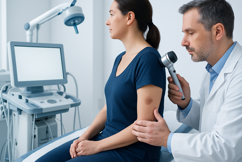
Seborrhoeic keratoses often appear as round or oval growths with a sharply defined edge. They may be tan, brown, black, or pale, and many have a slightly raised, warty, or greasy surface. Some people notice several lesions developing over time, especially with age.
Melanoma can look very different from one person to another, which is part of what makes it concerning. It may begin as a changing mole, a new dark spot, or an irregular patch that looks unlike the person’s other marks. The lesion may be asymmetric, have uneven borders, show more than one color, or enlarge over time.
Symptoms that should prompt a medical review include:
- A lesion that is new or changing in size, shape, or color
- Bleeding, crusting, or repeated irritation without a clear reason
- Itching, tenderness, or pain in a spot that was previously quiet
- A growth that looks different from the person’s other skin marks
- A dark lesion on the palm, sole, nail, or mucosal area
One challenge is that seborrhoeic keratoses can also be dark, raised, and irregular at the edges. In addition, some melanomas can appear deceptively bland. That is why a clinician’s eye, supported by dermoscopy and pathology when needed, matters more than a guess at home.
Causes and Risk Factors

Seborrhoeic keratosis is not caused by poor hygiene or skin injury. It is a common benign skin change that becomes more frequent with age and often runs in families. Sun exposure may play a role in some people, but it is not the same as a precancerous lesion.
Melanoma develops when pigment cells acquire DNA changes that allow them to grow abnormally. Risk increases with fair skin, a history of blistering sunburns, tanning bed use, many atypical moles, a personal or family history of melanoma, and weakened immune function. Some melanomas also arise in areas not strongly exposed to sunlight.
Several factors make the distinction especially important during a consultation:
- Older age, when seborrhoeic keratoses are common and new lesions may be overlooked
- Very dark or very light pigmented lesions that are hard to classify by color alone
- A lesion in a hidden location such as the back, scalp, under a nail, or on the foot
- Prior skin cancer or strong family history
Travel medicine and oncology teams often ask patients to bring photographs of changing lesions, previous pathology reports, and a history of how long the spot has been present. Those details can help narrow the possibilities before an in-person exam.
Diagnosis
The first step is a full skin assessment and a discussion of the lesion’s history: when it appeared, whether it has changed, and whether it bleeds or itches. A clinician may compare the spot with the person’s other moles and growths, which can be very revealing.
Dermoscopy is commonly used to look more closely at skin patterns that are not visible to the naked eye. Some seborrhoeic keratoses show characteristic surface features, while melanomas may reveal an irregular pigment network or other warning signs. Even so, dermoscopy does not replace pathology when the diagnosis is uncertain.
If a lesion cannot be confidently identified as benign, a biopsy may be recommended. This usually means removing all or part of the lesion under local anesthesia and sending the tissue to a pathology laboratory. The biopsy result is what confirms whether the growth is seborrhoeic keratosis, melanoma, or another skin condition.
For people traveling for evaluation, coordination matters. A team may plan the initial visit, same-day dermoscopic assessment, biopsy if indicated, and follow-up pathology review so that the patient does not need to make repeated international trips for a straightforward diagnostic step.
Treatment Options
When seborrhoeic keratosis is confirmed, treatment is often not medically necessary. Many people choose to leave the lesion alone if it is not bothersome. If removal is desired because of irritation, cosmetic concern, or repeated rubbing, common options include cryotherapy, curettage, electrocautery, or simple excision depending on the lesion and the clinician’s judgment.
Melanoma treatment is different because the goal is cancer control. The main treatment for many localized melanomas is surgical removal with an appropriate margin of healthy tissue. Depending on the depth and stage, the care plan may also involve lymph node assessment, imaging, or further oncology review.
In more advanced melanoma, treatment may include immunotherapy, targeted therapy, radiation in selected situations, or systemic cancer care tailored to the tumor’s biology and stage. The exact approach is individualized and guided by pathology, molecular testing in some cases, and specialist evaluation.
People sometimes hope that an over-the-counter cream or home remedy can settle an uncertain spot. That is not a safe approach when melanoma is in the differential diagnosis. A suspicious lesion should be evaluated before any attempt to treat or remove it at home.
Prevention and Self-care
There is no proven way to prevent every seborrhoeic keratosis. Because these growths are usually harmless, the focus is often on monitoring them and avoiding unnecessary irritation. Patients can reduce friction from clothing, jewelry, or shaving if a lesion is repeatedly rubbed.
Melanoma risk can be lowered by thoughtful sun protection. This includes shade, protective clothing, broad-spectrum sunscreen, and avoiding tanning beds. Regular skin self-checks are also useful, especially for people with many moles or a personal history of skin cancer.
A practical self-check habit is to look for the “ugly duckling” lesion — the spot that does not resemble the others. Digital photographs can help a person notice subtle changes over time, especially when a doctor has already identified which lesions are stable and which deserve closer monitoring.
For patients who live far from their specialist, self-care also includes keeping records: save clinical photos if permitted, keep biopsy reports, and note any new symptoms such as bleeding or rapid growth. That record can make remote follow-up much more efficient and clinically useful.
When to See a Doctor
Any new, changing, bleeding, or unusual skin lesion should be checked by a clinician, especially if it looks different from a person’s other spots. This is particularly important for lesions that are dark, irregular, rapidly enlarging, or located on the scalp, soles, palms, or under the nails.
People should seek prompt assessment if a lesion itches persistently, becomes painful, crusts repeatedly, or appears after middle age and changes quickly. A spot that seems to be a seborrhoeic keratosis but does not behave like one deserves careful review rather than reassurance alone.
For international patients, it can help to plan ahead: bring clear photos, a list of medications, any prior skin cancer history, and previous pathology if available. Acibadem Health Point’s multidisciplinary specialists and JCI-accredited hospitals diagnose and treat these conditions for international patients, with care pathways designed to support diagnosis, treatment, and follow-up across borders.
Ultimately, the safest rule is not to self-diagnose a suspicious pigmented lesion. A qualified doctor can determine whether reassurance is enough or whether biopsy and treatment are needed.
Living With a Benign Diagnosis or Recovering From Melanoma Care
If the lesion proves to be seborrhoeic keratosis, many people feel immediate relief. The main task after diagnosis is usually simple observation and routine skin awareness, unless the growth is being removed for comfort or appearance. Patients can ask whether any future changes would warrant another review.
After melanoma treatment, recovery may include wound care, pathology review, surveillance visits, and sometimes imaging or blood tests depending on stage and treatment plan. The follow-up schedule is individualized, because the aim is not only healing the skin but also detecting recurrence or new primary skin cancers early.
Good communication is especially helpful for patients who are returning home after treatment abroad. Before traveling, it is sensible to confirm who will manage wound checks, how pathology results will be shared, and what symptoms should trigger an earlier review. That planning can make follow-up safer and less stressful.
Frequently Asked Questions
Can seborrhoeic keratosis turn into melanoma?
Seborrhoeic keratosis itself is considered benign and does not transform into melanoma. The concern is that a melanoma can sometimes look similar at first glance, which is why uncertain lesions should be examined by a doctor.
Can a doctor tell the difference without a biopsy?
Sometimes yes, especially when the lesion has classic features on examination or dermoscopy. If there is any doubt, a biopsy is the most reliable way to confirm the diagnosis.
Are dark seborrhoeic keratoses more dangerous?
Dark color alone does not make a seborrhoeic keratosis dangerous. However, dark lesions can be harder to distinguish from melanoma, so changing or unusual spots deserve proper evaluation.
What does melanoma usually look like?
Melanoma often shows asymmetry, irregular borders, multiple colors, or growth over time. That said, not every melanoma follows the classic pattern, so a clinician should assess any concerning lesion rather than relying on a checklist alone.
Is removal always necessary for seborrhoeic keratosis?
No. If the lesion is confirmed to be benign and is not bothersome, it can usually be left alone. Removal is commonly chosen only if it irritates, catches on clothing, or causes cosmetic concern.
What should an international patient prepare before a skin lesion consultation?
Clear photos of the spot, dates showing when it changed, prior biopsy results, and a list of medications are useful. These details help the specialist decide whether dermoscopy, biopsy, or treatment should happen during the same visit.
This article is for general information only and is not a substitute for professional medical advice. Please consult a qualified doctor about your individual situation.
Oncology care in Turkey — second opinion and treatment plan
JCI-accredited · board-certified surgeons · reply within 24h
Add us as a Preferred Source to see more of our trusted health content across Google Search, AI Overviews and Discover.