Private Pregnancy Ultrasound Explained: Causes, Management, and When to See a Doctor

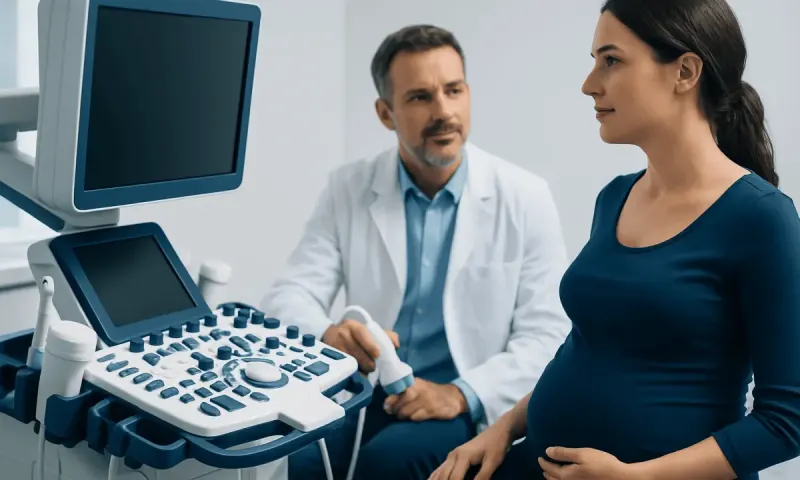
A private pregnancy ultrasound is an imaging appointment that can help confirm a pregnancy’s location, estimate gestational age and assess early development. The most useful timing depends on the reason for the scan, menstrual dates, symptoms and whether a transabdominal or transvaginal approach is used.
Private Pregnancy Ultrasound: an answer-first guide
A private pregnancy ultrasound is a clinical scan performed by qualified imaging and maternity professionals to assess an early pregnancy or monitor its progress. Depending on gestational age and the clinical question, it may help identify the pregnancy location, estimate how far along the pregnancy is, look for cardiac activity, check the number of embryos or fetuses, and assess pelvic structures.
It is important to know that an ultrasound does not always provide every answer at the first appointment. In the earliest weeks, normal development may simply be too small to see clearly. A clinician may recommend a repeat scan after an appropriate interval, sometimes together with blood tests for human chorionic gonadotropin (hCG), rather than drawing conclusions from a single early image.
A private scan should complement—not replace—routine antenatal care. The report should be reviewed with a qualified doctor or maternity professional, particularly if there is pain, bleeding, uncertain dates, a history of ectopic pregnancy, fertility treatment, or any concern about the result.
What an early pregnancy ultrasound can assess
Ultrasound uses sound waves rather than radiation to create images. In early pregnancy, a scan may be performed through the abdomen (transabdominal ultrasound) or internally using a slim vaginal probe (transvaginal ultrasound). The transvaginal method often produces more detailed images in the first trimester because the probe is closer to the uterus and early pregnancy structures.
Depending on timing, the sonographer may look for a gestational sac within the uterus, a yolk sac, an embryo or fetal pole, and cardiac activity. Measurements such as crown-rump length can be used to estimate gestational age once the embryo is visible. The scan may also identify whether there is more than one developing embryo.
Early imaging can be particularly important when symptoms raise concern for an ectopic pregnancy, meaning a pregnancy developing outside the uterus. A scan may also assess the ovaries and nearby pelvic area, although a diagnosis is not always possible immediately. For more information about concerning early-pregnancy symptoms, see ectopic pregnancy.
- Dating scan: estimates gestational age and helps establish an expected due date.
- Viability scan: checks development and whether cardiac activity is seen when it would be expected.
- Location scan: helps determine whether the pregnancy is inside the uterus.
- Reassurance scan: may be requested after symptoms or prior pregnancy concerns, but should still have a clear clinical purpose.
Is 4 weeks pregnant too early to see a doctor?

No. At 4 weeks pregnant, it is not too early to contact a doctor, midwife or maternity service after a positive home pregnancy test. Early contact supports advice about folic acid, current medicines, lifestyle, medical conditions and arranging appropriate antenatal care. It is especially important to seek advice promptly if the person has pain, bleeding, prior ectopic pregnancy, previous recurrent miscarriage, fertility treatment, or conditions that may affect pregnancy.
However, 4 weeks can be too early for an ultrasound to show a definite intrauterine pregnancy. Pregnancy weeks are usually counted from the first day of the last menstrual period, so at “4 weeks pregnant,” conception may have occurred only around two weeks earlier. A gestational sac may not yet be visible, particularly if ovulation occurred later than expected.
When there are no concerning symptoms, a clinician may advise waiting until around 6 to 7 weeks from the last menstrual period for a more informative early scan. If symptoms or risk factors are present, assessment should not be delayed; clinicians may use repeat ultrasound and serial hCG testing to understand what is happening safely.
What not to do before a pregnancy ultrasound?
Preparation depends on the type of ultrasound scheduled. For an early transabdominal scan, a moderately full bladder may improve visibility by creating an acoustic window. The imaging provider will usually give specific instructions, and it is best to follow those rather than drinking excessive amounts of water. For a transvaginal scan, an empty bladder is commonly preferred.
There is usually no need to fast, stop ordinary meals or change prescribed medication before a pregnancy ultrasound unless a clinician gives a specific instruction. A person should not stop prescribed medicines, supplements or treatment for a health condition based only on an upcoming scan. Questions about medication safety in pregnancy should be discussed with the prescribing clinician or maternity team.
It is also best not to rely on a scan alone for urgent symptoms. Pain, bleeding, dizziness or feeling unwell should be reported when booking or attending the appointment. Bringing the date of the last menstrual period, details of cycle regularity, previous scan reports, fertility treatment information and relevant medical history can help the clinician interpret findings accurately.
What will not show up on an ultrasound?
Not every pregnancy feature is visible at every stage. Before the pregnancy is far enough along, a scan may not show a gestational sac, yolk sac, embryo or cardiac activity—even in a normally developing pregnancy. Visibility also depends on the accuracy of pregnancy dates, the type of scan, image quality, the position of the uterus and individual anatomy.
An ultrasound cannot identify every cause of early-pregnancy symptoms or predict every future pregnancy outcome. It may not explain bleeding, cramping or pregnancy symptoms when the images appear reassuring. It also cannot detect all fetal conditions, and early scans are not a substitute for recommended first-trimester screening, anatomy scanning and other prenatal testing when appropriate.
Very early scans may also be unable to confirm the location of a pregnancy. This situation may be described as a pregnancy of unknown location. It requires careful clinical follow-up because it can represent a very early intrauterine pregnancy, a miscarriage in progress, or less commonly an ectopic pregnancy. Repeat testing is often the safest and most informative next step.
I am 7 weeks pregnant but my ultrasound showed only 5 weeks. Is this normal?
A scan measurement that appears two weeks behind menstrual dates can occur for several reasons and does not automatically mean there is a problem. The most common explanation is that ovulation or implantation happened later than assumed. This is particularly common with irregular cycles, recent hormonal contraception, breastfeeding, polycystic ovary syndrome, or uncertainty about the date of the last period.
At 7 weeks by menstrual dates, a transvaginal scan may often show an embryo and cardiac activity, but findings vary. If the scan appears closer to 5 weeks, the clinician may recommend a repeat ultrasound after an appropriate interval. The purpose is to look for expected growth and developmental change rather than to make a diagnosis too early.
Occasionally, measurements that do not progress as expected can indicate an early pregnancy loss. This should only be diagnosed using established ultrasound criteria and, when needed, repeat assessment by qualified clinicians. If there is bleeding, worsening cramps, one-sided pain, dizziness or fainting, urgent medical evaluation is needed.
When to seek medical care
Anyone with a positive pregnancy test should arrange routine maternity care, even if they feel well. A timely appointment allows a healthcare professional to review health history, medicines, nutrition, supplements and any individual risks. A private pregnancy ultrasound can be arranged when it has a clear clinical purpose and is integrated with appropriate medical follow-up.
Urgent medical care is needed for heavy vaginal bleeding, severe or increasing abdominal or pelvic pain, pain mainly on one side, shoulder-tip pain, fainting, marked dizziness, fever, or feeling severely unwell. These symptoms may have several causes, but they require prompt assessment in pregnancy because ectopic pregnancy and other complications need to be excluded.
For Myomectomy Recovery for International Patients: The Usual Travel Timeline" class="ahp-ilk">international patients, Acıbadem Health Point’s multidisciplinary specialists and JCI-accredited hospitals can assess pregnancy-related symptoms and provide diagnostic imaging and maternity care. When ultrasound findings are uncertain, the clinical team may arrange follow-up imaging and relevant laboratory testing rather than relying on a single result.
Frequently asked questions
01When is the best time for a private pregnancy ultrasound?
The best time depends on why the scan is needed. For a scan intended to confirm location, development and possible cardiac activity, around 6 to 7 weeks from the first day of the last menstrual period is often more informative than scanning earlier. Symptoms such as bleeding or pain should be assessed promptly regardless of gestational age.
02Is transvaginal ultrasound safe in early pregnancy?
Transvaginal ultrasound is widely used in early pregnancy and is generally considered safe when performed by trained professionals. It does not use ionising radiation and does not enter the uterus. It may provide clearer images than an abdominal scan in the earliest weeks.
03Can an ultrasound confirm pregnancy at 4 weeks?
It may be too early to confirm a pregnancy on ultrasound at 4 weeks, particularly if dates are based on the last menstrual period. A pregnancy test may be positive before a gestational sac can be seen. A clinician may recommend repeat ultrasound or hCG blood tests if confirmation is needed.
04Do I need a full bladder for an early pregnancy scan?
A full bladder may be requested for a transabdominal early-pregnancy ultrasound because it can improve the view of the uterus. For a transvaginal ultrasound, an empty bladder is generally preferred. The imaging centre should provide instructions before the appointment.
05Can stress or exercise cause an ultrasound to measure behind?
Everyday stress or usual activity does not typically explain a difference between menstrual dates and ultrasound measurements. Differences are more often related to variation in ovulation timing, implantation timing or uncertainty about menstrual dates. A follow-up scan can clarify whether development is progressing as expected.
06Can a private pregnancy ultrasound replace antenatal appointments?
No. An ultrasound is one part of pregnancy care and does not replace routine antenatal assessments, screening discussions, blood tests or medical review. Results should be shared with the person’s maternity provider so they can be interpreted in the context of the whole pregnancy.
This article is for general information only and is not a substitute for professional medical advice. Please consult a qualified doctor about your individual situation.
Heart care in Turkey — expert evaluation and treatment
JCI-accredited · board-certified surgeons · reply within 24h
Add us as a Preferred Source to see more of our trusted health content across Google Search, AI Overviews and Discover.