Private Breast Screening: Tests, Timing and Results
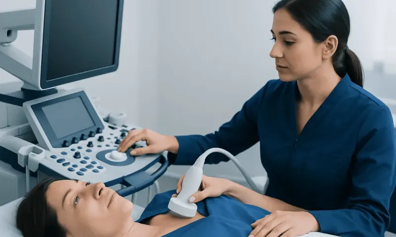
Private breast screening is an individualised assessment that may include a mammogram, review of personal risk factors and, when appropriate, ultrasound or further imaging. It is designed to identify breast changes early, but it does not replace prompt medical assessment of a new lump, nipple change or other symptom.
Private Breast Screening: An Overview
Private breast screening is a planned breast health assessment arranged outside a population screening programme. It commonly includes a discussion of health history and breast cancer risk, imaging such as mammography, and clear follow-up advice if an image needs closer review. The purpose is to find certain breast cancers or other changes before they cause symptoms, when Uterine fibroids Treatment Options: From Medication to Surgery" class="ahp-ilk">treatment options may be simpler.
Screening is intended for people who do not have a current breast concern. A person with a lump, persistent focal breast pain, skin dimpling, nipple discharge or a new nipple change needs diagnostic assessment rather than routine screening. A recent normal mammogram is reassuring, but it should not delay an appointment for a new symptom.
A private appointment can offer flexibility in scheduling and may allow a clinician to tailor imaging to factors such as age, breast density, previous breast procedures, family history and known genetic risk. It is important to choose an accredited imaging service where results are interpreted by appropriately trained breast radiologists and where further assessment can be arranged if needed.
What Happens During a Private Breast Screening Appointment?
Before imaging, the healthcare team usually asks about previous mammograms, breast symptoms, hormone use, pregnancies, breast operations, family history and any known inherited cancer-related gene variant. Bringing prior mammogram images and reports, if available, can help the radiologist compare changes over time and may reduce the chance of being called back for additional views.
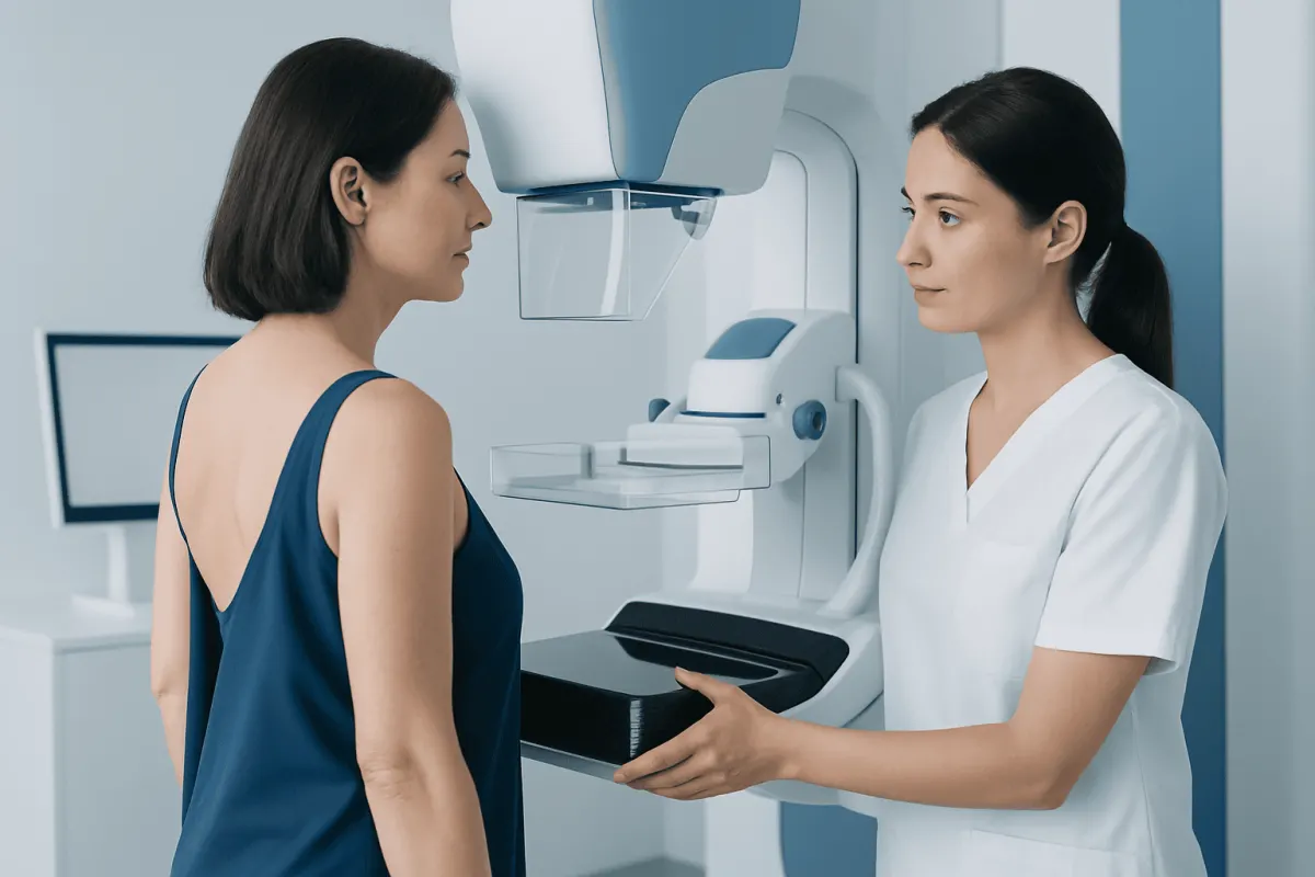
For most people, mammography is the main screening examination. The breast is positioned and briefly compressed between plates while X-ray images are taken. Compression can be uncomfortable, but it lasts only a short time and helps produce clear images using a low radiation dose. The test itself usually takes only a few minutes.
Some individuals may also be advised to have ultrasound or magnetic resonance imaging (MRI). These tests are not automatic replacements for mammography. They are selected according to the clinical situation, particularly for people with dense breasts, a significant family history, a genetic predisposition or an abnormality requiring clarification.
Results may be reported as normal, probably benign with planned monitoring, or needing further assessment. A recall for extra images or ultrasound is common and does not mean cancer is present. Most recalled findings prove not to be cancer, but completing the recommended assessment is important.
What Is the Difference Between a Mammogram and a Breast Screening?
A mammogram is a specific imaging test that uses low-dose X-rays to look for changes in breast tissue. It can identify some cancers before they can be felt and may show small calcium deposits, masses or distortions in breast structure. Mammograms may be performed as either screening tests or diagnostic tests.
Breast screening is the broader preventive process. It includes deciding whether a person should have screening, reviewing their level of risk, carrying out the appropriate test and arranging follow-up. For many average-risk adults, mammography is the central part of breast screening, but the screening plan may also include risk counselling and decisions about supplementary imaging.
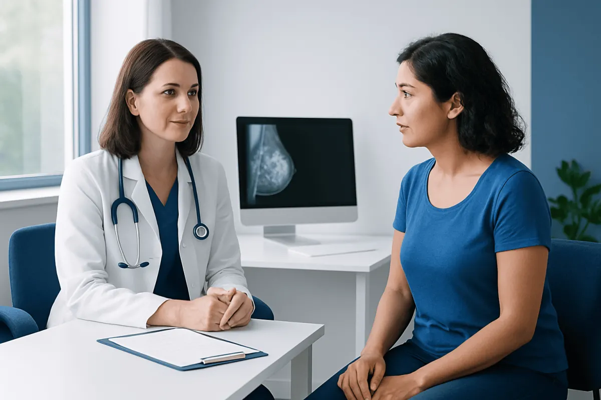
A diagnostic mammogram is different from a routine screening mammogram because it is used to investigate a symptom or an area seen on prior imaging. Diagnostic assessment may involve additional mammographic views, targeted ultrasound, MRI or a biopsy. If cancer is diagnosed, care may involve specialists in breast cancer and related treatment services.
What Are the New Guidelines for Mammogram Screening in 2026?
There is no single worldwide mammogram screening guideline in 2026. Recommendations differ between health authorities because they weigh potential benefits and possible harms, such as false-positive results, overdiagnosis and additional testing, somewhat differently. A person should follow the guidance applicable in their country while discussing individual risk factors with a clinician.
In the United States, the U.S. Preventive Services Task Force recommends mammography every two years from ages 40 to 74 for women at average risk. The American Cancer Society advises that women aged 45 to 54 have mammography every year, with the option to begin annual screening from age 40; from age 55, screening may continue yearly or change to every two years. Other professional organisations commonly support beginning annual or biennial screening at age 40.
People with a high risk of breast cancer may need earlier and more intensive surveillance, often involving annual mammography and breast MRI. High risk can include a known harmful genetic variant, prior chest radiation at a young age, certain high-risk biopsy findings, or a strong family history. A specialist can help establish a personalised plan rather than relying only on age-based guidance.
Screening decisions after age 74 are usually individualised. Overall health, life expectancy, prior screening history and personal preferences all matter. Screening should also be considered in transgender and gender-diverse people according to the breast tissue present, prior surgery, hormone exposure and individual risk.
Why Are Breast Exams No Longer Recommended?
Routine clinician breast examinations and formal monthly breast self-exams are no longer recommended as stand-alone screening methods for average-risk women by many major organisations. Studies have not shown that these approaches reduce breast cancer deaths when mammography is available, and they can lead to false alarms, unnecessary imaging and avoidable biopsies.
This does not mean people should ignore their breasts or avoid discussing changes. Many experts encourage breast awareness: knowing what is normal for the individual and reporting a new or persistent change without delay. Breast awareness is not a prescribed monthly examination technique; it is an ongoing familiarity with the appearance and feel of the breasts.
Healthcare professionals may still examine the breasts when a person reports symptoms, has a higher-risk history, attends a relevant clinical appointment or needs diagnostic evaluation. An examination can provide useful information, but it cannot reliably detect every early cancer and does not replace appropriate imaging.
Is a 3D Mammogram Covered by Insurance?
A 3D mammogram, also called digital breast tomosynthesis, creates multiple thin image slices of the breast. It may make it easier to assess overlapping tissue and can be useful for many patients, including some with dense breasts. Whether it is used depends on local availability, radiologist practice and the person’s clinical needs.
Insurance coverage for a 3D mammogram varies considerably. In some countries or insurance plans, it is covered as part of routine mammography screening; in others, it may require specific eligibility, prior authorisation or an additional payment. Coverage can also differ between screening and diagnostic imaging.
Before an appointment, the person can contact their insurer or benefits administrator to ask whether screening mammography, tomosynthesis, ultrasound, MRI, radiologist interpretation and any additional diagnostic imaging are covered. The imaging centre may also be able to explain the service offered, although it cannot determine an insurer’s final payment decision.
When to Seek Medical Care
A person should arrange prompt medical assessment for a new breast lump or thickened area, a change in breast shape, persistent swelling, skin puckering or redness, a newly inverted nipple, a rash around the nipple, or nipple discharge that is bloody or occurs without squeezing. Persistent pain in one specific area also deserves evaluation, especially when it is new or associated with another change.
Most breast changes are not caused by cancer. Hormonal changes, benign cysts, fibroadenomas, infections and other non-cancerous conditions can all affect the breasts. However, it is not possible to determine the cause reliably at home, so a clinician may recommend examination and diagnostic imaging.
Urgent care is appropriate for fever with a painful, red, hot or swollen breast, especially during breastfeeding, because infection may need timely treatment. People with a known high-risk genetic variant or a strong family history should also seek advice about a tailored surveillance plan, even if they have no symptoms.
Acıbadem Health Point’s multidisciplinary specialists and JCI-accredited hospitals can assess breast symptoms and coordinate imaging, biopsy and breast cancer treatment when required for Myomectomy Recovery for International Patients: The Usual Travel Timeline" class="ahp-ilk">international patients.
Frequently asked questions
01Who should consider private breast screening?
People who are due for routine mammography, have limited access to a public screening programme or want an individual risk review may consider private breast screening. It can be particularly useful when a person has dense breasts, a significant family history or previous breast imaging that needs comparison. A clinician can advise which tests are appropriate.
02Can breast screening detect every breast cancer?
No screening test detects every breast cancer. Mammography is effective at finding many early changes, but its sensitivity can be lower in dense breast tissue and some cancers develop between screenings. New symptoms should always be assessed, even after a normal result.
03Is mammography safe?
Mammography uses a small amount of radiation, and the expected benefit of recommended screening generally outweighs this exposure for eligible people. Compression is brief and may be uncomfortable, but it is necessary for clear images and accurate interpretation. Anyone who is pregnant or might be pregnant should tell the imaging team before the examination.
04Do dense breasts mean breast cancer is present?
No. Dense breasts are common and describe the proportion of glandular and fibrous tissue compared with fatty tissue on a mammogram. Density can slightly increase breast cancer risk and can make mammograms harder to interpret, so a clinician may discuss whether any additional imaging is appropriate.
05How often should high-risk people have breast screening?
The schedule depends on the reason for increased risk. Many high-risk people are advised to have yearly mammography and breast MRI, often starting earlier than average-risk screening. A breast specialist or genetics professional can determine the most suitable plan.
06What happens if a screening result is abnormal?
An abnormal screening result usually means more information is needed; it does not automatically mean cancer. The next steps may include additional mammogram views, ultrasound, MRI or a needle biopsy. The care team should explain the result, the purpose of each test and the expected timeframe for follow-up.
This article is for general information only and is not a substitute for professional medical advice. Please consult a qualified doctor about your individual situation.
Oncology care in Turkey — second opinion and treatment plan
JCI-accredited · board-certified surgeons · reply within 24h
Add us as a Preferred Source to see more of our trusted health content across Google Search, AI Overviews and Discover.