PET Imaging in Cancer Diagnosis, Staging, and Follow-Up
PET imaging for cancer is a nuclear medicine test that shows areas of increased biological activity, helping clinicians detect, stage, and monitor many cancers. It is usually interpreted together with CT, MRI, laboratory results, symptoms, and—when needed—a biopsy, rather than used as a stand-alone cancer diagnosis.
How PET Imaging Helps in Cancer Care
PET imaging for cancer is a scan that helps doctors see how tissues are functioning, not only how they look. Most cancer PET scans use a radiotracer called fluorodeoxyglucose (FDG), a glucose-like substance. Many cancer cells use glucose more actively than surrounding tissue, so they may appear as areas of increased tracer uptake on the images.
In practice, PET is often combined with computed tomography as a PET-CT scan. CT provides detailed anatomical information, while PET shows tracer activity in the same area. Some centers also use PET-MRI for selected situations. This combined approach can help the cancer team understand whether a finding is likely to be active disease and where it is located.
PET is an important tool, but it is not a substitute for clinical assessment or tissue diagnosis. A scan may raise or lower concern for cancer, but a biopsy is often needed to confirm the diagnosis and identify the cancer type. The value of PET depends on the individual cancer, the clinical question, and the timing of the scan.
When Doctors May Recommend a PET Scan
A doctor may request PET imaging at different points in a cancer pathway. It can help determine the extent of a newly diagnosed cancer, a process called staging. Staging identifies whether disease appears limited to one site or has involved lymph nodes or other organs, which supports treatment planning and discussion of outlook.
PET imaging may also be used when a symptom, blood test, or another scan suggests possible recurrence after treatment. In some cancers, it can evaluate how a tumor is responding during or after chemotherapy, immunotherapy, radiation therapy, or other systemic treatment. A reduction in tracer uptake can be encouraging, although the meaning of a result must be judged in context.
It is particularly useful in many cases of lymphoma, lung cancer, head and neck cancers, melanoma, colorectal cancer, esophageal cancer, and some gynecological cancers. It may also help in selected prostate cancer cases using prostate-specific membrane antigen (PSMA) tracers. However, not every cancer requires PET imaging, and some slow-growing tumors or very small lesions may be less visible on FDG PET.
- Clarifying suspicious findings seen on CT, MRI, or other imaging
- Staging a confirmed or strongly suspected cancer
- Identifying a suitable site for biopsy
- Planning surgery or radiation therapy
- Assessing treatment response or suspected recurrence
What a PET Scan Can and Cannot Show

Areas with increased uptake are often described as “PET-avid” or “hypermetabolic.” These descriptions indicate tracer activity; they do not, by themselves, establish that cancer is present. Infection, inflammation, recent surgery, radiation-related changes, and normal activity in some organs can also cause uptake. The brain, heart, kidneys, bladder, and bowel may show normal or variable tracer activity depending on the tracer and examination.
Likewise, a scan that does not show abnormal uptake is reassuring in some circumstances but cannot rule out all malignancies. Small tumors may be below the scan’s resolution, and some cancers do not use FDG intensely. Certain well-differentiated, slow-growing, mucin-producing, or low-grade tumors may be less conspicuous. Blood glucose levels, movement during the scan, and timing after treatment can also affect image interpretation.
Radiologists and nuclear medicine physicians assess the images alongside the CT or MRI component, prior scans, pathology results, and medical history. When a finding could change treatment, the team may recommend targeted imaging, repeat assessment after an interval, or a biopsy rather than making a decision from PET findings alone.
Preparing for PET Imaging and What Happens During the Scan
Preparation instructions vary by tracer and the reason for imaging. For a typical FDG PET-CT, patients are commonly asked to avoid strenuous exercise beforehand and to fast for several hours, while drinking plain water unless told otherwise. Exercise can increase muscle uptake, and food intake can influence blood glucose and tracer distribution. People with diabetes need individualized instructions because medication, meals, and glucose control require careful planning.
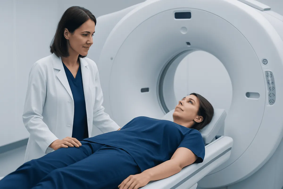
At the appointment, a small amount of radiotracer is injected into a vein. There is usually a quiet waiting period while the tracer circulates through the body. The scan itself is painless. The patient lies still on a padded table that moves through the scanner; the imaging portion often takes around 20 to 40 minutes, although total visit time is longer because of preparation and uptake time.
The radiation exposure from PET-CT is considered in relation to its expected clinical benefit. The care team avoids unnecessary imaging and selects the most appropriate test for the question being asked. Patients should tell the imaging team about pregnancy, possible pregnancy, breastfeeding, diabetes, recent infection or vaccination, surgery, radiation therapy, and any difficulty lying flat or remaining still.
PET Results, Cancer Outlook, and Treatment Decisions
PET findings can influence cancer outlook because they may provide a clearer picture of disease extent and biological activity. Still, a PET result is only one part of prognosis. Cancer type, stage, pathology, molecular features, general health, response to treatment, and available treatment options all matter. A scan should not be interpreted as a prediction for one individual without a full discussion with the treating specialist.
For some cancers, PET helps determine whether treatment is intended to remove or control localized disease, treat disease in multiple areas, or relieve symptoms. It may support planning for radiation therapy by identifying active areas that need careful targeting. It can also help clinicians determine whether systemic treatment, surgery, surveillance, or further investigation is appropriate.
When a scan suggests possible active cancer after treatment, clinicians consider the timing carefully. Inflammation from surgery or radiation can persist and may mimic disease on PET. For this reason, repeat imaging, comparison with earlier scans, or biopsy may be recommended before major treatment decisions are made. For patients with lymphoma, PET imaging is often integrated into staging and response assessment, though protocols differ by lymphoma subtype.
Understanding Common PET Report Terms
A PET report may use technical terms that are easy to misinterpret when read without clinical context. “Uptake” means that the tracer has collected in a tissue. “Avid” or “increased uptake” means more tracer activity than expected in that region, while “no evidence of PET-avid disease” means no clearly abnormal tracer-avid lesions were identified on that examination.
Reports may also mention a standardized uptake value, or SUV. This is a semi-quantitative measurement of tracer uptake. A higher SUV does not automatically mean a more aggressive cancer, because SUV can be influenced by tissue type, inflammation, scan technique, blood glucose, and other factors. Values are most useful when interpreted by specialists and, in some situations, compared over time under similar scanning conditions.
It is reasonable for patients to ask which finding is most important, whether it is new compared with prior imaging, and whether it changes the next step. The referring doctor can explain how the report fits with pathology and the overall treatment plan. Keeping copies of prior imaging reports and, when possible, image files can be helpful when care is shared between hospitals.
When to Seek Medical Care
Patients should contact their cancer specialist promptly if they develop new or worsening symptoms that may require assessment, such as persistent unexplained pain, increasing shortness of breath, new neurological symptoms, unexplained weight loss, prolonged fever, or a new lump. These symptoms have many possible causes and do not necessarily indicate cancer progression, but timely review is important.
Urgent medical assessment is appropriate for severe chest pain, sudden weakness or difficulty speaking, significant breathing difficulty, confusion, uncontrolled bleeding, or a high fever during chemotherapy or other treatment that can affect infection defenses. The local emergency service should be used for severe or rapidly worsening symptoms.
Questions about a PET scan result should be discussed with the clinician who ordered it, ideally before drawing conclusions from the written report alone. Acıbadem Health Point’s multidisciplinary specialists and JCI-accredited hospitals evaluate cancer imaging and coordinate diagnostic and treatment planning for international patients.
Frequently asked questions
01Can a PET scan diagnose cancer by itself?
No. PET imaging can identify areas that may need further investigation, but increased tracer uptake can also occur with infection, inflammation, or healing tissue. A biopsy is often required to confirm cancer and determine its specific type.
02What is the difference between PET and PET-CT?
PET shows metabolic or tracer activity in the body. PET-CT combines this information with CT images, which show anatomy and help localize an area of uptake more precisely. PET-CT is widely used in cancer assessment.
03Does a normal PET scan mean there is no cancer?
Not always. Some cancers are too small to detect or have low uptake of the tracer used, and scan sensitivity differs among tumor types. Doctors interpret a normal scan alongside symptoms, examination findings, laboratory tests, and other imaging.
04Can inflammation cause an abnormal PET scan?
Yes. Inflammation, infection, recent surgery, radiation-related changes, and some benign conditions can cause increased uptake. This is why an abnormal result may require comparison with other tests, follow-up imaging, or biopsy.
05How should a person prepare for an FDG PET scan?
The imaging center will provide individual instructions, which commonly include fasting for several hours and avoiding strenuous activity beforehand. People with diabetes, pregnancy concerns, or breastfeeding should notify the team in advance because preparation may need to be adjusted.
06How soon are PET scan results available?
Images are reviewed by a nuclear medicine physician or radiologist, and the report is sent to the referring clinician. Timing varies by facility and clinical urgency. The treating doctor is best placed to explain what the findings mean for the individual care plan.
This article is for general information only and is not a substitute for professional medical advice. Please consult a qualified doctor about your individual situation.
Oncology care in Turkey — second opinion and treatment plan
JCI-accredited · board-certified surgeons · reply within 24h
Add us as a Preferred Source to see more of our trusted health content across Google Search, AI Overviews and Discover.