Pancreas Ultrasound: How This Scan Evaluates the Abdomen
A pancreas ultrasound is a painless imaging examination that uses high-frequency sound waves to create images of the pancreas and surrounding upper abdominal organs. It can help investigate symptoms such as Abdominal Pain: Causes and Treatment" class="ahp-ilk">upper abdominal pain or jaundice, although other tests may be needed when the pancreas cannot be seen clearly.
What Is a Pancreas Ultrasound?
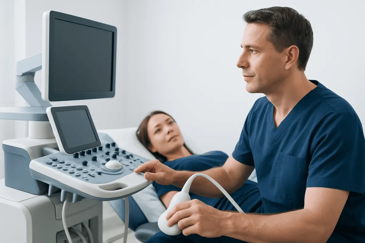
A pancreas ultrasound, usually performed as part of an abdominal ultrasound, is an imaging test that uses sound waves to produce pictures of organs inside the abdomen. A small handheld device called a transducer sends sound waves through the skin and receives the returning echoes. A computer converts these echoes into real-time images.
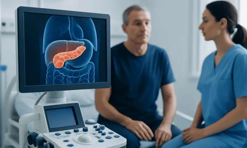
The pancreas is an organ located deep in the upper abdomen, behind the stomach. It makes digestive enzymes and hormones, including insulin, that help regulate blood glucose. Because the pancreas is partly hidden by the stomach and bowel, it may not always be fully visible on a standard ultrasound. Even so, ultrasound is often a useful first examination because it is quick, widely available, and does not involve ionising radiation.
During the same appointment, the sonographer or radiologist commonly examines nearby structures. These may include the liver, gallbladder, bile ducts, spleen, kidneys, aorta and other blood vessels. This broader view can help identify other possible explanations for abdominal symptoms.
Why Might a Doctor Request This Scan?
A doctor may recommend a pancreas ultrasound when a person has symptoms that could be related to the pancreas, gallbladder, liver or bile ducts. Common reasons include persistent upper abdominal discomfort, pain that may spread to the back, nausea, vomiting, unexplained Acid Reflux Weight Loss: A Complete Medical Overview" class="ahp-ilk">weight loss, or yellowing of the skin or eyes. The test may also be requested after abnormal liver or pancreatic blood test results.
Ultrasound is especially helpful for looking for gallstones, enlargement of the bile ducts, fluid collections, cyst-like changes, or changes in the appearance of the liver and gallbladder. Gallstones can sometimes obstruct bile flow and contribute to inflammation of the pancreas, known as pancreatitis. The examination may therefore help clinicians assess possible causes of symptoms rather than evaluate the pancreas in isolation.
In some situations, the scan is used to follow an earlier finding or to assess abdominal symptoms in people with a history of pancreatic, liver, or gallbladder conditions. It is not generally used as a routine screening test for pancreatic cancer in people without symptoms or a high-risk medical history.
How to Prepare for a Pancreas Ultrasound

Preparation instructions vary between imaging centres, so patients should follow the guidance provided with their appointment. Many centres ask adults not to eat for approximately six to eight hours beforehand. Fasting reduces food and gas in the stomach and bowel, which can make the pancreas and gallbladder easier to see.
Small amounts of water may often be permitted, and regular medicines can usually be taken with a sip of water. However, people who use insulin or other diabetes medicines, take medications that must be taken with food, are pregnant, or have other health concerns should ask the requesting clinician or imaging department for individual instructions before fasting.
It is helpful to wear comfortable, loose-fitting clothing. Patients may be asked to lift clothing around the upper abdomen or change into a gown. Bringing previous scan reports, a medication list, and relevant medical information can also help the care team compare findings and provide appropriate follow-up.
- Confirm the fasting instructions when booking the appointment.
- Tell the team about diabetes, pregnancy, recent surgery, or difficulty lying flat.
- Ask whether prescribed medicines should be taken as usual.
- Arrive in time to complete any required registration paperwork.
What Happens During the Examination?
A pancreas ultrasound is usually performed by a trained sonographer, with images reviewed by a radiologist or another qualified clinician. The patient lies on an examination couch, generally on the back. A clear water-based gel is placed on the skin of the upper abdomen to help sound waves travel effectively between the transducer and the body.
The sonographer moves the transducer over different areas of the abdomen and may apply gentle pressure. The patient may be asked to take a deep breath, hold their breath briefly, or turn onto one side. These changes can move bowel gas and improve the view of deeper organs. The procedure is usually painless, although pressure over a tender area may cause temporary discomfort.
The examination commonly takes about 15 to 30 minutes, depending on the reason for the scan and how clearly the organs can be viewed. There are no injections, no needles, and no recovery period for a routine external abdominal ultrasound. Most people can return to usual activities immediately afterwards.
What Can the Results Show?
A report may describe the size, shape and visible texture of the pancreas, as well as whether the pancreatic duct appears enlarged. It may also record whether there are visible cysts, fluid collections, calcifications, or a focal area that needs further assessment. A normal report can be reassuring, but it does not always exclude every pancreatic condition because some parts of the organ may be obscured by bowel gas or body tissue.
Findings involving the gallbladder and bile ducts are often important. For example, gallstones or a widened bile duct may help explain abdominal pain, jaundice, or abnormal liver blood tests. The report may also identify findings unrelated to the pancreas, such as fatty change in the liver, kidney cysts, or enlargement of the abdominal aorta. These findings vary in significance and should be discussed with the clinician who ordered the test.
An abnormal appearance on ultrasound does not by itself establish a diagnosis. Inflammation, infection, benign cysts, scarring, and tumours can sometimes have overlapping features. The doctor may consider symptoms, examination findings, blood tests such as pancreatic enzyme and liver tests, and previous imaging before explaining what the result means.
Limits of Ultrasound and Possible Next Steps
The usefulness of a pancreas ultrasound depends partly on how well the pancreas can be seen. Gas in the bowel is the most common obstacle, which is one reason fasting is often advised. Body shape, abdominal scarring, and the deep position of the pancreas may also limit detail. A report may state that the pancreas was partially visualised or not well seen; this does not necessarily mean that a problem is present.
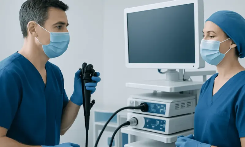
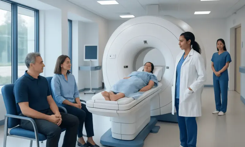
If symptoms continue, blood tests are abnormal, or the ultrasound identifies an area that requires clarification, the doctor may recommend another test. A CT scan can provide detailed cross-sectional images and may be useful in urgent abdominal conditions. MRI and magnetic resonance cholangiopancreatography (MRCP) can assess the pancreas and bile ducts without radiation. Endoscopic ultrasound, performed using a flexible tube passed through the mouth into the digestive tract under sedation, can produce close-up images and may allow tissue sampling when clinically appropriate.
The next step is individual. Some people need no further testing, while others may need repeat imaging, specialist review, or treatment for an identified cause such as gallstones. Patients should avoid interpreting a scan report alone and should arrange follow-up with the clinician who requested the examination.
When to Seek Medical Care
People should arrange medical assessment for ongoing or recurrent upper abdominal pain, especially when it affects eating, sleep, or daily activities. Evaluation is also appropriate for unexplained loss of appetite or weight, repeated nausea or vomiting, pale stools, dark urine, or new yellowing of the eyes or skin. These symptoms can have many causes, and timely assessment helps determine the appropriate tests.
Urgent medical care is important for severe or rapidly worsening abdominal pain, particularly if it occurs with fever, persistent vomiting, fainting, confusion, chest pain, or yellowing of the skin or eyes. These symptoms may require prompt evaluation and should not be managed only with an outpatient ultrasound appointment.
For international patients who require further assessment, Acıbadem Health Point’s multidisciplinary specialists and JCI-accredited hospitals can help diagnose and treat pancreatic and related digestive conditions. The most suitable care plan depends on the patient’s symptoms, scan findings, medical history, and any necessary additional tests.
Frequently asked questions
01Is a pancreas ultrasound painful?
A pancreas ultrasound is usually painless. The transducer is moved over gel on the skin, and the sonographer may apply gentle pressure to obtain clearer images. There may be mild temporary discomfort if the abdomen is already tender.
02Do patients need to fast before a pancreas ultrasound?
Many imaging centres ask patients to avoid food for about six to eight hours before an abdominal ultrasound. This can reduce bowel gas and improve visibility of the pancreas and gallbladder. Instructions differ, so patients should follow the advice from their imaging centre and ask about medicines if needed.
03Can an ultrasound detect pancreatitis?
Ultrasound can sometimes show signs that may support pancreatitis or identify gallstones that could be contributing to it. However, the pancreas may be difficult to see clearly, and blood tests and CT or MRI may be needed to confirm the diagnosis or assess severity. A clinician interprets all of these findings together.
04Can a pancreas ultrasound detect cancer?
An ultrasound may identify a mass or an indirect sign, such as widening of a bile duct, but it cannot reliably exclude pancreatic cancer. Small lesions and areas hidden by bowel gas may not be visible. If there is a concerning symptom, blood test, or scan finding, doctors may recommend CT, MRI, or endoscopic ultrasound.
05How long does it take to get pancreas ultrasound results?
The timing varies by healthcare setting. Images are often reviewed after the examination, and a formal report is sent to the requesting clinician, who discusses the result and any next steps. Urgent findings are generally communicated promptly through the relevant clinical pathway.
06Can patients eat normally after the scan?
Yes, most people can eat, drink, drive, and return to usual activities immediately after a routine external ultrasound. There are no sedatives or contrast injections involved in a standard scan. If the patient has been given different instructions for another test on the same day, those instructions should be followed.
This article is for general information only and is not a substitute for professional medical advice. Please consult a qualified doctor about your individual situation.
Medical weight-loss options in Turkey — costs & eligibility
JCI-accredited · board-certified surgeons · reply within 24h
Add us as a Preferred Source to see more of our trusted health content across Google Search, AI Overviews and Discover.