Ovarian Cyst Diagnosis: Exams, Ultrasound, and Tests
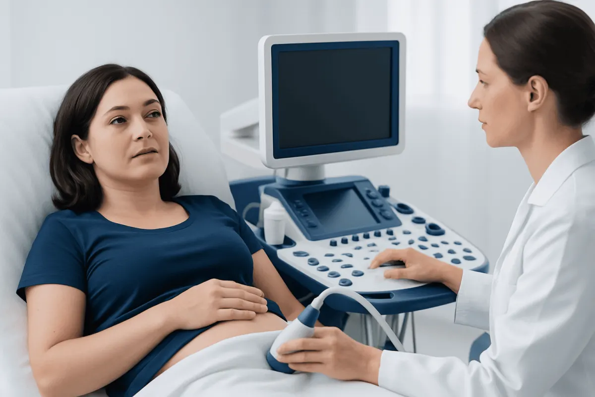
Ovarian cyst diagnosis commonly starts with a discussion of symptoms and a pelvic examination, followed by ultrasound imaging to identify the cyst’s size, location and appearance. Most cysts are benign and may resolve without treatment, but further tests can clarify whether monitoring, specialist review or treatment is appropriate.
Overview: How Ovarian Cyst Diagnosis Works
Ovarian cyst diagnosis is usually based on a combination of symptoms, medical history, pelvic examination and imaging. A pelvic ultrasound is the most important test because it can show whether a cyst is fluid-filled, contains solid areas, has internal partitions, and how large it is. These details help clinicians decide whether the cyst is likely to be a common benign type or needs closer assessment.
Ovarian cysts are sacs or pockets filled with fluid or other material that develop on or in an ovary. They are common during the reproductive years, and many form naturally as part of ovulation. Often, they cause no symptoms and are discovered during an examination or scan performed for another reason.
Diagnosis is not based on one result alone. The clinician considers the scan findings alongside a person’s age, menstrual history, pregnancy status, symptoms and family history. This careful approach helps avoid unnecessary procedures while identifying people who may benefit from follow-up or referral.
Symptoms and Findings That May Lead to Testing
Many ovarian cysts do not cause noticeable symptoms. When symptoms occur, they may include one-sided pelvic discomfort, a feeling of pressure or fullness in the lower abdomen, bloating, pain during sex, or changes in periods. These symptoms can also have other causes, so testing is used to understand what is happening rather than assuming a cyst is responsible.
A clinician may also investigate an ovarian cyst after feeling an enlargement during a pelvic examination or seeing an unexpected finding on imaging. Cysts may be identified during pregnancy scans or during imaging of the abdomen and pelvis for urinary, digestive or gynecological symptoms.
- Persistent or worsening pelvic pain
- Pelvic pressure, fullness or swelling
- Changes in menstrual bleeding or cycle pattern
- Pain with intercourse or certain movements
- A pelvic mass found during an examination or scan
Symptoms alone cannot tell whether a cyst is simple, complex or related to another condition. Assessment by a qualified clinician is especially important for new symptoms after menopause, persistent symptoms, or symptoms that interfere with daily life.
Medical History and Pelvic Examination

The first part of an assessment is a focused medical history. The clinician may ask when symptoms started, whether they change during the menstrual cycle, whether pregnancy is possible, and whether there is a history of ovarian cysts, endometriosis, pelvic infection or previous surgery. Information about close relatives with ovarian, breast or related cancers may also be relevant.
A pelvic examination may be recommended to check for tenderness, enlargement or a mass near an ovary. During a bimanual examination, the clinician gently assesses the uterus and ovaries through the vagina and abdomen. This examination can provide useful information, but it cannot reliably determine the exact type of cyst.
For this reason, a normal examination does not rule out an ovarian cyst, and an abnormal examination does not by itself establish a diagnosis. Imaging is generally needed to characterize a suspected cyst and guide next steps.
Ultrasound and Other Imaging Tests
Pelvic ultrasound is the primary imaging test for ovarian cyst diagnosis. It uses sound waves rather than radiation and is generally painless. A transabdominal ultrasound is performed over the lower abdomen, usually with a comfortably full bladder. A transvaginal ultrasound uses a slim probe placed in the vagina and often provides more detailed views of the ovaries and nearby structures.
During the scan, the sonographer or radiologist assesses the cyst’s size, shape, wall thickness and contents. A simple cyst is usually thin-walled and filled with clear fluid. Other cysts may contain blood, tissue or internal structures. Doppler ultrasound may be used to assess blood flow in and around the cyst, which can add useful information in selected situations.
MRI may be recommended when ultrasound does not clearly identify the type of cyst or when more detailed tissue characterization is helpful. CT is not usually the first test for an ovarian cyst, but it may identify a pelvic mass incidentally or be used when clinicians need to evaluate the abdomen more broadly. Timely access to appropriate ultrasound and MRI imaging can help reduce uncertainty and support a clear follow-up plan.
Imaging findings are interpreted in context. A cyst that appears uncomplicated may only need a repeat scan, while a larger, persistent or complex-looking cyst may require gynecological review. For information on the broader condition and management pathways, see ovarian cyst care.
Blood Tests and Additional Assessment
Blood tests are not required for every ovarian cyst. A pregnancy test is often performed in people of reproductive age with pelvic pain or a missed period, because early pregnancy-related conditions can cause similar symptoms and may need urgent assessment.
Other blood tests may be used to evaluate anemia when there has been heavy bleeding, signs of inflammation or infection, or other possible causes of symptoms. In selected cases, clinicians may request tumor marker tests, including CA-125. These tests cannot diagnose ovarian cancer on their own, because levels can rise for many non-cancerous reasons, including menstruation, endometriosis, fibroids and pelvic inflammation.
When a tumor marker is considered, it is interpreted alongside imaging findings, menopausal status and clinical history. The aim is to assess risk appropriately, not to make conclusions from a single blood test. If there is concern about a cyst’s features, referral to a gynecologist or specialist gynecologic oncology team may be advised.
What to Expect: Step-by-Step Diagnosis and Follow-Up
The diagnostic process often begins with a consultation, symptom review and examination. If a cyst is suspected, a pelvic ultrasound may be arranged on the same day or at a later appointment depending on symptoms, local availability and the clinical situation. The scan itself commonly takes a short time, and preliminary information may sometimes be available immediately, although a formal report may follow.
After reviewing the results, the clinician explains whether the cyst appears likely to be functional, simple or more complex. Many simple cysts in people who have not reached menopause can be monitored with repeat ultrasound after an interval recommended by the treating clinician. This allows time for a cycle-related cyst to resolve naturally.
If symptoms are significant, the cyst is persistent or enlarging, or imaging shows features that need further evaluation, additional imaging, blood tests or referral may follow. When surgery is considered, the choice depends on factors such as cyst type, size, symptoms, fertility wishes and concern for complications. Ovarian cyst treatment options may include observation, medication for symptom control, or surgery when clinically appropriate.
Recovery after diagnostic testing is usually immediate. There is typically no recovery period after ultrasound or routine blood tests, and normal activities can usually continue. If a transvaginal scan causes brief discomfort, it generally settles quickly; patients can discuss any concerns or preferences with the imaging team beforehand.
Benefits, Limits and Possible Risks of Testing
The main benefit of ovarian cyst testing is that it helps clinicians make a more informed plan. Ultrasound can often distinguish a cyst that is suitable for observation from one that needs closer review. It can also provide a baseline measurement for future comparison, which is useful when monitoring a cyst over time.
Ultrasound does not use ionizing radiation and is considered safe, including during pregnancy when clinically indicated. Transvaginal ultrasound may feel uncomfortable for some people, but it should not be painful; patients should tell the clinician or sonographer if they experience pain. MRI also does not use ionizing radiation, though it may not be suitable for everyone and can involve a longer scan time.
No test is perfect. Some cysts cannot be fully characterized from a single scan, and an indeterminate result does not automatically mean a serious problem. Repeat imaging or specialist interpretation may be the safest way to obtain a clearer answer while avoiding unnecessary intervention.
When to Seek Medical Care
It is appropriate to arrange a routine medical appointment for persistent pelvic discomfort, bloating, changes in periods, pain during sex, or a new feeling of pressure in the lower abdomen. People who are postmenopausal should seek assessment for new pelvic symptoms, even when they seem mild, because the evaluation approach differs after menopause.
Urgent medical assessment is needed for sudden severe pelvic or abdominal pain, especially if it occurs with nausea, vomiting, fever, dizziness, fainting, weakness or rapid breathing. These symptoms can occur with several urgent conditions, including a ruptured cyst or ovarian torsion, and should not be managed at home.
Acıbadem Health Point’s multidisciplinary specialists and JCI-accredited hospitals assess and treat ovarian cysts for international patients, using appropriate gynecological evaluation and imaging. A clinician can explain which tests are suitable and help interpret results in light of individual symptoms, reproductive plans and medical history.
Frequently asked questions
01What is the main test for ovarian cyst diagnosis?
Pelvic ultrasound is the main test used to diagnose and assess an ovarian cyst. It can show the cyst’s size, location and internal appearance, helping the clinician determine whether observation or further assessment is appropriate.
02Can an ovarian cyst be diagnosed with a blood test?
Blood tests alone cannot diagnose an ovarian cyst. They may be used alongside ultrasound to check for pregnancy, anemia, inflammation, or selected markers when imaging findings and clinical circumstances warrant further assessment.
03Is a transvaginal ultrasound necessary for an ovarian cyst?
A transvaginal ultrasound is often helpful because it provides detailed images of the ovaries and pelvic organs. However, the approach is individualized, and an abdominal ultrasound may be used alone or in combination depending on the person’s circumstances and preferences.
04How long does it take to get ovarian cyst ultrasound results?
The scan is completed during the appointment, and the imaging team may be able to share general information at that time. A formal interpretation and follow-up plan may take longer, depending on the healthcare setting and whether further review is needed.
05Does a complex ovarian cyst mean cancer?
No. Complex means the cyst has features beyond simple clear fluid, such as internal material or divisions, and many complex cysts are benign. It does mean that the clinician may recommend follow-up imaging, blood tests or specialist review to clarify the finding.
06When is MRI used for an ovarian cyst?
MRI may be used when ultrasound does not provide a clear enough answer or when more detailed information about the cyst’s tissue characteristics is needed. It is usually an additional test rather than the first investigation.
07Can an ovarian cyst disappear on its own?
Yes. Many functional ovarian cysts develop during the menstrual cycle and resolve without treatment over subsequent cycles. A clinician may recommend follow-up ultrasound to confirm that a cyst is decreasing in size or has disappeared.
This article is for general information only and is not a substitute for professional medical advice. Please consult a qualified doctor about your individual situation.
Persistent digestive symptoms? Get evaluated in Turkey
JCI-accredited · board-certified surgeons · reply within 24h
Add us as a Preferred Source to see more of our trusted health content across Google Search, AI Overviews and Discover.