Occipital Lobe: Function and Disorders

Key Takeaways
- The occipital lobe processes visual information and helps interpret color, shape, movement, and depth.
- Problems in this brain region may cause blurred vision, visual field loss, flashes, hallucinations, or difficulty recognizing what is seen.
- Common causes include stroke, head injury, seizures, tumors, infections, and some degenerative conditions.
- Diagnosis often combines neurological examination, eye assessment, and brain imaging.
- Treatment depends on the underlying cause and may include emergency care, medication, rehabilitation, or surgery.
- Early medical evaluation is important when visual symptoms begin suddenly or with other neurological changes.
The occipital lobe is the brain’s main center for vision, helping the eyes and brain turn light into meaningful images. When this area is affected by injury, stroke, seizure, or disease, changes in sight can be sudden, confusing, and sometimes subtle.
Overview
The occipital lobe sits at the back of the brain and serves as the main processing center for vision. While the eyes collect light and send signals onward, the occipital lobe helps the brain organize those signals into what a person recognizes as shape, color, motion, contrast, and visual detail.
Because vision is so deeply tied to this region, even a small problem in the occipital lobe can alter the way someone sees the world. Some changes are dramatic, such as losing part of the visual field after a stroke. Others are more puzzling, such as seeing flashes, misreading objects, or having difficulty identifying what the eyes are looking at despite normal eye examination findings.
For international patients, this can be especially unsettling if symptoms begin while traveling or after returning home from a trip. Understanding the occipital lobe’s role can make the next steps clearer: identify whether the symptom is likely coming from the eyes, the brain, or both, and get the right evaluation without delay.
Symptoms
Occipital lobe problems often show up as visual changes rather than pain. A person may notice that one side of the visual field seems missing, that reading becomes harder, or that straight lines appear distorted. Some people have trouble seeing movement, while others find colors less vivid or objects unexpectedly hard to identify.
Symptoms can vary depending on the exact area affected. Possible signs include:
- Loss of vision in part of the visual field
- Blurred or distorted sight
- Seeing flashes of light, shapes, or patterns
- Visual hallucinations
- Difficulty recognizing faces, objects, or written words
- Sensitivity to light or unusual visual discomfort
In some cases, the brain adapts and the person may not immediately realize there is a problem. Family members, travel companions, or colleagues may notice missed objects on one side, bumping into things, or confusion when reading signs, screens, or documents.
Causes & Risk Factors

The occipital lobe can be affected by many conditions, and the symptoms often reflect the underlying cause. A stroke is one of the most important causes because blood flow to the visual center may be interrupted suddenly. Head trauma, including concussion or more severe injury, may also damage the area.
Other causes can include epilepsy or focal seizures that begin in the visual cortex, brain tumors, infections, inflammation, migraines with visual aura, and certain degenerative neurological diseases. Less commonly, toxic or metabolic disorders may interfere with how the region functions.
Risk factors depend on the cause. Vascular risk factors such as high blood pressure, diabetes, smoking, high cholesterol, and Neurology for Stroke Prevention: Which Patients May Benefit?" class="ahp-ilk">Stroke Prevention in Atrial Fibrillation: How Doctors Choose Between Drugs and Procedures" class="ahp-ilk">atrial fibrillation can increase the risk of stroke. A history of seizures, prior head injury, cancer, immune suppression, or recent infection may also raise concern for occipital lobe involvement.
Diagnosis
Evaluation usually begins with a careful history of the visual symptom: when it started, whether it came on suddenly or gradually, whether one or both sides are affected, and whether other neurological changes are present. A clinician may also ask about headache, speech changes, weakness, seizure activity, recent injury, fever, or cancer history.
A neurological exam and a focused eye assessment help determine whether the issue is likely in the eye itself, the optic nerve, or the brain’s visual pathways. Formal visual field testing can map missing areas of vision and provide important clues about where the problem may be located.
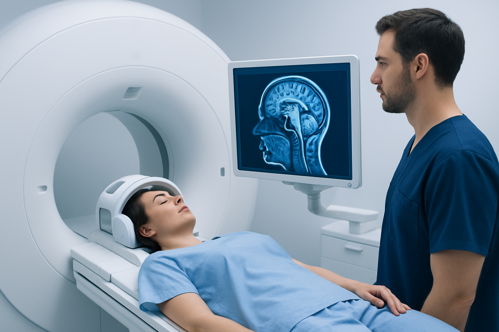
Brain imaging is often needed, especially if symptoms are new or sudden. Magnetic resonance imaging is commonly used to look for stroke, tumor, inflammation, or structural injury, while computed tomography may be used more quickly in urgent settings. Depending on the case, additional tests such as blood work, EEG, or vascular studies may help identify seizures, infection, or a circulation problem.
Treatment Options
Treatment depends entirely on the cause. A stroke involving the occipital lobe is a medical emergency and may require time-sensitive hospital treatment, followed by secondary prevention to reduce the chance of another event. If a seizure disorder is responsible, medication may be used to reduce or prevent further episodes.
When the problem is related to a tumor, infection, inflammation, or another structural condition, care may involve surgery, medication, radiation, antibiotics, or immune-based treatment depending on the diagnosis. If head injury is involved, management may include observation, rest, rehabilitation, and close monitoring for complications.
Recovery often includes support beyond the initial diagnosis. People with occipital lobe damage may benefit from visual rehabilitation, occupational therapy, adaptive strategies for reading and mobility, and follow-up with neurology and ophthalmology. For international patients, coordinated care is particularly helpful because imaging, specialist review, and follow-up planning may need to be organized around travel and recovery at home.
Prevention & Self-care
Not every occipital lobe problem can be prevented, but some causes can be reduced through general brain and vascular health. Managing blood pressure, blood sugar, and cholesterol, avoiding smoking, staying physically active when appropriate, and treating heart rhythm problems can all support stroke prevention.
People with seizure disorders or migraines may be advised to follow their treatment plans consistently and avoid known triggers when possible. Protective headgear in high-risk activities and road safety measures can also help reduce injury-related causes.
In daily life, self-care is often about safety and adaptation. Good lighting, large-print materials, screen magnification, mobility support, and arranging the environment to reduce falls can make a meaningful difference. If symptoms are being evaluated after travel, keeping a written timeline of when they started and what changed can help specialists interpret the situation more accurately.
When to See a Doctor
Sudden vision loss, new blind spots, flashes with neurological symptoms, or any abrupt change in sight should be assessed promptly. The same is true if visual symptoms occur together with headache, weakness, numbness, trouble speaking, confusion, seizure activity, or loss of balance.
Medical review is also important if vision changes are persistent, worsening, or unexplained even when the eyes seem otherwise healthy. People should not assume that a normal eye exam rules out a brain-related cause; occipital lobe disorders can be present even when the eye itself looks normal.
If symptoms begin while traveling, it is reasonable to seek urgent local assessment first rather than wait for return home. When further specialist care is needed, centers with coordinated neurology, imaging, and rehabilitation services can help confirm the cause and plan next steps; Acibadem Health Point supports international patients with multidisciplinary specialists and JCI-accredited hospitals for diagnosis and treatment.
Living With Occipital Lobe Problems
Living with an occipital lobe condition often means adapting to changes in how the brain sees and interprets the environment. Some people recover partially or fully after treatment of the underlying cause, while others learn long-term strategies to compensate for visual field loss or visual processing difficulties.
Practical support may include mobility training, home adjustments, workplace accommodations, and regular follow-up to reassess vision and neurological status. Family members can help by learning how the condition affects scanning, reading, navigation, and fatigue, so the person has the right support without unnecessary alarm.
A calm, organized care plan usually makes a meaningful difference. Clear instructions from the care team, attention to medication and appointments, and a thoughtful return-to-travel plan can all reduce stress and help recovery stay on track.
Frequently asked questions
01What does the occipital lobe do?
The occipital lobe processes visual information from the eyes and helps the brain interpret what is being seen. It plays a central role in recognizing color, shape, motion, and visual detail.
02Can occipital lobe problems cause blindness?
They can cause partial or complete loss of vision depending on how much of the area is affected. In some cases, the eyes themselves are healthy, but the brain cannot properly process visual signals.
03What are common signs of an occipital lobe stroke?
A person may notice sudden loss of part of the visual field, blurred sight, visual distortion, or trouble seeing on one side. Some people also have headache, confusion, or other neurological symptoms.
04Can seizures start in the occipital lobe?
Yes. Occipital lobe seizures may produce visual symptoms such as flashing lights, patterns, or brief visual loss. A neurologist may use an EEG and imaging to help confirm the diagnosis.
05How is an occipital lobe problem diagnosed?
Doctors usually combine a neurological exam, eye testing, visual field testing, and brain imaging. The exact tests depend on the symptoms and how quickly they appeared.
06Is recovery possible after occipital lobe damage?
Recovery depends on the cause, how quickly treatment begins, and the extent of the injury. Some people improve significantly, while others need rehabilitation and long-term visual adaptations.
This article is for general information only and is not a substitute for professional medical advice. Please consult a qualified doctor about your individual situation.
Explore treatments in Turkey — costs, top hospitals & a free quote
JCI-accredited · board-certified surgeons · reply within 24h
Add us as a Preferred Source to see more of our trusted health content across Google Search, AI Overviews and Discover.