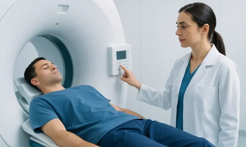
Normal vs Abnormal HIDA Scan Pictures: Key Differences
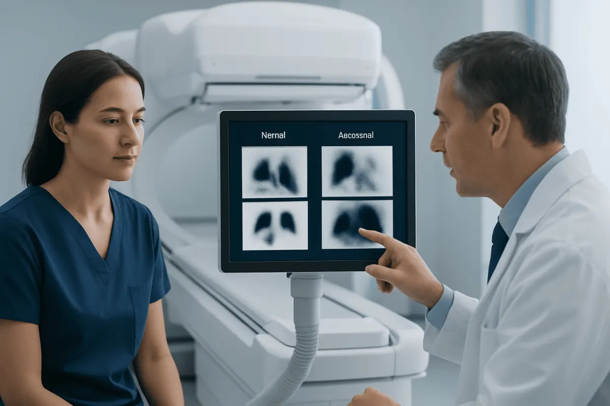
Normal vs abnormal HIDA scan pictures are best understood as a series showing the movement of a small amount of radioactive tracer through the liver, bile ducts, gallbladder and small intestine. A normal study shows expected tracer flow and gallbladder emptying, while an abnormal pattern may suggest inflammation, a blockage, a bile leak or reduced gallbladder function.
Normal vs abnormal HIDA scan pictures: a side-by-side comparison
Normal vs abnormal HIDA scan pictures do not usually look like a single photograph. They are a timed sequence of nuclear medicine images. After a tracer is injected into a vein, the camera records how it is taken up by the liver and released into bile. The scan can help assess the gallbladder, bile ducts and the passage of bile into the small intestine.
The comparison below describes common patterns. Timing and interpretation can vary according to the reason for the scan, whether medicines were used during testing and the imaging protocol. A radiologist or nuclear medicine physician makes the final interpretation.
| Feature | Typical normal pattern | Possible abnormal pattern |
|---|---|---|
| Liver uptake | Tracer appears in the liver soon after injection. | Uptake or clearance is delayed, which can occur with significant liver dysfunction or severe illness. |
| Bile ducts and bowel | Tracer progresses through the ducts and reaches the small intestine. | Markedly delayed or absent bowel activity can raise concern for impaired bile drainage or obstruction. |
| Gallbladder filling | The gallbladder becomes visible within the expected imaging period. | The gallbladder does not become visible despite appropriate delayed imaging, a pattern that can support acute cholecystitis in the right setting. |
| Gallbladder emptying | After stimulation, the gallbladder contracts and releases a measured amount of bile. | Low ejection fraction may indicate reduced gallbladder motility when symptoms and other findings fit. |
| Tracer location | Tracer remains within the expected liver, ducts, gallbladder and bowel pathway. | Tracer seen outside the biliary system may indicate a bile leak, particularly after surgery or injury. |
Brightness alone does not determine whether a scan is normal or abnormal. The key information is where the tracer appears, where it does not appear, and how these findings change across the series of images.
How to tell if a HIDA scan is abnormal?
A HIDA scan may be considered abnormal when the tracer does not follow its expected path or does not do so within the expected timeframe. For example, non-visualization of the gallbladder after appropriate imaging can indicate that the cystic duct is blocked. In a person with persistent right-upper abdominal pain, fever, tenderness and supportive blood tests, this may help confirm acute gallbladder inflammation.
Another abnormal finding is a low gallbladder ejection fraction. This value measures how much tracer-containing bile leaves the gallbladder after it is stimulated, often with a medication that mimics a digestive hormone. A low result can support gallbladder dyskinesia, also called a functional gallbladder disorder, but it needs careful clinical interpretation.
Delayed delivery of tracer to the small intestine can suggest an obstruction affecting bile flow, although it may also occur with liver disease, severe illness, prolonged fasting or medication effects. Tracer outside the bile ducts or bowel may suggest a bile leak. The report should state the imaging findings, their likely meaning and any limitations of the examination.
How to read HIDA scan images?

Patients should not try to diagnose a condition from HIDA scan images alone. The images are grayscale and are often displayed as a series, so a bright area simply indicates where more tracer has collected at that moment. The scan is designed to assess function over time, not to provide a detailed picture of gallstones or surrounding organs.
In a typical sequence, the liver appears first because it extracts the tracer from the blood. The tracer then enters the bile ducts. It should subsequently be seen in the gallbladder and in the first part of the small intestine. The exact order and timing can be influenced by the protocol, so the report is more reliable than comparing images online.
When gallbladder function is being measured, the report may include an ejection fraction and describe the medication or meal used to stimulate contraction. Different protocols may use different reference ranges. Therefore, an ejection fraction should not be labelled normal or abnormal without the laboratory’s stated standard and the clinician’s assessment of symptoms.
What does a bad HIDA scan look like?
“Bad HIDA scan” is an informal term rather than a medical diagnosis. It may refer to several distinct patterns, and each has a different possible explanation. In some studies, the tracer enters the liver and bowel but the gallbladder remains unseen; this can be consistent with cystic duct obstruction and acute cholecystitis when the clinical picture supports it.
In other studies, the gallbladder fills but does not empty adequately after stimulation. This can be reported as reduced gallbladder ejection fraction and may be seen in functional gallbladder disorders. It is not the same as finding gallstones, and it should not be used as the sole reason for treatment decisions.
A different concerning pattern is delayed tracer passage through the ducts into the bowel, which may point to impaired biliary drainage. If tracer accumulates outside the expected pathway, the images may suggest a bile leak. A clinician will consider additional testing, including ultrasound, blood tests, magnetic resonance imaging or endoscopic assessment, depending on the suspected cause.
What are the differences between normal and abnormal HIDA scan images?
The main difference between normal and abnormal HIDA scan images is the pattern of bile movement over time. In a normal study, tracer is processed by the liver and travels through the bile system in an orderly way. The gallbladder is seen when appropriate, bile reaches the intestine, and the gallbladder contracts adequately if a function test is performed.
Abnormal images may show a missing step, delayed movement or tracer in an unexpected location. A gallbladder that does not appear may reflect a blocked cystic duct. Tracer that reaches the bowel only very late may indicate slowed bile flow. A filled gallbladder that empties poorly may suggest reduced motility, while tracer outside the normal pathway can indicate leakage.
Importantly, the same image pattern may have more than one explanation. Eating too recently, fasting for an extended period, taking opioid pain medicine, having serious systemic illness or having liver impairment can alter scan findings. This is why radiology specialists review preparation details and compare the results with symptoms and other tests.
What to do after normal or abnormal HIDA scan results
A normal HIDA scan can make certain gallbladder and bile-flow problems less likely, but it does not explain every cause of upper abdominal pain, nausea or indigestion. A doctor may evaluate other digestive conditions, liver conditions, pancreatic disorders, reflux, ulcers or non-digestive causes based on the symptoms and examination.
If the scan is abnormal, the next step depends on the pattern and the person’s condition. Suspected acute cholecystitis or bile duct obstruction may require prompt assessment and treatment. Reduced gallbladder emptying may lead to a discussion of whether symptoms are typical, whether other causes have been excluded and whether observation, dietary adjustments or surgery is appropriate. Information about gallbladder diseases can help patients understand the range of possible conditions.
When gallstones or gallbladder inflammation are confirmed and treatment is needed, a surgeon may discuss gallbladder surgery. The decision should be individualized; a scan result is one part of the overall assessment, not a replacement for clinical care.
Preparing for a HIDA scan and supporting accurate results
Following preparation instructions is important because food intake and some medicines can change bile flow or gallbladder contraction. Patients are commonly asked to fast for a specified period before the test, but fasting too long can also affect results. The imaging department will provide the correct instructions for the planned examination.
Before the scan, patients should tell the care team about pregnancy, breastfeeding, allergies, recent imaging tests and all medicines or supplements they use. In particular, opioid pain medicines and certain medications that affect digestion may influence gallbladder function. A clinician can advise whether any medicine should be adjusted; patients should not stop prescribed medicine on their own.
Most people can return to usual activities after a HIDA scan unless their care team gives different advice. Drinking fluids after the examination may help the body clear the tracer. The amount of radiation used is generally small and is selected for the diagnostic test, but individual questions should be discussed with the nuclear medicine team.
When to seek medical care
Anyone with ongoing or recurrent pain in the upper right or middle abdomen should arrange a medical review, especially if pain occurs after meals or is associated with nausea, vomiting, bloating or reduced appetite. A doctor can determine whether a HIDA scan, ultrasound or another test is appropriate.
Urgent medical assessment is important for severe or worsening abdominal pain, fever, repeated vomiting, yellowing of the skin or eyes, dark urine, pale stools, fainting, confusion or an inability to keep fluids down. These symptoms can have several causes and should not be managed by relying on scan images online.
Acıbadem Health Point’s multidisciplinary specialists and JCI-accredited hospitals can assess gallbladder and biliary concerns for international patients, using clinical evaluation and appropriate imaging to guide care.
Frequently asked questions
01Is a HIDA scan more accurate than an ultrasound for gallbladder problems?
The tests provide different information. Ultrasound is usually the first test because it can identify gallstones, gallbladder wall changes and bile duct enlargement. A HIDA scan is often useful when ultrasound findings are unclear or when clinicians need to assess bile flow and gallbladder function.
02Can a low HIDA scan ejection fraction mean cancer?
A low ejection fraction generally reflects reduced gallbladder emptying and is not, by itself, a sign of cancer. The result must be interpreted with symptoms, medical history and other imaging. A doctor may recommend further evaluation if there are other concerning findings.
03Can medications affect HIDA scan results?
Yes. Opioid pain medicines and some other drugs can alter bile flow or gallbladder contraction. Patients should provide a complete medication list before the test and follow instructions from the imaging team about preparation.
04Does an abnormal HIDA scan always mean the gallbladder must be removed?
No. The appropriate treatment depends on the specific scan pattern, symptoms, examination findings and other <a href="https://www.acibademhealthpoint.com/blog/blood-test-results-explained-what-cbc-hemoglobin-and-platelets-mean/" title="Blood Test Results Explained: What CBC, Hemoglobin, and Platelets Mean" class="ahp-ilk">test results. Some causes need urgent treatment, while reduced gallbladder function may require a more individualized discussion before surgery is considered.
05How long does a HIDA scan take?
Many HIDA scans take about one to several hours, depending on how quickly the tracer moves and whether delayed images or a gallbladder ejection fraction measurement are needed. The imaging department can explain the expected schedule before the appointment.
06Can a HIDA scan miss gallstones?
A HIDA scan evaluates bile flow and gallbladder function rather than directly visualizing stones. Ultrasound is generally better for detecting gallstones. Both tests may be used because they answer different clinical questions.
This article is for general information only and is not a substitute for professional medical advice. Please consult a qualified doctor about your individual situation.
Persistent digestive symptoms? Get evaluated in Turkey
JCI-accredited · board-certified surgeons · reply within 24h
Add us as a Preferred Source to see more of our trusted health content across Google Search, AI Overviews and Discover.