A Normal Knee MRI Does Not Always Explain Knee Pain
Your knee still hurts, but the MRI report says everything looks fine. That combination is confusing, and it is more common than you might think.
A normal knee MRI usually means the scan did not show a significant tear, fracture, mass, or other visible structural problem. It is reassuring, but it does not always identify the source of knee pain, stiffness, instability, or symptoms that occur only during movement.
What does a normal knee MRI mean?
A normal knee MRI means that the radiologist did not identify a significant abnormality in the structures visible on the scan. In practical terms, this often means there is no obvious major ligament rupture, meniscus tear, bone fracture, large cartilage defect, infection, tumor, or substantial joint fluid collection. This is generally reassuring and can help rule out several important causes of knee symptoms.
A normal result does not mean your pain is imaginary, and it does not always mean there is no knee-related problem. The MRI is only one piece of the assessment. A doctor considers why the scan was ordered, where the pain occurs, whether there was an injury, how the knee performs during daily activities, and what is found during a physical examination.
The wording of the report matters. Reports may describe a scan as normal, unremarkable, or showing no acute abnormality. They may also note minor findings that are common and may not be the cause of symptoms, such as very mild wear-related changes or a small amount of fluid. The clinician who requested the test can explain what the report means in the context of the individual situation.
What a knee MRI can and cannot show
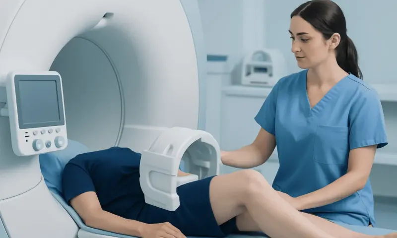
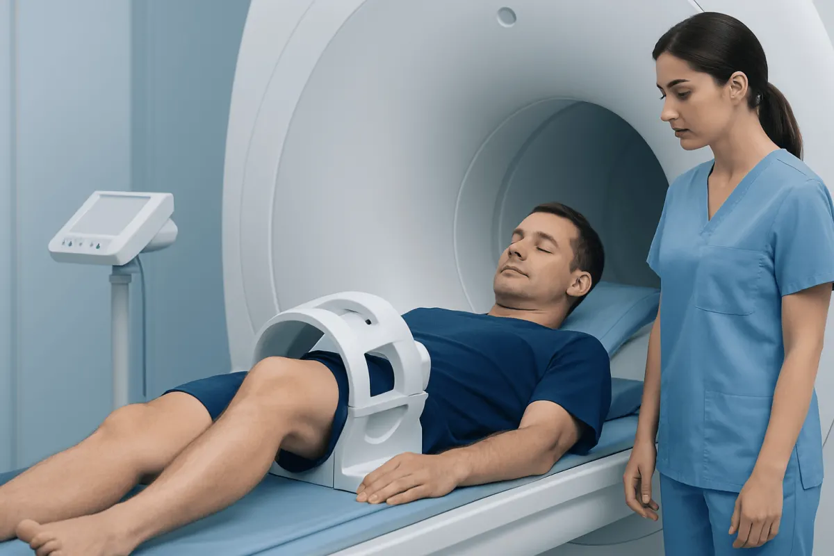
MRI uses magnetic fields and radio waves to create detailed images of soft tissues and bone. It is particularly useful for assessing the menisci, cruciate and collateral ligaments, cartilage surfaces, tendons, muscles, bone marrow, and the lining of the joint. It can also show inflammation, fluid, certain stress injuries, and many changes associated with arthritis.
Even a high-quality MRI has limits. You are lying perfectly still inside the scanner, so it may not reveal symptoms that happen only during running, squatting, stair climbing, pivoting, or prolonged standing. Problems related to movement patterns, kneecap tracking, muscle control, joint loading, or subtle instability may be more apparent during a physical assessment than on static images.
Some conditions may be too early, too small, or too subtle to be confidently seen. The scan may also focus on the knee joint itself, while pain may originate from nearby structures, including the hip, lower back, nerves, blood vessels, or tissues around the knee. So the images always need to be read alongside everything else.
A normal MRI also does not measure pain severity. Pain is influenced by tissue sensitivity, inflammation, biomechanics, sleep, stress, prior injury, and other health factors. A normal scan can therefore be compatible with symptoms that still deserve careful assessment and treatment.
Why knee pain can continue despite a normal MRI
One common reason is an overuse or load-related problem. A sudden increase in walking, running, cycling, sport, kneeling, or squatting can irritate tissues without causing a visible structural tear. Pain around or behind the kneecap, often called patellofemoral pain, may be related to how the hip, thigh, knee, and foot work together during movement.
Tendon irritation can also cause pain while leaving no major abnormality on MRI, especially in the early stages. Examples include discomfort around the patellar tendon below the kneecap or the quadriceps tendon above it. Symptoms may be most noticeable during jumping, climbing stairs, getting up from a chair, or returning to activity after rest.
Other possible explanations include mild sprains that are healing, temporary inflammation of tissues around the joint, muscle weakness or imbalance, reduced flexibility, or altered walking mechanics after an injury. In some people, pain is referred from the hip or lower back. A clinician may examine these areas if the knee findings do not fully explain the symptoms.
Less commonly, the timing of imaging can affect what is seen. For example, a very recent injury, a problem that is intermittent, or symptoms that arise only under specific physical demands may require follow-up assessment. Persistent symptoms do not automatically mean that something serious was missed, but they do justify a review when recovery is not progressing as expected.
How doctors interpret a normal result
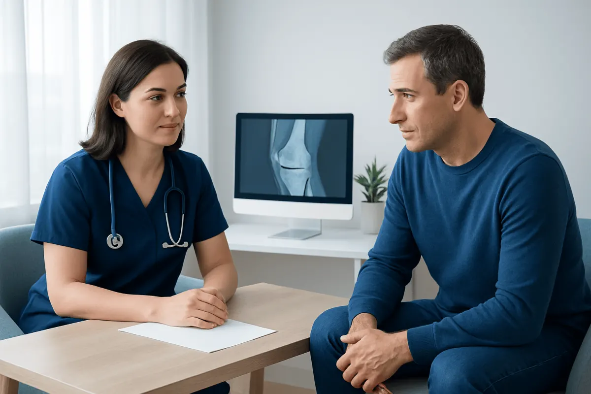
Doctors begin with the person’s story. They may ask when symptoms began, whether there was a twist, fall, impact, or increase in activity, and whether the knee swells, gives way, catches, locks, clicks, or feels unstable. The exact location of pain and the activities that trigger it can provide useful clues.
A physical examination may include checking knee alignment, range of motion, tenderness, swelling, strength, balance, walking pattern, and stability. Tests of the ligaments, menisci, kneecap, hip, and lower back may be performed. These findings can identify functional or movement-related problems that do not necessarily appear on an MRI.
Depending on the symptoms, further testing may or may not be helpful. Plain X-rays can sometimes better show joint alignment, certain types of arthritis, or bone-related changes. Blood tests may be considered if there are signs of inflammatory disease or infection. Ultrasound can be useful for selected tendon, bursa, or superficial soft-tissue concerns, particularly when symptoms are focused outside the joint.
Often the next step is not another scan but a period of targeted care, then a fresh look. A clinician may recommend physiotherapy, activity modification, or treatment directed at the likely source of pain. Progress over time is an important part of confirming whether the working diagnosis is correct.
What treatment and self-care may involve
Care after a normal knee MRI depends on the symptoms, examination, activity demands, and individual health needs. For many non-urgent knee problems, a gradual return to comfortable movement is encouraged rather than complete rest. Temporarily reducing activities that consistently aggravate pain, such as deep squats, jumping, or running hills, can allow irritated tissues to settle while maintaining general fitness through suitable alternatives.
Physiotherapy or a supervised exercise program may focus on restoring knee motion, building strength in the quadriceps, hamstrings, hips, and calf muscles, and improving balance and movement control. Exercise should be tailored to the person. Mild temporary discomfort can occur during rehabilitation, but pain that is sharp, escalating, or associated with swelling or instability should be discussed with a healthcare professional.
Simple measures may help some people, including ice or heat according to preference, supportive footwear, and pacing activities throughout the day. Over-the-counter pain medicines may be appropriate for some adults, but they are not suitable for everyone. People with kidney disease, stomach ulcers, heart disease, liver disease, pregnancy, allergies, or regular medicines should seek advice from a pharmacist or clinician before using pain medication.
If symptoms do not improve, a doctor can reconsider the diagnosis and care plan. Acıbadem Health Point’s multidisciplinary specialists and JCI-accredited hospitals assess knee symptoms for international patients using clinical examination, imaging review, and rehabilitation planning when appropriate.
When to seek medical care
Medical assessment is important if knee pain persists beyond a reasonable recovery period, repeatedly limits work, sleep, exercise, or daily activities, or returns whenever activity is resumed. A review is also sensible when the knee feels unstable, gives way, catches, or cannot fully bend or straighten, even if the MRI report was normal.
More urgent medical care is needed after a significant injury if the person cannot bear weight, the knee looks deformed, or there is rapidly increasing swelling. Prompt assessment is also important for a hot, red, very swollen joint, particularly when fever, chills, or feeling unwell are present, as these symptoms can indicate an infection or inflammatory condition requiring timely treatment.
Emergency care should be sought for severe pain with a cold, pale, blue, or numb foot, or for sudden calf swelling and pain accompanied by shortness of breath or chest pain. These symptoms may not be caused by a routine knee problem and should not be managed at home.
Bring the MRI report to your appointment, and the image files too if you have them. It also helps to jot down where the pain sits, when it swells, what sets it off, and whether the knee ever locks or gives way. This information helps the clinician connect the scan findings with the real-life pattern of symptoms.
Questions to ask after receiving the report
A normal knee MRI can leave people uncertain about what to do next. Useful questions include: What structures were assessed? Is there any minor finding that may relate to the symptoms? Does the examination suggest a problem involving the kneecap, tendons, muscles, hip, back, or walking pattern? Knowing the likely explanation can make a rehabilitation plan feel clearer and more manageable.
It is also reasonable to ask which activities should be modified, which movements are safe to continue, and what signs would suggest the need for reassessment. A clinician can advise on an appropriate timeframe for follow-up based on the severity and duration of symptoms, the person’s age, level of activity, and any previous injuries.
For people who play sport or have physically demanding jobs, return to activity should usually be gradual. Being able to walk comfortably is not always the same as being ready to sprint, pivot, jump, climb, or lift repeatedly. Strength, control, confidence, and the ability to perform job- or sport-specific movements should be considered.
A normal scan is usually good news. With a careful clinical review and a practical treatment plan, many people can tackle what is driving the knee pain and work their way back to their usual activities.
Frequently asked questions
01Can an MRI miss a knee injury?
MRI is highly useful for many knee injuries, but no test identifies every possible cause of symptoms. Very subtle, early, intermittent, or movement-related problems may not be clearly visible. The report should be reviewed alongside the physical examination and symptom pattern.
02Is a normal knee MRI good news?
Yes, it is generally reassuring because it makes major structural damage less likely. It does not mean that pain or reduced function should be ignored. A clinician can help identify causes that may not be obvious on the scan.
03Why does my knee hurt when my MRI is normal?
Pain may be related to overuse, tendon irritation, kneecap movement, muscle weakness, altered movement patterns, or pain referred from another area. Some of these issues are best identified through history-taking and examination rather than imaging. A tailored rehabilitation plan may be helpful.
04Should I get a second MRI if my knee still hurts?
A second MRI is not always necessary and may not change treatment. A doctor may first reassess the symptoms, review the original images, examine the hip and back, or consider other tests if indicated. Repeat imaging is more likely to be considered when symptoms change, worsen, or new concerning signs develop.
05Can I exercise with knee pain and a normal MRI?
Many people can continue some form of exercise, but the type and intensity should be adjusted to symptoms. Low-impact activities and guided strengthening are often used while more provocative activities are reduced temporarily. A physiotherapist or clinician can provide individual guidance.
06What symptoms need urgent attention even if the MRI was normal?
Seek prompt medical care for inability to bear weight after injury, a deformed knee, marked swelling, or a hot and red joint with fever. Emergency assessment is needed for a cold, pale, blue, or numb foot, or calf symptoms with chest pain or shortness of breath. These signs may indicate a problem requiring immediate evaluation.
This article is for general information only and is not a substitute for professional medical advice. Please consult a qualified doctor about your individual situation.
Joint and spine care in Turkey — expert assessment & treatment
JCI-accredited · board-certified surgeons · reply within 24h
Add us as a Preferred Source to see more of our trusted health content across Google Search, AI Overviews and Discover.