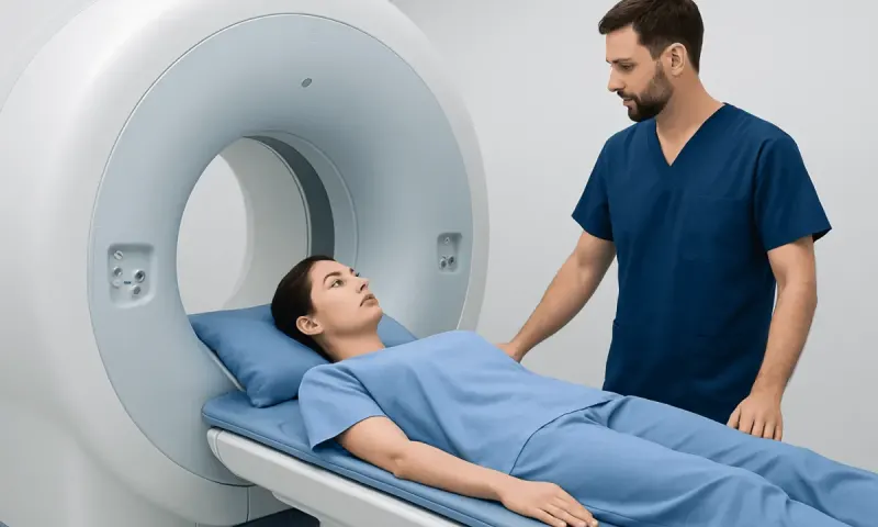
Myelogram: What to Expect

Key Takeaways
- A myelogram uses contrast material and imaging to show the spinal canal and nerve pathways more clearly.
- It may help identify herniated discs, spinal stenosis, nerve compression, or other structural causes of pain or numbness.
- Preparation often includes discussing medications, allergies, and any history of bleeding problems with the care team.
- Most people go home the same day, but rest, hydration, and following aftercare instructions are important.
- A doctor may recommend it when MRI or CT alone does not give enough information or cannot be performed safely.
A myelogram is a specialized imaging test that helps doctors evaluate the spinal cord, nerve roots, and the spaces around them. It is often used when symptoms suggest pressure, narrowing, or another structural problem in the spine and more detail is needed than a standard scan can provide.
Overview
A myelogram is a diagnostic imaging study that gives doctors a closer look at the spinal canal, spinal cord, and the nerves that branch away from it. It is not a treatment; it is a way to map the anatomy when symptoms suggest that something in the spine may be crowding or irritating the nerve structures.
The test combines a contrast dye with X-ray imaging or CT scanning. The contrast outlines the flow of fluid around the spinal cord, making certain problems easier to see, especially those involving narrowing, pressure, or blockage. For many patients, the decision to have a myelogram comes after symptoms have persisted, other tests have been reviewed, and the care team needs more detailed information before choosing the next step.
For international patients, a myelogram may be part of a broader spine evaluation that includes consultation, imaging, and a same-day recovery plan. The process is usually organized so that preparation, the procedure, and follow-up instructions are clearly explained in advance, which can make the experience more manageable when care is being arranged from another country.
Symptoms That May Lead to a Myelogram

Doctors do not order a myelogram simply because someone has back pain. It is usually considered when symptoms suggest that a nerve or the spinal canal itself may be involved and when more precise anatomical detail is needed.
Common reasons include pain that spreads into an arm or leg, numbness or tingling, weakness, or signs that spinal nerves may be compressed. Some people also have balance problems, changes in walking, or symptoms that suggest spinal stenosis, disc herniation, or another structural issue affecting the flow of spinal fluid.
A myelogram may also be helpful when MRI is not possible, such as in some patients with certain implanted devices, or when prior imaging results do not fully explain a patient’s symptoms. In those cases, the test can add a more detailed view that supports a safer, more informed treatment plan.
Causes & Risk Factors

There is no single cause for needing a myelogram; rather, the test is used to investigate conditions that may be affecting the spine. These can include degenerative changes that develop over time, herniated discs, spinal canal narrowing, spinal tumors, scar tissue after surgery, or other sources of nerve compression.
Risk factors for spine problems vary widely. Aging, previous spine injury, repetitive strain, arthritis, and a history of spine surgery can all increase the chance that a doctor will want a closer look at the spinal canal. In some people, a myelogram is also considered when symptoms are complex and standard imaging does not answer the clinical question.
Because the procedure involves a needle placed into the spinal fluid space, the care team also reviews factors that can affect safety. These include blood-thinning medicines, bleeding disorders, contrast reactions, pregnancy, and a personal history of certain neurologic conditions. Careful screening helps the doctor decide whether the test is appropriate and how to reduce avoidable risk.
Diagnosis: How the Test Works
During a myelogram, a doctor places contrast material into the fluid space around the spinal cord, usually through a needle in the lower back. The contrast mixes with the cerebrospinal fluid and outlines the spinal canal on imaging, making it easier to identify areas where something may be pressing on or narrowing the pathway.
After the contrast is introduced, X-rays or a CT scan are taken while the dye spreads through the spinal canal. The images can show the shape of the nerve sleeves, the degree of spinal narrowing, and any blockages or irregularities in the flow of contrast. This can be especially useful when the anatomy is complex or when surgical planning requires very detailed information.
Before the test, the medical team usually reviews symptoms, prior imaging, medication use, allergies, and any conditions that could affect the procedure. This step matters because the value of the myelogram depends not only on the image itself, but also on matching the imaging plan to the patient’s medical history and current concerns.
Treatment Options
A myelogram does not treat a spine problem, but it can help guide treatment by showing where the problem is and how extensive it may be. The next step depends on what the images reveal and on the patient’s symptoms.
Possible treatments after a myelogram may include observation, physical therapy, pain management, medication adjustments, injections, or surgery for conditions such as significant nerve compression or spinal stenosis. In many cases, the imaging results help the doctor decide whether conservative care is reasonable or whether a more targeted intervention is likely to help.
For patients traveling for care, the test may be linked to a larger treatment pathway: imaging review, consultation with a spine specialist, and discussion of options before returning home. That coordinated approach can be particularly useful when the goal is to avoid repeated trips and to make follow-up plans clear before travel ends.
Prevention & Self-care
People cannot usually prevent the need for a myelogram if they already have symptoms that require a closer spinal assessment. What they can do is prepare thoughtfully so the test is as smooth and safe as possible.
The care team may ask the patient to review all medicines, including blood thinners, anti-inflammatory drugs, and supplements. It is also important to mention allergies, kidney problems, prior reactions to contrast material, and any chance of pregnancy. Following instructions about eating, drinking, and arranging a ride home can make the day of the procedure less stressful.
- Drink fluids as advised after the test to help with recovery.
- Rest for the period recommended by the clinical team.
- Watch for unusual symptoms such as severe headache, fever, new weakness, or drainage at the puncture site.
- Keep follow-up appointments so the images can be reviewed in context.
Self-care after the test usually focuses on hydration, short-term activity modification, and paying attention to the body’s response. The goal is not to overthink every sensation, but to notice anything that seems unusual and report it promptly if needed.
When to See a Doctor
Medical attention is appropriate when back or neck symptoms persist, worsen, or begin to affect daily function. A myelogram may be recommended when the doctor needs more information than other imaging tests can provide, especially if pain, numbness, weakness, or walking difficulty suggests nerve involvement.
After the procedure, the patient should contact the care team if there is a severe or persistent headache, fever, increasing pain, confusion, weakness, seizures, or fluid leaking from the needle site. These symptoms are uncommon, but they deserve prompt medical review.
For people planning care abroad, it is especially useful to clarify before travel who will review the images, how results will be shared, and what symptoms should trigger a return call or urgent evaluation. Acibadem Health Point’s multidisciplinary specialists and JCI-accredited hospitals can diagnose and treat this condition for international patients in a coordinated setting.
Recovery and Follow-up
Most patients recover from a myelogram without needing an overnight stay. The main aftercare focus is usually rest, hydration, and a gradual return to normal activity according to the instructions given by the medical team.
Some people experience a temporary headache or mild discomfort at the puncture site. These symptoms often improve with time and supportive care, but the doctor should be informed if they become intense, do not ease, or are accompanied by neurologic changes.
Follow-up matters because the images are most helpful when they are interpreted alongside the patient’s symptoms, examination findings, and treatment goals. For international patients, this may include a final review before departure and a clear plan for sharing reports with a home-country physician if ongoing care is needed.
Frequently asked questions
01What is a myelogram used for?
A myelogram is used to look at the spinal canal, spinal cord, and nerve roots in more detail. Doctors may order it to help evaluate spinal stenosis, herniated discs, nerve compression, or other structural problems that may be causing pain or neurologic symptoms.
02Is a myelogram the same as an MRI?
No. MRI and myelogram are different tests, although both can help evaluate the spine. A myelogram uses contrast and X-ray or CT imaging, and it may be preferred when MRI is not possible or when a different view of the spinal canal is needed.
03Does a myelogram hurt?
Most people feel pressure or brief discomfort during the needle placement, but the procedure is usually manageable. The team takes steps to keep the patient comfortable and explains each stage beforehand so there are fewer surprises.
04How should a patient prepare for a myelogram?
Preparation usually involves sharing medication lists, allergies, and relevant medical history with the care team. The doctor may also give instructions about eating, drinking, and arranging transportation home after the test.
05What are the possible side effects after a myelogram?
Temporary headache, mild back discomfort, or soreness at the puncture site can occur. More serious problems are uncommon, but severe headache, fever, weakness, or fluid leakage should be reported promptly.
06How soon are the results available?
That depends on the facility and whether the scan is interpreted immediately or after a radiologist review. In many settings, the imaging results are discussed soon after the test so the next step can be planned without unnecessary delay.
This article is for general information only and is not a substitute for professional medical advice. Please consult a qualified doctor about your individual situation.
Joint and spine care in Turkey — expert assessment & treatment
JCI-accredited · board-certified surgeons · reply within 24h
Add us as a Preferred Source to see more of our trusted health content across Google Search, AI Overviews and Discover.