MRI vs X-Ray: Which Scan Shows What?
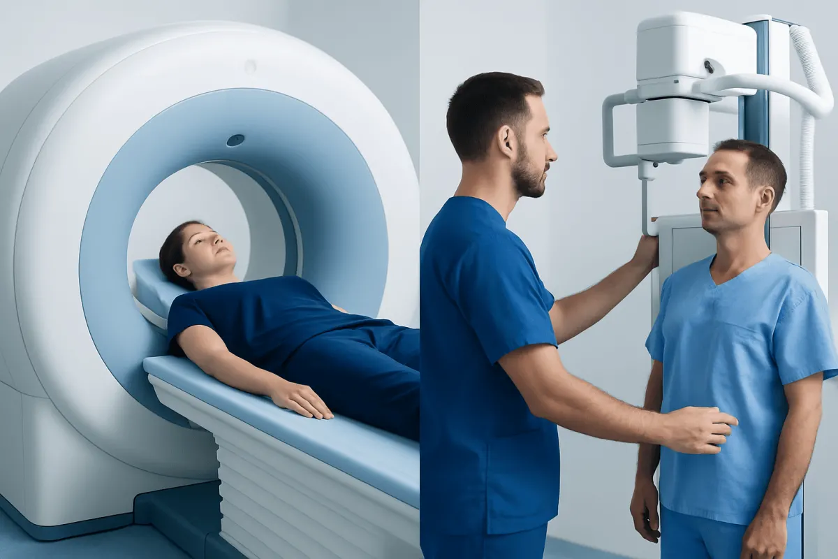
MRI and X-rays are imaging tests used for different clinical questions. X-rays are fast and especially useful for bones and the chest, while MRI provides highly detailed images of soft tissues such as the brain, spinal cord, muscles, ligaments and internal organs without using ionizing radiation.
MRI vs Xray: the main difference
The main MRI vs Xray difference is the type of information each examination provides. An X-ray is a quick test that uses a small dose of ionizing radiation to show dense structures, especially bones. It is commonly used to assess suspected fractures, joint alignment, some types of arthritis, and conditions affecting the lungs or chest.
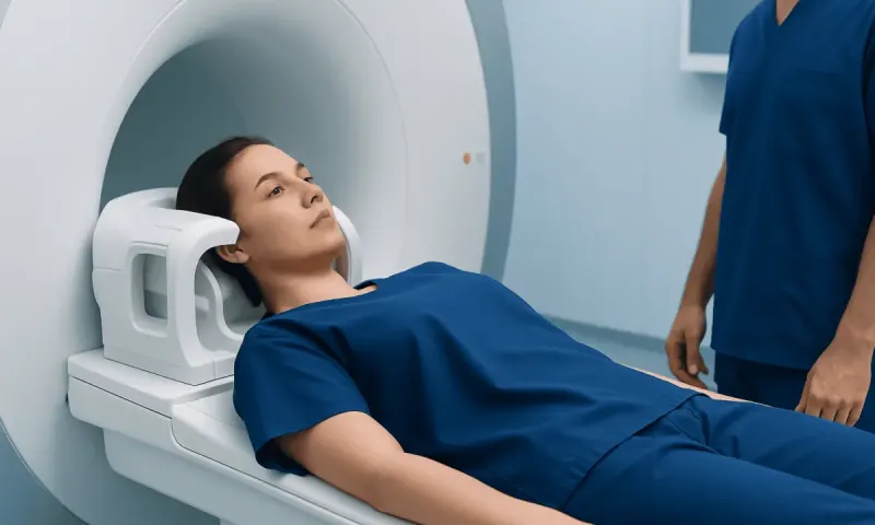
MRI, or magnetic resonance imaging, uses a strong magnetic field and radio waves rather than radiation. It produces detailed pictures of soft tissues, including the brain, spinal cord, nerves, muscles, tendons, ligaments, cartilage and many organs. It may be selected when an X-ray is normal or unclear but symptoms suggest an injury or condition involving soft tissue.
Neither test is universally better. The appropriate examination is the one most likely to answer the clinical question safely and efficiently. A clinician considers the location of symptoms, possible diagnosis, urgency, previous imaging and a person’s individual circumstances before recommending a scan.
How X-rays and MRI scans work
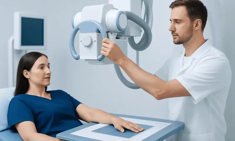
During an X-ray, a machine directs a controlled beam of radiation through the body onto a detector. Dense materials absorb more radiation and appear lighter on the image; this is why bones usually appear white. Air appears dark, while many soft tissues have similar shades of gray and may be less clearly distinguished.
An MRI scanner contains a powerful magnet. The magnet temporarily aligns hydrogen particles in the body, and radiofrequency pulses alter that alignment. As the particles return to their usual state, they release signals that a computer converts into detailed cross-sectional images. Different MRI sequences can emphasize fluid, fat, blood flow or other tissue characteristics.
In some situations, contrast material is used to make certain structures or abnormalities easier to assess. MRI contrast is usually gadolinium-based, while many X-ray-based examinations use iodine-containing contrast. The care team reviews kidney function, allergy history and other relevant factors when contrast may be needed.
What does an MRI show that other scans don't?

MRI can provide a level of soft-tissue detail that plain X-rays cannot. For example, it can show the spinal cord, brain tissue, intervertebral discs, ligaments, tendons, bone marrow and many types of muscle injury. It can also help evaluate subtle changes within joints and organs.
For a painful knee, an X-ray may identify a fracture, joint narrowing or bone changes, whereas MRI may show a meniscus tear, ligament injury, cartilage damage or bone marrow swelling. In the spine, an X-ray can show alignment and some degenerative changes, while MRI can assess discs, nerves and the spinal cord.
MRI does not replace every other scan. CT often provides better detail of complex fractures, calcification and certain urgent chest or abdominal concerns, and it is usually much faster. Ultrasound is particularly useful for real-time assessment of structures near the body surface, pregnancy-related imaging, blood flow and some abdominal or pelvic concerns.
When comparing MRI vs Xray vs ultrasound, ultrasound does not use radiation and is portable, but image quality can be affected by air, bone, body habitus and the area being examined. In MRI vs Xray vs CT, CT uses radiation but may be preferred in emergencies because it is fast and gives detailed images of bone, lungs and many internal injuries.
Can you see inflammation on an MRI?
Yes. MRI can often show signs that are consistent with inflammation, especially swelling or increased fluid in tissues. Depending on the body area and MRI technique, it may help identify inflammation involving joints, tendons, muscles, the spine, bowel, brain or other organs.
For example, MRI may demonstrate fluid in a joint, inflamed tissue around a tendon, swelling in bone marrow, or changes in the tissues around the spine. Contrast-enhanced MRI can sometimes provide additional information about active inflammation or blood supply to tissues.
However, an MRI image alone does not always establish the cause of inflammation. Infection, autoimmune disease, injury and other conditions may produce overlapping appearances. Clinicians interpret the scan alongside symptoms, examination findings, laboratory tests and other relevant information.
Why don't doctors recommend MRI?
Doctors do recommend MRI when it is likely to provide information that will influence diagnosis or treatment. However, it is not always the first or most appropriate test. For many suspected fractures, a standard X-ray can answer the question quickly, while for some urgent conditions a CT scan may be faster and more practical.
MRI examinations generally take longer than X-rays and require the person to remain still in a confined scanner space. The test may be difficult for people with significant claustrophobia, severe pain that prevents stillness, or some medical devices and metallic fragments. Many implanted devices are MRI-conditional rather than automatically unsafe, so the exact device details must be checked in advance.
Doctors also avoid unnecessary imaging because incidental findings can lead to anxiety, follow-up tests or procedures that may not improve health outcomes. The goal is not to order the most detailed scan in every situation, but to use the right test at the right time.
What are two major disadvantages of MRI scans?
Two major disadvantages of MRI scans are the time required and limitations related to the magnetic environment. A scan often lasts longer than an X-ray, and remaining very still is important for clear images. The machine can also be noisy and feel enclosed, although staff can provide communication, hearing protection and, in appropriate cases, support for anxiety.
A second limitation is that MRI is not suitable without careful assessment for every person with implanted medical devices, metal fragments or certain metal-containing materials in the body. Many modern implants can be scanned safely under specified conditions, but this decision must be made by trained imaging professionals using accurate device information.
MRI may also involve contrast in selected cases. Serious reactions are uncommon, but contrast decisions should account for kidney disease, previous reactions and the expected benefit of additional imaging detail. A radiologist and referring clinician can explain whether contrast is likely to be useful.
What to expect: candidacy, steps and recovery
Before an imaging test, patients should tell the care team about pregnancy, possible pregnancy, kidney disease, allergies, surgeries, implanted devices, metal injuries and any difficulty lying flat or remaining in enclosed spaces. X-rays are often appropriate in urgent care, while MRI may be scheduled when more detailed soft-tissue imaging is needed.
For an X-ray, the patient may change into a gown and remove jewelry or other objects from the area being examined. A radiographer positions the body, briefly asks the patient to stay still or hold their breath, and takes one or more images. The imaging part commonly takes only a few minutes.
For MRI, metal objects and electronic items must be removed before entering the scanner room. The patient lies on a padded table that moves into the scanner, and staff remain in contact throughout the examination. The scanner makes repeated tapping or knocking sounds while images are acquired. If contrast is needed, it may be given through a vein during the test.
There is usually no recovery time after a routine X-ray or MRI. Most people can return to normal activities immediately. If sedation is used for anxiety or another reason, a responsible adult may need to accompany the patient home and driving should be avoided until the effects have fully worn off.
Benefits, risks and when to seek medical care
The key benefit of imaging is that it can help clarify a diagnosis, guide treatment and monitor certain conditions. X-rays offer speed and excellent basic bone assessment. MRI provides detailed soft-tissue evaluation without ionizing radiation. Both tests are generally safe when used appropriately and interpreted by qualified professionals.
X-rays and CT scans involve ionizing radiation, but imaging teams use the lowest practical exposure needed to obtain useful images. MRI does not use ionizing radiation, yet its strong magnetic field requires thorough safety screening. Patients should ask questions if they are unsure why a scan has been recommended or how to prepare.
Medical care should be sought urgently for symptoms such as sudden weakness, difficulty speaking, severe headache unlike usual headaches, chest pain, trouble breathing, major injury, uncontrolled bleeding, loss of consciousness, or new loss of bladder or bowel control. Prompt assessment is also important for persistent or worsening pain, fever with severe localized pain, unexplained weakness, or numbness after an injury.
For international patients who need coordinated imaging and specialist assessment, Acıbadem Health Point’s multidisciplinary teams at JCI-accredited hospitals diagnose and treat a broad range of bone, joint, neurological and other health concerns.
Frequently asked questions
01Is MRI better than an X-ray?
MRI is not always better; it is better for specific questions involving soft tissues such as ligaments, tendons, nerves, the brain and spinal cord. X-rays are often better as a quick first test for fractures, bone alignment and some chest problems. The best option depends on the symptoms and suspected condition.
02Does an MRI use radiation like an X-ray?
No. MRI uses magnetic fields and radio waves to create images and does not use ionizing radiation. X-rays use a controlled, low amount of ionizing radiation to produce images.
03Why might an X-ray be done before an MRI?
An X-ray is quick, widely available and can identify many bone problems, including obvious fractures and joint changes. If the X-ray does not explain symptoms, or if a soft-tissue injury is suspected, an MRI may then be recommended. This stepwise approach can avoid unnecessary testing.
04Can an MRI detect a fracture that an X-ray misses?
Yes, MRI can sometimes identify stress fractures or subtle fractures that are not visible on an initial X-ray. It can also show bone marrow swelling associated with recent injury. A clinician will decide whether MRI, CT or repeat X-rays are most appropriate.
05Is MRI safe for people with metal implants?
Safety depends on the type of implant, when it was placed and the scanning conditions. Many modern implants are MRI-conditional and can be scanned under specific protocols, but some devices or metal fragments require extra precautions or may prevent MRI. Patients should provide all available implant information before the examination.
06How long do MRI and X-ray results take?
The images are available soon after the test, but they must be reviewed and reported by a radiologist. Timing varies according to the urgency of the examination and local workflow. The referring clinician explains the findings and what they mean for next steps.
This article is for general information only and is not a substitute for professional medical advice. Please consult a qualified doctor about your individual situation.
Joint and spine care in Turkey — expert assessment & treatment
JCI-accredited · board-certified surgeons · reply within 24h
Add us as a Preferred Source to see more of our trusted health content across Google Search, AI Overviews and Discover.