Why a Maisonneuve Fracture Is Easy to Miss on Ankle X-Rays

You twist your ankle, it swells, and every step hurts. The ankle is what you feel — so that’s what gets looked at. But sometimes the break is somewhere else entirely.
In a Maisonneuve fracture, a break near the top of the fibula happens alongside damage to the ligaments that stabilize the ankle. It can be missed because ankle pain may be much more noticeable than pain in the upper leg. That’s exactly why getting checked promptly matters.
Overview: What Is a Maisonneuve Fracture?
A Maisonneuve fracture is a distinctive and potentially unstable injury involving the ankle and lower leg. It typically includes a spiral fracture of the upper part of the fibula, the thinner bone on the outside of the lower leg, together with injury to the syndesmosis. The syndesmosis is a strong set of ligaments connecting the tibia and fibula just above the ankle joint.
Here is the catch: the fracture may sit close to the knee while the loudest symptoms are down at the ankle. In some cases, the medial ankle ligament, called the deltoid ligament, is also torn. Instead of a ligament tear, there may be a fracture of the medial malleolus, the bony prominence on the inner side of the ankle. These associated injuries can allow the ankle bones to move out of their normal relationship.
This is not simply a broken fibula. It is an ankle stability injury, and treatment decisions rest on the position and stability of the ankle joint as much as on the fibular fracture itself. With an accurate diagnosis, stabilization where it is needed, and rehabilitation, many people get back to daily activities and sport over time.
How the Injury Happens
Most Maisonneuve fractures result from a forceful twisting mechanism. A common pattern is external rotation, in which the foot turns outward while the leg remains relatively fixed. This may happen during a fall, a misstep on uneven ground, a collision in contact sport, skiing, or a road traffic accident.
The rotating force may begin at the ankle and travel upward through the syndesmotic ligaments and the membrane between the tibia and fibula, called the interosseous membrane. The force can eventually cause the upper fibula to fracture. This explains why a person may not remember a direct blow to the outer upper leg.
Anyone can sustain this injury, but it turns up most often after sudden turns, falls, or high-energy impacts. It can also follow a lower-energy fall in older adults or in people with reduced bone strength. What matters is usually the direction of the force and the ligament damage that goes with it, not how dramatic the accident looked.
Symptoms and Signs to Notice

Symptoms often start with pain, swelling, and tenderness around the ankle. The inner ankle may be particularly sore if the deltoid ligament has been injured or if there is a medial malleolus fracture. Bruising, difficulty putting weight through the foot, and a feeling that the ankle is unstable are also possible.
Some people have pain or tenderness along the outside of the lower leg or near the upper fibula, just below the knee. However, that pain can be mild or missing altogether. So do not assume an ankle injury is just a sprain if you have a lot of swelling, cannot walk normally, are tender over the inner ankle, or feel pain higher up the leg.
Visible ankle deformity, marked swelling, numbness, tingling, a cold or pale foot, or worsening pain require urgent assessment. These signs can indicate a dislocation, circulation problem, nerve involvement, or another complication that needs prompt care. Do not keep testing the ankle, and never try to force it back into position yourself.
Why Diagnosis Can Be Challenging
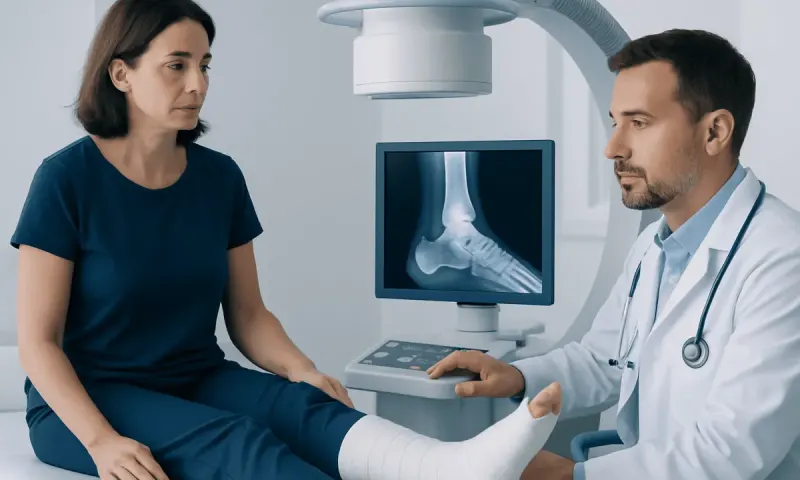
A Maisonneuve fracture can be overlooked if evaluation focuses only on the area of greatest pain. Standard ankle X-rays may show widening between the tibia and fibula, an inner ankle fracture, or subtle signs of instability. However, the upper fibula can remain outside the image unless the clinician examines the whole leg and requests full-length tibia-fibula X-rays.
During the examination, a clinician checks the ankle, lower leg, knee, skin, circulation, sensation, and ability to move the toes. Tenderness along the fibula and pain with certain carefully performed ankle stress maneuvers may raise concern for a syndesmotic injury. Clinicians also consider other injuries that may occur after twisting trauma, including a ankle sprain, but a suspected unstable injury needs more detailed assessment.
Imaging may include ankle and lower-leg X-rays taken from different angles. Weight-bearing or stress views may be used in selected cases to assess ankle stability. CT scans can provide a clearer view of small fractures and joint alignment, while MRI can help evaluate ligaments, cartilage, and the interosseous membrane when the diagnosis remains uncertain. Which scans you need depends on your injury and what the examination shows.
Treatment Options and Recovery Planning
Initial treatment aims to protect the ankle and control discomfort. This may include a splint or boot, elevation, ice wrapped in a cloth for short periods, and pain relief recommended by a clinician. Weight-bearing should be avoided until the injury has been assessed and the care team advises that it is safe. If the ankle is dislocated, it may need urgent realignment before definitive treatment.
Management depends mainly on whether the ankle joint is stable and properly aligned. A small number of stable injuries without meaningful displacement may be managed without surgery, using immobilization and close follow-up imaging. However, many Maisonneuve fractures involve syndesmotic instability and require an operation to restore and maintain the normal relationship between the tibia and fibula at the ankle.
Surgery may involve fixation across the syndesmosis with screws or a flexible fixation device. Associated ankle fractures may also need fixation. The high fibula fracture itself often heals without direct surgery once the ankle is stabilized, although each injury is assessed individually. Orthopedic surgeons tailor the approach to the fracture pattern, soft-tissue condition, activity needs, and overall health of the patient.
Recovery generally includes a period of protection followed by progressive movement, strengthening, balance training, and walking rehabilitation. The time before weight-bearing and return to driving, work, or sport varies widely. Follow-up appointments and imaging help confirm that the ankle remains well aligned and that healing is progressing as expected.
Self-Care, Rehabilitation, and Preventing Further Injury
After diagnosis, the most helpful self-care step is following the treatment plan closely. This includes using crutches, a walker, a boot, or a cast as instructed; attending follow-up visits; and avoiding weight-bearing or high-impact activity until cleared. Keeping the leg elevated when resting may help reduce swelling, particularly in the early phase of recovery.
Rehabilitation is an important part of treatment. A physiotherapist may guide exercises to restore ankle range of motion, calf and leg strength, coordination, and balance. Progression should be gradual. Pushing through sharp pain, returning to pivoting sports too soon, or stopping support devices without medical advice can increase the risk of delayed healing or recurrent instability.
It is not possible to prevent every twisting injury, but supportive footwear, activity-specific training, strength and balance exercises, and a gradual return to sport may reduce risk. People with repeated ankle injuries may benefit from a professional assessment of movement patterns, footwear, and rehabilitation needs. Maintaining bone health through adequate nutrition, regular safe weight-bearing exercise, and management of conditions that affect bone density is also valuable.
When to Seek Medical Care
Anyone with significant ankle pain after a twist, fall, or collision should seek medical assessment if they cannot take several steps, have substantial swelling or bruising, or feel that the ankle is unstable. Assessment is particularly important when there is pain on the inner side of the ankle or tenderness along the outer leg near the knee, as these findings can occur with a Maisonneuve fracture.
Emergency care is appropriate for an obvious deformity, an open wound, severe or rapidly increasing pain, loss of sensation, weakness in the foot, or a foot that appears pale, blue, or unusually cold. Until help is available, the person should keep the leg still, avoid food or drink if surgery may be needed, and not attempt to straighten a deformed ankle.
Even once the swelling settles, see a qualified clinician if ankle pain lingers, walking stays difficult, or the ankle feels like it gives way. At Acıbadem Health Point, our orthopedic teams and JCI-accredited hospitals assess and treat complex bone and joint injuries for patients from abroad.
Frequently asked questions
01Can a Maisonneuve fracture be missed on an ankle X-ray?
Yes. The fibular fracture is usually near the knee, so it may not appear on ankle X-rays alone. When a syndesmotic injury is suspected, clinicians examine the full leg and may order X-rays of the tibia and fibula as well as additional ankle imaging.
02Can a person walk with a Maisonneuve fracture?
Some people may still be able to take a few steps, particularly early after the injury. However, walking does not rule out a significant fracture or unstable ankle injury. Pain, swelling, or difficulty bearing weight after a twisting injury should be assessed by a healthcare professional.
03Does every Maisonneuve fracture require surgery?
Not every case requires surgery, but many do because the syndesmotic ligaments and ankle alignment are often unstable. The decision is based on examination findings, imaging, fracture displacement, and whether the ankle remains properly aligned. An orthopedic specialist can explain the most suitable approach.
04How long does recovery from a Maisonneuve fracture take?
Recovery time varies according to the severity of ligament injury, associated ankle fractures, treatment method, and rehabilitation progress. Bone and soft tissues need time to heal, and return to impact activity or sport may take longer than return to basic walking. Follow-up guidance from the treating team is important.
05Where is the pain with a Maisonneuve fracture?
Pain is commonly felt around the ankle, often on the inner side, with swelling and bruising. Some people also have tenderness near the upper outer part of the lower leg, below the knee, where the fibula is fractured. Upper-leg pain may be subtle, which is one reason this injury can be overlooked.
06What happens if a Maisonneuve fracture is not treated properly?
If ankle instability is not recognized and corrected, the ankle may heal in poor alignment. This can lead to ongoing pain, weakness, a feeling of instability, reduced function, and earlier wear of the joint cartilage. Prompt evaluation helps clinicians identify the injury and plan appropriate treatment.
This article is for general information only and is not a substitute for professional medical advice. Please consult a qualified doctor about your individual situation.
Joint and spine care in Turkey — expert assessment & treatment
JCI-accredited · board-certified surgeons · reply within 24h
Add us as a Preferred Source to see more of our trusted health content across Google Search, AI Overviews and Discover.