Lung Function Test: Obstructive Vs Restrictive Patterns
Obstructive and restrictive patterns on lung function tests describe different breathing problems: obstruction limits how quickly air leaves the lungs, while restriction limits how much air the lungs can hold. Spirometry can suggest either pattern, but complete pulmonary function testing and clinical assessment are often needed to confirm the cause.
Lung Function Test Obstructive vs Restrictive: A Side-by-Side Comparison
A lung function test obstructive vs restrictive comparison helps explain why a person may feel breathless, cough, wheeze, or have reduced exercise tolerance. An obstructive pattern means air has difficulty moving out through narrowed or collapsible airways. A restrictive pattern means the lungs, chest wall, or breathing muscles cannot expand normally, reducing total lung capacity.
These are test patterns rather than final diagnoses. A clinician considers test quality, age, height, biological sex, symptoms, smoking and exposure history, medicines, examination findings, and imaging before identifying the underlying condition.
- Obstructive pattern: the FEV1/FVC ratio is below the lower limit expected for that person. FEV1 is commonly reduced, and air trapping may increase residual volume.
- Restrictive pattern: FVC is reduced while the FEV1/FVC ratio is normal or increased. True restriction is confirmed when total lung capacity is below the lower limit of normal.
- Mixed pattern: both airflow obstruction and reduced total lung capacity are present. Further testing is particularly important in this situation.
- Normal pattern: results may be within expected limits even when symptoms are present, so some people need additional assessment.
Breathing tests are most useful when performed carefully with strong, repeatable effort. A low value can sometimes reflect cough, pain, fatigue, poor seal around the mouthpiece, or not inhaling or exhaling fully, rather than a permanent lung problem.
How Do You Interpret Spirometry Results to Determine if They Are Obstructive or Restrictive?
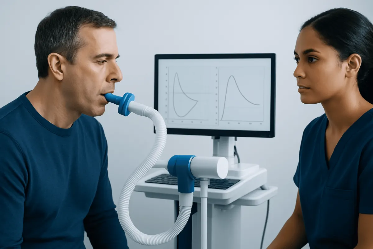
Spirometry measures how much air a person can forcefully exhale and how quickly this happens after taking a full breath in. Two central measurements are FEV1, the volume exhaled in the first second, and FVC, the total volume forcefully exhaled. The FEV1/FVC ratio compares these values.
An obstructive pattern is suggested when the FEV1/FVC ratio falls below the lower limit of normal for the individual. This indicates that the first-second airflow is disproportionately reduced. Clinicians may also repeat spirometry after an inhaled bronchodilator to see whether airflow improves, which can help characterize conditions such as asthma.
A restrictive pattern cannot be diagnosed by spirometry alone. Spirometry may show a low FVC with a preserved or elevated FEV1/FVC ratio, which is sometimes called a possible restrictive pattern. Full lung-volume testing is then used to measure total lung capacity. A low total lung capacity confirms restriction.
Results are usually compared with reference values that account for personal factors such as age, height, sex, and ancestry where applicable. A percentage of predicted value may appear on a report, but clinicians increasingly rely on the lower limit of normal and z-scores because they better account for expected variation between people.
How to Differentiate Restrictive vs Obstructive Lung Disease?

The main physiological difference is whether the central problem is airflow or lung expansion. In obstructive disease, the airways are narrowed, inflamed, filled with mucus, or prone to collapse during exhalation. A person may take a long time to empty the lungs and can develop air trapping or hyperinflation.
In restrictive disease, the total amount of air the lungs can hold is reduced. This can occur because the lung tissue is stiff, the pleura is affected, the chest wall cannot move freely, the breathing muscles are weak, or excess body weight limits chest expansion. People may breathe with smaller volumes and often notice shortness of breath during activity.
Common obstructive conditions include asthma, chronic obstructive pulmonary disease (COPD), bronchiectasis, and some forms of bronchiolitis. COPD includes chronic bronchitis and emphysema; readers can learn more about chronic obstructive pulmonary disease. Common causes of restriction include interstitial lung diseases, previous lung surgery, severe obesity, certain neuromuscular disorders, and chest wall abnormalities.
Symptoms alone cannot reliably separate the two patterns. Wheezing may occur with obstruction but is not always present, while dry cough and exertional breathlessness may occur with restriction but also occur in many other conditions. Testing provides objective information, and a chest X-ray or CT scan may be needed when the cause remains unclear.
How Do I Interpret My PFT Results?
Complete pulmonary function tests, often called PFTs, may include spirometry, lung volumes, and diffusing capacity testing. Lung volumes show whether total lung capacity is low and whether residual volume is high from air trapping. Diffusing capacity, often reported as DLCO, estimates how efficiently gases move from the lungs into the bloodstream.
A low FEV1 may occur in both obstruction and restriction, so it should not be interpreted alone. A low FEV1/FVC ratio supports obstruction. A low FVC with a normal ratio prompts lung-volume measurement, while a low total lung capacity establishes restriction. If both the ratio and total lung capacity are low, a mixed pattern may be present.
DLCO can add useful context but does not make a diagnosis on its own. It may be reduced in emphysema, some interstitial lung diseases, pulmonary vascular conditions, anemia, and other situations. It may be normal in some airway disorders or chest wall and neuromuscular causes of restriction.
A report may include terms such as mild, moderate, or severe impairment. These descriptions refer to test measurements, not necessarily to how unwell a person feels or what treatment they need. The ordering clinician should review the report in the context of the whole clinical picture and explain whether follow-up tests are appropriate.
What Happens After an Obstructive or Restrictive Pattern?
For an obstructive pattern, the next step commonly includes reviewing smoking, vaping, workplace exposures, allergies, infections, and current inhaler use. Depending on the likely cause, clinicians may consider bronchodilator response testing, peak-flow monitoring, imaging, allergy assessment, or referral to a respiratory specialist. Treatment may include avoiding triggers, stopping smoking, vaccinations, pulmonary rehabilitation, and inhaled medicines selected for the individual.
Asthma treatment is tailored to symptoms, exacerbation risk, and lung function, often using inhaled anti-inflammatory medicines and reliever therapy. COPD care may include inhalers, rehabilitation, activity support, vaccination, and help to stop smoking. Some people may be evaluated for bronchoscopy when airway obstruction, infection, bleeding, or an unexplained finding requires closer assessment.
For a confirmed restrictive pattern, clinicians look for the location and cause of reduced expansion. They may request high-resolution chest CT, blood tests, oxygen measurement, sleep assessment, neuromuscular evaluation, or cardiac testing when indicated. Management can range from treating inflammation or an underlying systemic disease to rehabilitation, weight management support, breathing assistance, or specialist monitoring.
Acıbadem Health Point’s multidisciplinary specialists at JCI-accredited hospitals assess respiratory symptoms and pulmonary test abnormalities for international patients, coordinating respiratory medicine, imaging, rehabilitation, and other services when needed.
Can Restrictive Lung Disease Get Better?
Whether restrictive lung disease can improve depends on its cause. Some causes are reversible or partly reversible, including temporary inflammation, fluid around the lungs, poorly controlled obesity-related restriction, certain medication effects, and reduced effort or incomplete inhalation during a test. Treating the cause and improving physical conditioning may improve symptoms and measurements in appropriate cases.
Other causes, such as some forms of lung scarring, may not be fully reversible. Even in these situations, careful treatment can help slow progression, address complications, preserve daily function, and improve quality of life. The outlook varies considerably between different interstitial lung diseases and should be discussed with a respiratory specialist.
People with restriction related to weakened breathing muscles or chest wall conditions may benefit from targeted rehabilitation, airway clearance where needed, vaccination, nutrition support, and assisted ventilation in selected cases. If a clinician suspects interstitial lung disease, prompt specialist evaluation can clarify the diagnosis and management plan.
Follow-up PFTs may be scheduled to monitor change over time. A change in numbers does not always mean a condition has progressed or improved, because results can also vary with technique, acute illness, and the equipment or reference equations used.
Practical Preparation, Prevention and Self-Care
Before spirometry or PFTs, patients should follow the instructions provided by their testing center. They may be asked to avoid smoking, vigorous exercise, large meals, or certain inhalers for a specified period. They should not stop prescribed medicines unless the clinician or test center specifically advises doing so.
For long-term lung health, avoiding tobacco smoke and vaping is among the most important protective steps. Using appropriate workplace respiratory protection, reducing exposure to dusts and fumes, staying physically active within personal limits, and keeping recommended vaccinations up to date can also support respiratory health.
People with diagnosed asthma, COPD, or another chronic lung condition should use medicines as prescribed and ask for a clear written plan for worsening symptoms. Correct inhaler technique matters; a pharmacist, nurse, or respiratory clinician can check it. Pulmonary rehabilitation may help selected people build exercise tolerance, learn breathing strategies, and manage breathlessness safely.
When to Seek Medical Care
A person should arrange medical assessment for ongoing or recurrent breathlessness, wheezing, chest tightness, a cough lasting several weeks, reduced ability to exercise, or unexplained fatigue that affects daily life. Assessment is also important after significant exposure to smoke, occupational dust, chemicals, or air pollution, particularly if respiratory symptoms develop.
Urgent medical care is needed for severe or rapidly worsening shortness of breath, bluish or gray lips or face, new confusion, fainting, chest pain, coughing up more than a small streak of blood, or difficulty speaking in full sentences because of breathlessness. These symptoms can have causes beyond lung disease and should not be managed by relying on a PFT report alone.
People who already have a respiratory diagnosis should seek timely advice if their usual treatment is no longer controlling symptoms, they need reliever medicine more often than advised, or they develop fever, a marked change in sputum, or worsening exercise tolerance. A clinician can determine whether infection, an exacerbation, a medication issue, or another problem is responsible.
Frequently asked questions
01Does a low FEV1 always mean obstructive lung disease?
No. FEV1 can be low in both obstructive and restrictive patterns because it reflects the amount of air exhaled during the first second. The FEV1/FVC ratio, lung volumes, test quality, and clinical context are needed to determine the likely pattern.
02Can spirometry confirm restrictive lung disease?
Spirometry can suggest restriction when FVC is low and the FEV1/FVC ratio is normal or high. However, restriction is confirmed by measuring a low total lung capacity with full pulmonary function testing. A low FVC can also occur because of poor test effort or air trapping.
03What is the typical spirometry pattern in asthma?
Asthma commonly produces an obstructive pattern, with a reduced FEV1/FVC ratio during periods of airway narrowing. Spirometry may improve after a bronchodilator, although a normal test between episodes does not exclude asthma. Clinicians interpret the results alongside symptom history and other tests when needed.
04Can a person have both obstructive and restrictive lung disease?
Yes. A mixed pattern can occur when airflow obstruction and reduced total lung capacity are both present. Full lung-volume testing is important because spirometry alone may not reliably distinguish mixed disease from obstruction with air trapping.
05Why might my PFT result be abnormal if I do not feel short of breath?
Mild abnormalities may be detected before symptoms become noticeable, especially in people with smoking or occupational exposure histories. Results can also be influenced by testing technique, recent illness, anemia, body size, or other non-lung factors. A clinician can advise whether the finding needs repeat testing or further evaluation.
06How often should pulmonary function tests be repeated?
The timing depends on the reason for testing and the suspected or diagnosed condition. Tests may be repeated after treatment changes, after an exacerbation has settled, or periodically to monitor <a href="https://www.acibademhealthpoint.com/blog/life-expectancy-and-chronic-disease-how-conditions-change-the-outlook/" title="Life Expectancy and Chronic Disease: How Conditions Change the Outlook" class="ahp-ilk">chronic disease. A respiratory clinician can recommend an interval that is appropriate for the individual.
This article is for general information only and is not a substitute for professional medical advice. Please consult a qualified doctor about your individual situation.
Explore treatments in Turkey — costs, top hospitals & a free quote
JCI-accredited · board-certified surgeons · reply within 24h
Add us as a Preferred Source to see more of our trusted health content across Google Search, AI Overviews and Discover.
More from the Health Library

Central Sleep Apnea: How It Differs From Obstructive Sleep Apnea

Over the Counter Meds for Cough: Common Causes, Related Conditions, and When to See a Doctor