Laminotomy Removes a Small Piece of Bone to Free a Nerve
Does your leg start burning or going heavy after a few minutes of walking, easing only when you sit down or lean on a shopping trolley? That pattern often points to a nerve in your spine being squeezed.
One way surgeons take the pressure off is a laminotomy. It removes a small portion of the bony arch at the back of a vertebra, called the lamina, to create more room for compressed spinal nerves. When non-surgical care has not given you enough relief, it may help reduce leg or arm pain, numbness, weakness, or walking difficulty.
What Is a Laminotomy?
Laminotomy is one of the spinal decompression operations. The surgeon removes or reshapes a small section of the lamina, the thin plate of bone forming part of the back wall of the spinal canal. That extra space gives a compressed spinal cord or nerve root room to breathe.
The goal is to reduce symptoms caused by nerve pressure, such as pain traveling into an arm or leg, tingling, numbness, weakness, or difficulty walking. A laminotomy does not treat every type of back or neck pain. It is generally considered when symptoms and examination findings suggest that a specific area of nerve compression is responsible.
A laminotomy may be performed in the lumbar spine, the lower back, or in the cervical spine, the neck. The operation is often combined with another procedure when needed, such as removal of part of a herniated disc or enlargement of the nerve opening, known as a foraminotomy.
How Laminotomy Differs From Other Spine Procedures
Neurosurgeon or Orthopedic Surgeon?" class="ahp-ilk">Spine surgery terms sound alike, and that trips people up. What separates them is how much bone comes out, and what kind. In a laminotomy, only a limited portion of the lamina is removed, often on one side. A laminectomy involves removal of a larger section or all of the lamina at one or more vertebral levels. Both procedures can decompress nerves, but the most appropriate choice depends on the location and extent of narrowing.
Some surgeons use a technique called bilateral decompression through a unilateral approach. This means the surgeon works from one side while decompressing both sides of the spinal canal, with the aim of preserving supportive structures where appropriate. Minimally invasive approaches may use smaller incisions and specialized instruments, although they are not suitable for every spinal problem.
Spinal fusion is different from decompression. Fusion joins two or more vertebrae to improve stability and may be recommended when there is spinal instability, deformity, significant vertebral slippage, or when decompression would require removal of structures essential for stability. Fusion is not automatically necessary with every laminotomy.
Why a Laminotomy May Be Recommended
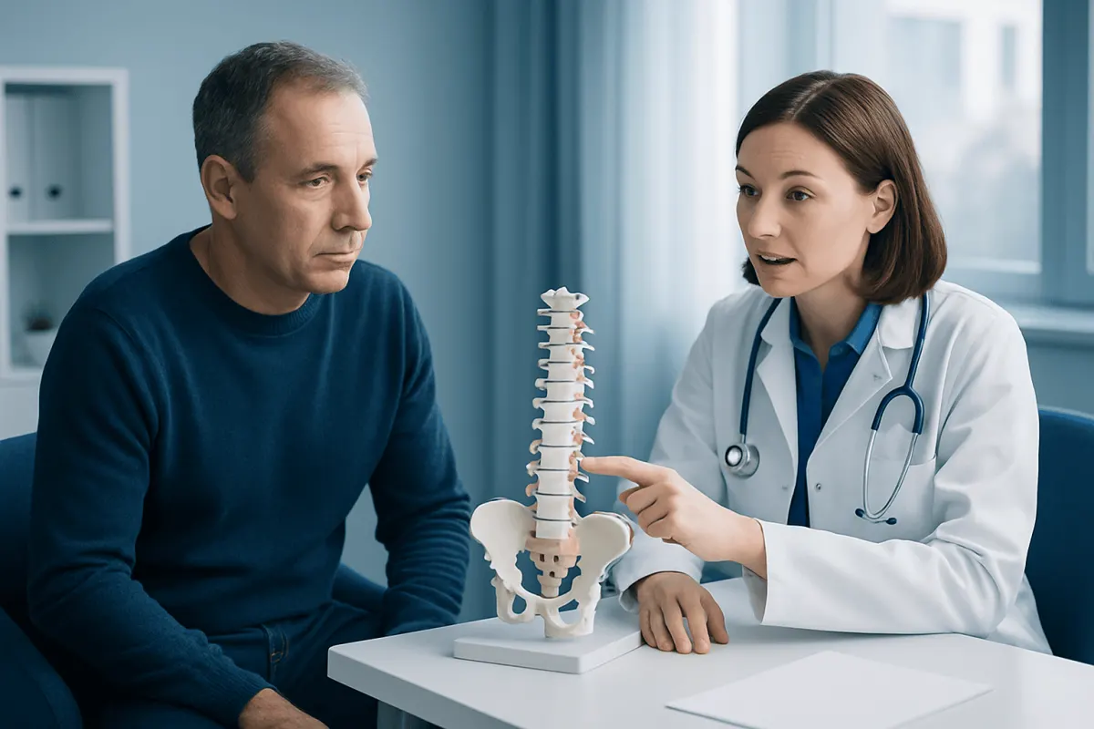
A common reason for laminotomy is spinal stenosis, a narrowing of the spinal canal or spaces around the nerves. In the lower back, this may cause neurogenic claudication: leg pain, heaviness, tingling, or weakness that develops with standing or walking and improves with sitting or leaning forward. Age-related changes in discs, joints, and ligaments are frequent contributors.
Other possible reasons include a herniated disc pressing on a nerve root, thickened spinal ligaments, bone spurs, or a synovial cyst arising near a facet joint. In the neck, nerve compression can cause pain, numbness, or weakness in an arm or hand. When spinal cord compression is present, symptoms can also include reduced hand coordination, balance difficulties, or changes in walking.
No surgeon decides on a scan alone. The location of symptoms, neurological examination, day-to-day function, imaging findings, overall health, and response to non-surgical treatment all help determine whether surgery is likely to be useful. Many people with imaging signs of spinal narrowing do not need an operation if they have no meaningful symptoms.
Assessment and Planning Before Surgery
Before recommending laminotomy, a clinician takes a detailed history and performs a physical and neurological examination. This may include checking strength, reflexes, sensation, balance, walking, and whether particular movements reproduce symptoms. The evaluation also looks for other possible sources of pain, including hip disease, peripheral nerve disorders, or vascular circulation problems.
MRI is commonly used to show discs, nerves, ligaments, and narrowing within the spinal canal. CT scans can provide more detail about bone, and X-rays taken while bending may help identify instability or vertebral movement. Imaging findings are most helpful when they match the person’s symptoms and examination.
When it is safe, you will usually try non-surgical treatment first. Options can include activity modification, guided exercise or physical therapy, pain-relieving medicines when appropriate, and selected spinal injections. Surgery may be discussed sooner if there is progressive weakness, severe loss of function, or evidence of significant spinal cord or nerve compression.
What Happens During Laminotomy
Laminotomy is performed in an operating room under anesthesia. The patient is positioned carefully to protect pressure points and allow the surgeon to access the affected part of the spine. Through an incision in the back of the neck or lower back, the surgeon gently moves muscles aside and uses imaging guidance to confirm the correct spinal level.
Small instruments, a drill, or both may be used to remove a targeted portion of bone and, if needed, part of a thickened ligament. The surgeon then relieves pressure around the nerve or spinal cord. If a disc fragment is compressing a nerve, it may be removed during the same operation. The incision is closed after bleeding is controlled.
How the operation is done, and how long you stay, differs from person to person. Some patients can go home the same day or after a short stay, while others need more observation because of the procedure performed, their symptoms, medical conditions, or mobility needs. The surgical team should explain the planned approach, alternatives, and expected recovery before consent is given.
Recovery, Results, and Possible Risks
After surgery, patients are usually encouraged to begin gentle walking as advised by their care team. Pain around the incision and temporary muscle soreness are common early on. Leg or arm symptoms may improve quickly when nerve pressure has been relieved, but numbness and weakness can take longer to improve because nerves recover gradually and sometimes incompletely.
Your plan will be your own, but most include wound care, walking a little more each day, keeping away from heavy lifting or hard bending for a while, and coming back for follow-up appointments. Physical therapy may be recommended to rebuild movement, strength, and confidence with everyday activity. The surgeon provides individualized guidance about driving, work, exercise, and travel.
Potential complications include infection, bleeding, blood clots, a tear in the covering around the nerves that can cause spinal fluid leakage, nerve injury, and reactions to anesthesia. Some people continue to have symptoms, develop symptoms at another spinal level, or require additional treatment in the future. Rarely, removing bone can contribute to instability, which is why careful surgical planning is important.
When to Seek Medical Care
Book an appointment if back or neck pain keeps travelling into an arm or leg, if you notice new numbness or tingling, weakness, less walking tolerance, or if symptoms are getting in the way of your sleep, work, or daily life. A clinician can determine whether symptoms may be coming from spinal nerve compression and whether imaging or specialist assessment is needed.
Urgent medical care is needed for new difficulty controlling the bladder or bowel, numbness in the saddle area around the groin and buttocks, rapidly worsening leg weakness, or sudden major problems walking. These symptoms can indicate severe nerve compression and should not be managed with home care alone.
After laminotomy, patients should contact their surgical team promptly for fever, worsening wound redness or drainage, increasing severe pain, new weakness, Neurology May Be Urgently Needed" class="ahp-ilk">severe headache that is worse when upright, or other unexpected symptoms. At Acıbadem Health Point, our multidisciplinary specialists and JCI-accredited hospitals assess and treat spinal conditions for patients coming from abroad, and we build each care plan around what you clinically need.
Frequently asked questions
01Is laminotomy a major surgery?
Laminotomy is a surgical procedure on the spine, so it requires careful planning and anesthesia. However, it is usually less extensive than a full laminectomy because only part of the lamina is removed. The complexity depends on the spinal level, the cause of compression, whether other procedures are needed, and the patient’s overall health.
02What is the difference between laminotomy and laminectomy?
A laminotomy removes a limited section of the lamina to access and decompress nerves. A laminectomy removes a larger portion or the entire lamina at one or more levels. Both can be effective decompression procedures, and the choice is based on the anatomy and treatment goal.
03How long does recovery from laminotomy take?
Recovery varies considerably between individuals and surgical approaches. Many people begin walking soon after surgery and gradually resume light activities over the following weeks. Return to physically demanding work, sports, or heavy lifting may take longer and should follow the surgeon’s advice.
04Will laminotomy cure back pain?
Laminotomy is mainly intended to relieve symptoms caused by identifiable nerve compression, especially radiating arm or leg pain, numbness, weakness, or walking limitation. It may not fully resolve pain that comes from muscles, joints, discs, or other causes. Expected benefits should be discussed in relation to the person’s specific symptoms and imaging findings.
05Is spinal fusion always needed with a laminotomy?
No. Many laminotomy procedures are performed without fusion when the spine is stable and enough supporting anatomy can be preserved. Fusion may be considered if there is instability, vertebral slippage, deformity, or a need for more extensive bone removal.
06Can spinal stenosis return after laminotomy?
Symptoms can recur or develop later, either at the treated level or at another level of the spine. This may happen because spinal degeneration can continue over time, scar tissue can form, or a different condition may contribute to symptoms. Follow-up is appropriate if pain, numbness, weakness, or walking problems return.
This article is for general information only and is not a substitute for professional medical advice. Please consult a qualified doctor about your individual situation.
Joint and spine care in Turkey — expert assessment & treatment
JCI-accredited · board-certified surgeons · reply within 24h
Add us as a Preferred Source to see more of our trusted health content across Google Search, AI Overviews and Discover.