Keratosis Treatment Options, From Freezing to Field Creams
Noticed a rough, scaly patch on your scalp that never quite goes away? Or a waxy, raised spot that keeps catching on your collar? What happens next depends on the type of lesion, where it sits, and whether there is any concern about precancerous change.
A dermatologist can often treat individual lesions right there in the clinic. When several areas of sun-damaged skin are involved, creams or field treatments may be used instead.
Keratosis treatment: how it works
Keratosis treatment aims to remove, destroy or reduce abnormal skin cells, but the best approach depends on the diagnosis. Seborrheic keratoses are common, benign growths that often look waxy, raised or “stuck on”. They do not usually need treatment unless they itch, catch on clothing, bleed after irritation, or a clinician needs to confirm the diagnosis.
Actinic keratoses are rough, scaly patches caused by long-term ultraviolet exposure. They are considered precancerous because a small proportion may progress to squamous cell carcinoma over time. Treatment can address visible lesions and, in some cases, the wider area of sun-damaged skin where early changes may not yet be obvious.
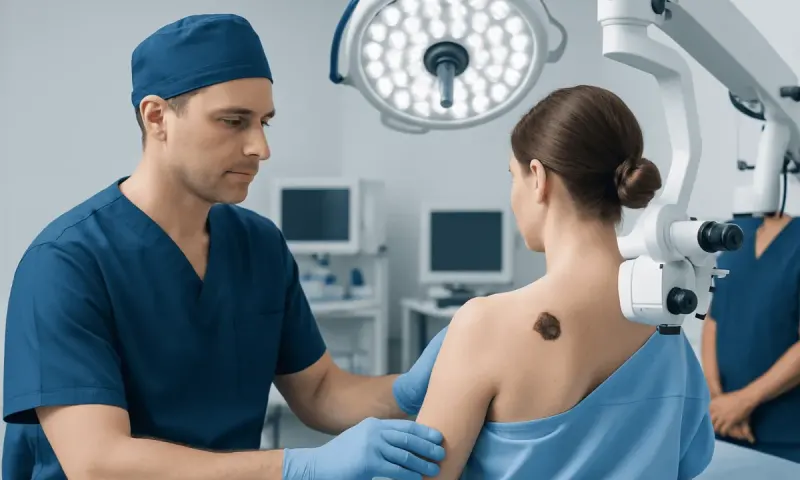
Never skip the skin examination before removal. Warts, eczema, melanoma and skin cancer can all look like keratosis. If a lesion has an uncertain appearance, your dermatologist may suggest a biopsy before or during treatment.
Choosing a treatment: candidacy and assessment
People may be candidates for keratosis removal when a lesion is diagnosed as benign but causes discomfort or cosmetic concern, or when actinic keratosis is present. The number, size, thickness and body location of lesions help guide the choice. A single thick lesion may be managed differently from many fine, rough spots across the face, scalp, ears, chest or forearms.
During assessment, the clinician considers skin tone, sun exposure history, previous skin cancers, immune system health, medicines and healing risks. People with a weakened immune system, including some transplant recipients or people taking immune-suppressing medicines, may need closer surveillance because sun-related lesions can behave more aggressively.
Pregnancy, breastfeeding, allergies and the use of certain medicines can affect which topical treatments are appropriate. Tell your doctor about any past reactions to local anaesthetics, the medicines you take now, including blood thinners, and whether you have ever scarred badly.
- Lesion-directed treatment targets one or a few defined growths.
- Field treatment treats a broader sun-damaged area and may uncover or clear early, less visible lesions.
- Biopsy may be recommended if a lesion looks atypical, has changed, or does not respond as expected.
Keratosis removal procedure: step by step
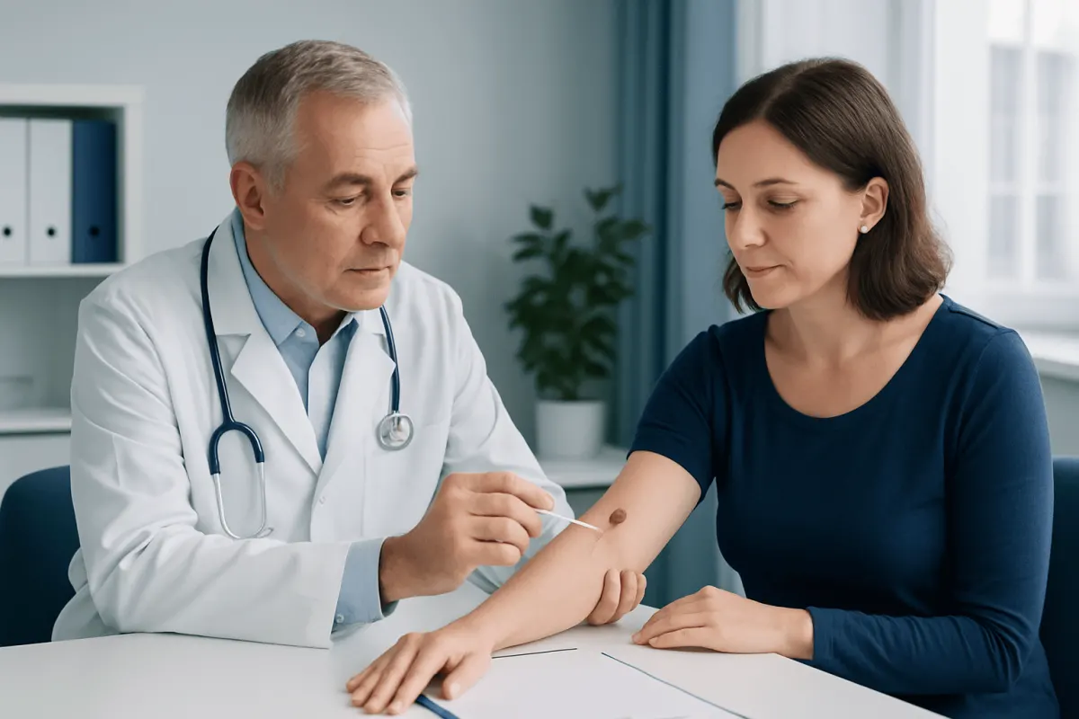
For a limited number of lesions, cryotherapy is one of the most common options. A clinician applies liquid nitrogen briefly to freeze the lesion. The treated area may sting during application, then become red, swollen or blistered before it dries, crusts and heals. More than one session may be needed for thicker lesions.
Curettage and electrosurgery are other office-based methods. After numbing the skin, the clinician may gently scrape the lesion with a curette and use heat or electrical current to control bleeding or destroy remaining abnormal cells. Shave removal may be used for selected raised seborrheic keratoses, and the removed tissue can be sent for laboratory examination when appropriate.
For widespread actinic keratoses, a dermatologist may prescribe topical treatment such as 5-fluorouracil, imiquimod, diclofenac or tirbanibulin, depending on the individual situation and local availability. These treatments are applied at home for a clinician-directed course and commonly cause temporary inflammation in the treatment area. Photodynamic therapy applies a light-sensitive medicine followed by controlled light exposure, selectively damaging abnormal cells.
Treatment gives you clearer skin and gets rid of the abnormal cells. It does not make you immune to new ones. New keratoses can still appear, because the cause, above all the sun exposure you have built up over the years, has not gone anywhere.
Recovery timeline, results and possible risks
Healing after treatment varies with the method, lesion depth, location and an individual’s skin. After cryotherapy, mild soreness or redness may last a few days. A blister or crust can develop, and many treated areas heal within one to three weeks. Lesions on the lower legs may heal more slowly than those on the face or scalp.
After curettage, electrosurgery or shave removal, the site usually forms a shallow wound. Gentle cleansing, petrolatum-based ointment if advised, and a simple dressing can support healing. Follow the aftercare instructions you are given, and leave the crusts alone. Picking raises the risk of infection and scarring.
Topical field therapies often produce redness, scaling, burning, tenderness and crusting while they work. The visible reaction can be more noticeable than after single-lesion treatment and may continue for days to weeks after the treatment course ends. Photodynamic therapy can cause stinging during light exposure and temporary redness or sensitivity afterward.
Possible risks include pigment changes, blistering, infection, incomplete clearance, recurrence and scarring, although serious complications are uncommon when treatment is performed appropriately. Contact the treating clinic promptly for spreading redness, pus, fever, severe pain, persistent bleeding or a wound that is not improving.
How long does it take for keratosis to go away?
The time depends on the kind of keratosis and the treatment used. A small seborrheic keratosis treated with freezing may crust and separate within one to three weeks, while a scraped or shaved lesion may take a similar period to heal. Larger, thicker lesions and areas below the knee can take longer.
Actinic keratoses may clear after one treatment, but some need repeat cryotherapy or a course of field treatment. Prescription creams can produce a treatment reaction over several weeks, followed by additional healing time. A dermatologist can set realistic expectations after examining the skin and selecting a treatment plan.
Even when a treated spot resolves, new lesions may develop in sun-exposed areas. Ongoing skin checks and sun protection are therefore an important part of long-term care.
What is the fastest way to get rid of keratosis?
For a diagnosed individual lesion, office-based removal such as cryotherapy, curettage or shave removal is often the quickest way to treat it. However, the fastest option is not always the most suitable one. A lesion must first be accurately identified, and a biopsy may be safer than immediate cosmetic removal if the diagnosis is uncertain.
For multiple actinic keratoses across a larger area, field treatment may be more effective than repeatedly treating only the visible spots. Although this approach takes longer and may cause temporary redness or peeling, it can address broader sun damage that contributes to new lesions.
Over-the-counter acids, wart removers and home freezing products should not be used on an undiagnosed lesion. They can injure healthy skin, delay diagnosis of skin cancer and may not remove the lesion completely.
How painful is keratosis removal?
Most keratosis removal procedures are well tolerated. Liquid nitrogen can cause a brief sharp sting, burning or aching sensation during and shortly after treatment. The area may remain tender for a day or two, particularly if a blister develops.
Procedures that involve scraping, shave removal or electrosurgery are generally performed with local anaesthetic. Patients may feel the initial anaesthetic injection and pressure or movement during the procedure, but should not feel sharp pain. Mild soreness afterward is common and can usually be managed with clinician-approved simple measures.
Topical medicines and photodynamic therapy can cause burning, sensitivity or discomfort as inflammation develops. Call your clinician if the discomfort is severe, keeps you awake, or comes with noticeable swelling, drainage or other signs that something is wrong.
How long does it take to get rid of actinic keratosis?
Individual actinic keratoses treated with cryotherapy may resolve over one to three weeks as the skin heals. Some lesions need a second treatment, especially if they are thick or located in an area that heals slowly. The doctor may reassess the site after healing to confirm it has cleared.
Field treatments generally take longer. Depending on the medicine or photodynamic therapy plan, treatment and recovery may extend over several weeks. During this time, redness, flaking, crusting and sensitivity can be expected to varying degrees; these changes often reflect the intended effect on abnormal cells.
Actinic keratosis is a marker of cumulative sun damage, so clearance of current lesions does not eliminate future risk. Regular follow-up is especially important for people with many lesions, a past skin cancer, extensive sun exposure or reduced immune function.
Prevention, self-care and when to seek medical care
Daily sun protection helps reduce new actinic keratoses and supports long-term skin health. This includes seeking shade when practical, wearing a broad-brimmed hat and protective clothing, and using broad-spectrum sunscreen with an SPF of 30 or higher on exposed skin. Sunscreen should be reapplied as directed, particularly after swimming or sweating.
After a procedure, stick to the wound-care plan you were given. Don’t pick, scratch, or put unapproved products on healing skin. If your clinician has asked for remote follow-up, clear photos before treatment and during healing help a lot.
When to seek medical care: Book a dermatology assessment if a lesion is new, changing quickly, asymmetric, unusually dark or multicoloured, painful, persistently itchy, bleeding without an obvious injury, ulcerated, or simply not healing. Get it looked at too if a keratosis comes back after treatment, or if several rough, scaly patches turn up on sun-exposed skin.
Acıbadem Health Point’s multidisciplinary specialists and JCI-accredited hospitals can assess suspicious skin lesions and provide individualized dermatology care for international patients. A qualified dermatologist can explain whether observation, biopsy, lesion removal or field treatment is the safest next step.
Frequently asked questions
01Can keratosis disappear without treatment?
Seborrheic keratoses usually do not disappear on their own, although they may remain unchanged for years. Actinic keratoses can occasionally regress, but they can also persist, recur or progress. Don’t wait for a rough or changing lesion to sort itself out; have it checked.
02Is keratosis treatment necessary for seborrheic keratosis?
Treatment is usually not medically necessary once a seborrheic keratosis has been confidently diagnosed. Removal may be considered if it is irritated, repeatedly injured, itchy, or bothersome in appearance. Any lesion with an uncertain diagnosis should be examined before removal.
03Can actinic keratosis come back after treatment?
A treated actinic keratosis can recur if abnormal cells remain, and new lesions can develop in nearby sun-damaged skin. This does not necessarily mean treatment failed, but it does mean ongoing follow-up and sun protection are important. A clinician may recommend repeat treatment or field therapy when lesions are numerous.
04Will keratosis removal leave a scar?
Many lesions heal with little or no visible scarring, especially when they are small and treated carefully. However, any procedure that disrupts the skin can leave a mark or cause lighter or darker pigmentation. Scarring risk can be higher with deeper lesions, infection, picking, delayed healing and certain skin types.
05Can you exercise after keratosis removal?
Light activity is usually fine after a minor procedure, though your clinician may ask you to hold off on swimming, heavy sweating, friction or anything that could disturb a dressing for a short while. It depends on the method used and where on the body it was. Follow the instructions you were given.
06How often should skin checks be done after actinic keratosis?
The appropriate schedule depends on the number of lesions, previous skin cancer history, immune status and degree of sun damage. Some people need periodic professional skin examinations, while others may need closer monitoring. A dermatologist can recommend a personal follow-up plan and explain how to perform regular self-checks.
This article is for general information only and is not a substitute for professional medical advice. Please consult a qualified doctor about your individual situation.
Hair transplant in Turkey — costs, graft counts & a free assessment
JCI-accredited · board-certified surgeons · reply within 24h
Add us as a Preferred Source to see more of our trusted health content across Google Search, AI Overviews and Discover.