Jejunostomy Tube vs PEG: How Feeding Routes Differ
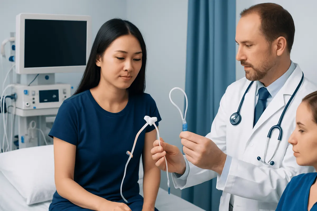
A jejunostomy tube and a PEG tube are both feeding tubes, but they deliver nutrition to different parts of the digestive tract. A PEG enters the stomach, while a jejunostomy reaches the jejunum, a section of the small intestine; the best option depends on swallowing ability, stomach function, aspiration risk, and the underlying medical condition.
Jejunostomy Tube vs PEG: Side-by-Side Comparison
A jejunostomy tube versus a PEG tube is mainly distinguished by where the tube ends inside the digestive system. A percutaneous endoscopic gastrostomy (PEG) tube passes through the abdominal wall into the stomach. A jejunostomy tube, often called a J-tube, delivers feed directly into the jejunum, which is the middle portion of the small intestine.
Both are forms of enteral nutrition, meaning they use the digestive tract rather than a vein to provide nutrition, fluids, and sometimes medicines. Neither option is automatically better for every person. The choice is based on the person’s medical needs, digestive function, anticipated length of feeding support, and ability to safely swallow.
- PEG tube location: stomach.
- Jejunostomy tube location: jejunum in the small intestine.
- PEG feeding pattern: may allow larger scheduled feeds when tolerated.
- Jejunal feeding pattern: is often given slowly and continuously using a pump.
- Typical reason for PEG: long-term difficulty swallowing with a functioning stomach.
- Typical reason for J-tube: a need to bypass the stomach because of poor stomach emptying, obstruction, reflux-related aspiration concerns, or certain upper digestive conditions.
Some tubes combine both routes. For example, a gastrojejunostomy tube enters through the stomach but extends into the jejunum. This is not the same as a standard PEG or a surgically placed J-tube, and the care instructions may differ.
How Clinicians Tell a Jejunostomy Tube From a PEG
Clinicians do not rely on appearance alone to identify a feeding tube. The safest approach is to review the procedure record, tube label, care plan, and prior imaging when available. The tube type, size, placement date, and intended feeding route should be documented in the medical record and, in many cases, in the patient’s discharge information.
The position on the abdomen can offer clues but is not definitive. A standard PEG commonly enters the upper central or upper left portion of the abdomen because it accesses the stomach. A J-tube may be positioned lower or farther to one side, depending on anatomy and the method used. However, placement sites vary, and a tube’s external location cannot reliably confirm its internal endpoint.
Healthcare professionals may also assess the external tube and its components. A PEG often has an external bumper or retention device and may have a port for gastric feeding. A jejunal system may have a smaller tube, a dedicated jejunal port, or pump-feeding equipment. If there is any uncertainty about position, a clinician may order imaging with contrast or another appropriate test before the tube is used.
A feeding tube should never be assumed to be correctly positioned after it has been pulled, replaced, become loose, or caused new symptoms. Confirmation is especially important before giving feeds, fluids, or medication through a tube when displacement is possible.
What a PEG Tube Is Used For

A PEG tube is generally considered when a person cannot take enough nutrition, fluid, or medication by mouth for an extended period but can still use the stomach for digestion. It may be needed after conditions that affect swallowing, such as stroke, neurological disease, head and neck cancer treatment, or severe injury. It can also be used when temporary nutritional support is needed during recovery from illness or treatment.
Because the stomach can act as a reservoir, gastric feeding through a PEG may be delivered as intermittent feeds or as a continuous infusion, depending on tolerance and clinical guidance. A dietitian and medical team select the formula, timing, fluid plan, and medication approach for the individual.
A PEG does not necessarily prevent all oral intake. Some people can still eat or drink safely in limited amounts, while others need to avoid oral intake because of swallowing-related aspiration risk. A swallowing assessment may help guide this decision. For people with swallowing problems related to neurological conditions, assessment and care should address the underlying cause as well as nutrition support.
PEG placement and ongoing care require attention to skin hygiene, tube security, flushing instructions, and monitoring for complications. A clinician should advise whether a tube can be used for medicines and how each medicine should be prepared; not all medications are suitable for crushing or tube administration.
What a Jejunostomy Tube Is Used For
A jejunostomy tube is used when feeding into the stomach is unsuitable, poorly tolerated, or considered less safe. It sends formula directly into the small intestine, bypassing the stomach. A J-tube may be placed during abdominal surgery, through a radiologic or endoscopic procedure, or through a stomach access with an extension into the jejunum.
Jejunal feeding may be considered for people with severe delayed stomach emptying, some types of gastric outlet obstruction, significant vomiting or reflux despite treatment, pancreatitis in selected circumstances, or a high concern that stomach contents could enter the airway. The underlying condition and the person’s overall nutrition needs determine whether this route is appropriate.
Because the jejunum has less capacity to hold a large volume at once, feeds are commonly delivered gradually by pump over many hours. This can help reduce cramping, diarrhea, nausea, or intolerance, although symptoms can still occur and should be discussed with the care team. Feed formulas and rates should not be changed independently without advice.
Medication administration through a J-tube needs particular care. Some drugs are absorbed differently when delivered beyond the stomach, and some formulations may clog the tube or should not be crushed. A pharmacist, doctor, or nutrition support nurse can provide medication-specific instructions.
Choosing the Right Tube for Each Situation
The decision between a jejunostomy tube and a PEG is made by considering the reason nutrition support is needed and whether the stomach and intestines are working adequately. A person with persistent swallowing difficulty and normal stomach emptying may be a candidate for gastric feeding through a PEG. Someone who cannot tolerate stomach feeds or needs the stomach bypassed may benefit from jejunal feeding instead.
Doctors also consider whether feeding support is expected to be short-term or long-term, whether abdominal surgery is already planned, past abdominal operations, nutritional status, and the person’s goals of care. In some situations, a temporary nasal feeding tube is used first to assess tolerance before a longer-term feeding tube is placed.
Conditions affecting the esophagus or stomach can influence this decision. For example, an obstruction, severe reflux, or impaired stomach emptying may require further investigation and a tailored plan. People receiving care for esophageal cancer or other upper digestive tract conditions may need coordinated input from gastroenterology, surgery, oncology, speech and language therapy, and clinical nutrition.
Tube feeding supports nutrition but does not treat the condition causing eating or swallowing difficulties. The care plan may also include swallowing rehabilitation, treatment of digestive symptoms, cancer therapy, neurological care, or surgery, depending on the diagnosis.
Daily Care and Preventing Tube Problems
Daily tube care can reduce skin irritation, blockage, and accidental displacement. The person or caregiver should follow the written instructions supplied by the healthcare team, including how to clean the skin around the site, secure the tube, flush it, and store feeding equipment. Hands should be cleaned before handling the tube or formula.
Feeding tubes should be flushed at the times advised by the clinical team, commonly before and after feeds and medications. This helps keep the tube open. Only approved fluids, formulas, and medicines should go through the tube. Household remedies, carbonated drinks, or improvised tools should not be used to clear a blockage unless specifically advised by a healthcare professional.
Common concerns can include minor leakage, redness, granulation tissue, discomfort, clogging, or loose external fittings. These issues are often manageable, but new or worsening symptoms should be assessed. It is important not to rotate, advance, remove, or replace a tube unless the person has been trained and instructed to do so.
A dietitian can help ensure the feeding regimen meets calorie, protein, hydration, and micronutrient needs. Regular review is useful because nutritional requirements may change with recovery, weight changes, treatment side effects, activity level, or changes in digestive tolerance.
When to Seek Medical Care
Prompt medical advice is appropriate if a feeding tube becomes dislodged, visibly moves, cracks, leaks heavily, cannot be flushed, or causes repeated vomiting, persistent diarrhea, increasing pain, or inability to tolerate feeds. A tube that comes out may need urgent attention because some feeding tube tracts can begin to narrow or close relatively quickly, particularly when the tube is new.
Urgent assessment is needed for severe abdominal pain, a swollen or rigid abdomen, fever, rapidly spreading redness around the site, pus-like drainage, significant bleeding, breathing difficulty, confusion, fainting, or signs of dehydration. These symptoms do not always mean a serious complication, but they should not be managed at home without clinical guidance.
For non-urgent questions, patients can contact the team that placed the tube, a gastroenterology service, a surgical team, or a nutrition support nurse. Acıbadem Health Point’s multidisciplinary specialists and JCI-accredited hospitals can assess feeding-tube needs and support international patients with diagnosis and treatment planning.
Keeping a current copy of the tube type, placement date, feeding prescription, medication instructions, and emergency contact details can make it easier to obtain appropriate care when traveling or attending an urgent care service.
Frequently asked questions
01Is a jejunostomy tube the same as a PEG tube?
No. A PEG tube enters the stomach, while a jejunostomy tube delivers feed into the jejunum of the small intestine. Both are feeding tubes, but they are chosen for different clinical situations.
02Can a PEG tube be used to feed into the jejunum?
Sometimes. A gastrojejunostomy tube may enter through a gastrostomy opening and extend into the jejunum. This is different from a standard PEG and should be managed according to the specific instructions from the healthcare team.
03Which is better, a PEG or a jejunostomy tube?
Neither is universally better. A PEG may be suitable when the stomach works well, while jejunal feeding may be preferred when the stomach needs to be bypassed. The decision depends on the person’s diagnosis, symptoms, nutritional needs, and risk of feeding complications.
04Can someone eat by mouth with a PEG or J-tube?
Some people can continue to eat or drink by mouth, while others cannot do so safely or comfortably. This depends on the reason for the feeding tube, swallowing safety, and the care team’s recommendations. A swallowing evaluation may be helpful when aspiration is a concern.
05How can a caregiver know whether a tube has moved?
A change in the visible tube length, new leakage, pain during feeding, difficulty flushing, or unexpected digestive symptoms can suggest a problem. The tube should not be used until the placement has been assessed if displacement is suspected. The care team can advise whether imaging or other confirmation is needed.
06What should be done if a feeding tube falls out?
The person or caregiver should contact the tube-placement team or seek urgent medical care promptly, especially if the tube is recently placed. They should not attempt reinsertion unless they have been specifically trained and instructed to do so. Covering the site with a clean dressing may be advised while arranging care.
This article is for general information only and is not a substitute for professional medical advice. Please consult a qualified doctor about your individual situation.
Persistent digestive symptoms? Get evaluated in Turkey
JCI-accredited · board-certified surgeons · reply within 24h
Add us as a Preferred Source to see more of our trusted health content across Google Search, AI Overviews and Discover.