Hip X-Rays Show Bone Clearly but Not Soft Tissue
You slipped on the stairs, or maybe the ache in your groin has been creeping up on you for months. Either way, the first test your doctor is likely to order is a hip xray.
It’s a common imaging examination that creates pictures of the hip bones and joint. It can help clinicians identify fractures, arthritis, dislocation, bone changes and some other causes of hip symptoms. What it can’t do is show every problem affecting the hip — and that distinction matters more than most people expect.
What is a hip xray?
The test uses a controlled, low dose of radiation to create pictures of your upper thigh bone, pelvic bones and hip joint. It is one of the most established ways to assess bone structure and joint alignment. Clinicians commonly request it to investigate hip pain, reduced movement, an injury, or concerns about arthritis or another bone-related condition.
The hip is a ball-and-socket joint: the rounded head of the femur fits into a cup-shaped part of the pelvis called the acetabulum. An xray can show the shape and position of these bones, the space between them, and changes in the bone near the joint. It can also include nearby parts of the pelvis when this is clinically useful.
Think of it as a first step, not the whole story. Bone shows up very clearly, but soft tissues — muscles, tendons, ligaments, cartilage and the labrum — do not. That is why your clinician reads the images alongside your symptoms, your medical history and a physical examination.
Why a clinician may recommend a hip xray
Hip xrays are often used after a fall, road traffic collision or sports injury, particularly when there is pain in the groin, buttock or outer hip, difficulty standing, or an inability to bear weight. They can quickly identify many fractures and dislocations, which may need prompt treatment. In older adults, even a seemingly minor fall can lead to a fracture, especially when bone strength is reduced.
For pain that develops gradually, the test may help identify changes associated with osteoarthritis. These can include narrowing of the joint space, bony growths called osteophytes, changes in bone density beneath the cartilage, or altered joint shape. A hip xray can also help evaluate structural differences, prior surgery, joint replacement components, certain bone infections or tumours, and some childhood or developmental hip conditions.
Not all pain felt near the hip starts in the hip joint. The lower back, sacroiliac joints, muscles, tendons, nerves and abdominal or pelvic organs can sometimes cause pain in this region. If the xray does not explain the symptoms, the clinician may consider other causes and decide whether further assessment is appropriate.
- Sudden pain after injury or a fall
- Persistent pain, stiffness or reduced walking tolerance
- Suspected fracture, dislocation or arthritis
- Follow-up after hip surgery or joint replacement
- Assessment of selected bone or joint abnormalities
What a hip xray can and cannot show

A hip xray is particularly useful for identifying broken bones, joint dislocation and changes in bone alignment. It may show signs of osteoarthritis, such as loss of joint space and bone spurs. It can also detect some conditions that alter bone shape or density, including certain longstanding structural abnormalities.
However, an xray cannot directly show cartilage, the labrum, tendons, muscles or most ligaments in detail. It may therefore be normal in people with tendon irritation, bursitis, labral injuries or early cartilage damage. Xrays can also miss some small, nondisplaced or stress fractures, particularly early after an injury.
When a fracture is strongly suspected despite normal initial xrays, further imaging should not be delayed simply because the xray appears reassuring. MRI is often used to detect hidden fractures and to examine soft tissues. CT can provide more detailed cross-sectional views of bone and may be useful for clarifying a complex fracture or planning treatment.
What shows on the image and what you feel do not always line up. Some people have visible arthritic changes and barely notice them; others hurt a great deal with only subtle findings. That is why a radiology report should be read by the clinician caring for you, not interpreted on its own.
How the hip xray procedure is performed
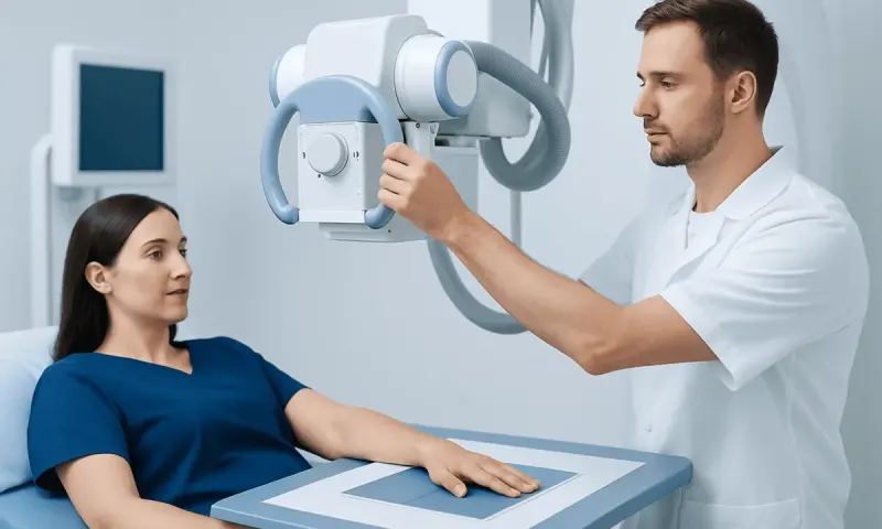
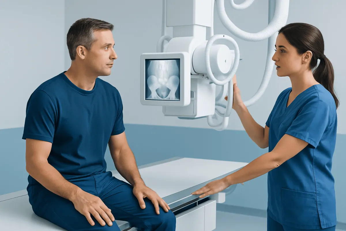
A radiographer performs the examination in an xray department, outpatient imaging centre, emergency department or hospital. Before the images are taken, the person may be asked to change into a gown and remove metal items around the waist or hip area, such as belts, jewellery or clothing with zips. Metal can obscure important details on an image.
The person is positioned carefully on an xray table or, in some situations, remains on a trolley or in a wheelchair. Common views include an image taken from the front of the pelvis and an angled or side view of the affected hip. Positioning may need to be adjusted so that the joint can be seen clearly. After a recent injury, the team will avoid unnecessary movement and prioritize comfort and safety.
The xray itself is painless and usually takes only a few minutes. You just need to hold still briefly while each image is taken. There are no injections and no contrast material is routinely needed. In many settings, the full appointment is short, although timing can vary if emergency imaging or additional views are required.
A radiologist or another appropriately trained clinician reviews the images and prepares a report for the referring healthcare professional. The referring clinician explains what the findings mean in the context of the person’s symptoms and discusses whether any treatment or further testing is needed.
Radiation safety and pregnancy considerations
Hip xrays use ionizing radiation, but the imaging team uses the lowest exposure that is reasonably achievable while obtaining diagnostically useful images. Modern equipment, careful positioning and avoiding unnecessary repeat images all help limit radiation exposure. For most people, the expected clinical benefit of an appropriately requested xray outweighs the small radiation risk.
Anyone who is pregnant, thinks they may be pregnant, or is trying to conceive should tell the radiographer before the examination. This does not automatically mean the xray cannot be performed. Instead, the referring clinician and imaging team consider how urgent the test is, whether another method might answer the question, and how to perform imaging as safely as possible.
Children are also imaged with particular care because they are more sensitive to radiation over their lifetime. Paediatric xray techniques use tailored settings, and imaging is requested when it is expected to provide useful information for care. A parent or caregiver may be allowed to stay with a child during the examination, depending on local safety procedures.
Understanding hip xray results and possible next steps
A report may describe the bones as normal, note evidence of a fracture, or identify degenerative changes consistent with arthritis. It may also mention joint alignment, changes around a previous hip replacement, bone density, calcifications or findings in the visible pelvis. Words such as “mild,” “moderate” and “severe” usually describe the appearance on imaging, rather than predicting exactly how a person feels or functions.
If the xray identifies a fracture or dislocation, the next steps depend on its location, stability and the person’s overall health. Treatment may include pain management, protected weight-bearing, rehabilitation, reduction of a dislocation, or surgery. A displaced fracture or inability to bear weight after trauma needs urgent medical assessment.
For osteoarthritis, care often begins with a combination of education, activity adjustments, exercise-based physiotherapy, weight management when relevant, and clinician-guided pain relief. When symptoms remain severe and substantially affect daily life despite non-surgical care, hip replacement surgery may be considered after a thorough orthopedic evaluation.
If your symptoms carry on but the images look normal or unclear, your clinician may arrange MRI, CT, ultrasound, blood tests or a specialist referral. At Acıbadem Health Point, our multidisciplinary specialists and JCI-accredited hospitals assess hip conditions and coordinate the right imaging and treatment for international patients.
When to seek medical care
Urgent medical care is appropriate after a fall or injury when there is severe hip pain, a visible change in leg position, inability to stand or bear weight, numbness, weakness, or a cold or pale foot. These symptoms can occur with a fracture, dislocation or injury affecting nerves or blood supply. It is safest not to force the hip to move or attempt to walk on it.
A prompt medical appointment is also advisable for persistent hip pain, progressive stiffness, limping, pain that disrupts sleep, or symptoms that do not improve with a reasonable period of rest and self-care. A clinician can determine whether a hip xray or another test is appropriate and can assess for causes outside the hip joint.
Fever, feeling unwell, marked redness or warmth around the hip, or new hip pain in a person with a weakened immune system should be assessed urgently. Although these symptoms have several possible explanations, they can occasionally indicate infection or another condition requiring timely treatment.
After a significant injury and until you have been assessed, stay away from anything that makes the pain worse and follow the advice you were given about moving and putting weight on the leg. And do not let a normal xray reassure you if severe pain, difficulty walking or worsening symptoms continue.
Frequently asked questions
01What does a hip xray show?
A hip xray shows the bones forming the hip joint, their alignment and the space between the joint surfaces. It can help identify fractures, dislocation, arthritis-related changes and some structural bone abnormalities. It is less effective for assessing muscles, tendons, cartilage and other soft tissues.
02Do hip xrays show arthritis?
Yes. A hip xray can show features commonly associated with osteoarthritis, including joint-space narrowing, bone spurs and changes in the bone beneath the joint surface. The image is only one part of diagnosis, because symptoms and physical examination findings are also important.
03Can a hip xray miss a fracture?
Yes, some fractures may not be visible on initial xrays, especially small, nondisplaced or stress fractures. If clinical suspicion remains high after a normal xray, a clinician may recommend MRI, CT or repeat imaging. Ongoing pain or inability to bear weight after injury should be reassessed promptly.
04How long does a hip xray take?
The images themselves are usually taken within a few minutes, although the whole visit may take longer for registration, changing and positioning. Additional views can be needed depending on the symptoms and the reason for imaging. Results may be available quickly in emergency care, while routine reporting times vary by facility.
05Is a hip xray painful?
The xray does not cause pain because it does not involve needles or injections. However, positioning the leg or pelvis can be uncomfortable when the hip is injured or very painful. The radiographer can adapt positioning where possible and should be told about discomfort.
06Is it safe to have a hip xray during pregnancy?
A hip xray involves radiation near the pelvis, so it is important to tell the healthcare team about a known or possible pregnancy before imaging. The team will assess whether the examination is necessary, whether another test may help, and how to minimize exposure. Urgent imaging should not be avoided when it is needed to assess a potentially serious condition.
This article is for general information only and is not a substitute for professional medical advice. Please consult a qualified doctor about your individual situation.
Joint and spine care in Turkey — expert assessment & treatment
JCI-accredited · board-certified surgeons · reply within 24h
Add us as a Preferred Source to see more of our trusted health content across Google Search, AI Overviews and Discover.