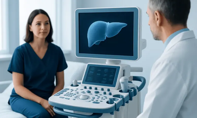
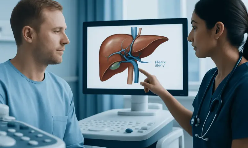
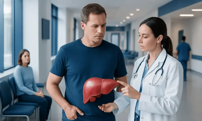
Hepatic Hypodensities on CT: Understanding Liver Findings
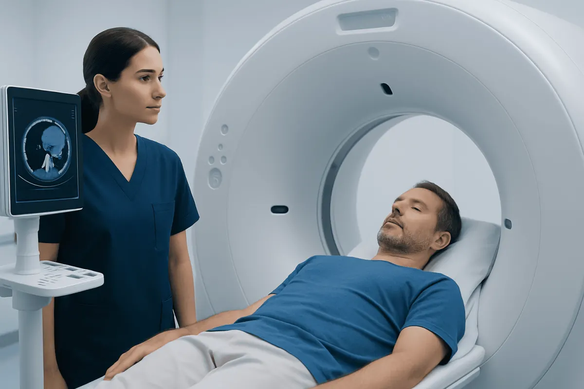
Hepatic hypodensities are areas of the liver that appear darker than surrounding tissue on a CT scan. They are an imaging description rather than a diagnosis, and many are harmless; further assessment depends on their appearance, size, number, symptoms, and the person’s health history.
What does hepatic hypodensity mean?
Hepatic hypodensities are areas within the liver that look darker than the surrounding liver tissue on a computed tomography (CT) scan. “Hepatic” means related to the liver, and “hypodense” means less dense on CT imaging. The term describes how an area looks on a scan; it does not, on its own, identify a disease or determine whether a finding is serious.
These findings are common because CT scans are frequently performed for many reasons, including abdominal pain, injury, kidney stones, bowel symptoms, or follow-up of another condition. A small liver finding may be discovered incidentally in a person with no liver symptoms. In many cases, the radiology report can identify a likely benign cause, while in others it may recommend comparison with older scans or further imaging.
The meaning of a hepatic hypodensity depends on the complete picture. Important details include whether contrast dye was used, whether the area has a simple fluid-like appearance, its margins and size, how many areas are present, and whether it changes over time. A clinician also considers a person’s age, liver health, history of cancer, infections, medication use, and relevant blood test results.
Why a CT scan can show darker areas in the liver
CT uses X-rays to create detailed cross-sectional images. Different tissues absorb X-rays to different degrees, creating variations in brightness on the image. Normal liver tissue has a relatively uniform appearance, while fluid, fat, blood vessels, inflammation, and some types of tissue growth can look less dense or darker.
Contrast-enhanced CT adds another layer of information. Contrast material travels through blood vessels and can make some liver lesions become brighter at particular times after injection. The pattern of contrast enhancement is often more informative than the word “hypodense” alone. For this reason, a finding described on a non-contrast scan may need a dedicated contrast CT or MRI for more confident characterization.
Scan timing and technical factors can also influence appearance. For example, a region may look different during arterial, portal venous, or delayed imaging phases. Radiologists interpret these images in context and may use standardized reporting language to communicate whether a finding appears clearly benign, needs routine follow-up, or requires more timely assessment.
Common causes of hepatic hypodensities
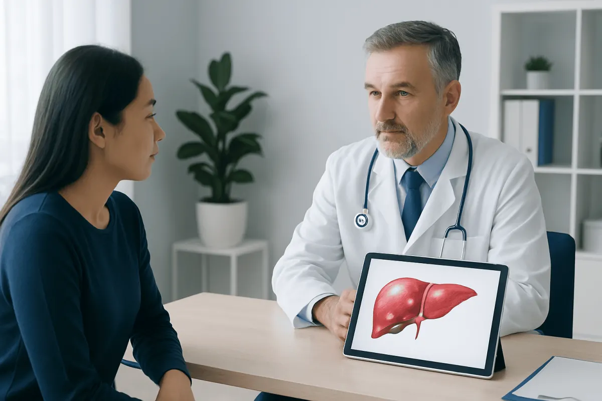
Many hepatic hypodensities are non-cancerous. A simple liver cyst is a common example. It is a thin-walled, fluid-filled sac that usually causes no symptoms and often needs no treatment. Another common benign lesion is a hemangioma, a collection of small blood vessels within the liver. Hemangiomas are often found incidentally and can usually be recognized by their characteristic behavior on contrast imaging.
Focal fat variation may also create a darker area. Fat can accumulate in or spare particular parts of the liver, especially in people with Liver Disease: Symptoms, Causes and Treatment" class="ahp-ilk">fatty liver disease, metabolic conditions, or changes in weight. These areas are not necessarily masses and may be clarified with MRI or by their typical location and appearance. For broader information about liver fat, see fatty liver disease.
Other possible explanations include inflammation, infection, prior injury, localized changes in blood supply, or benign growths such as focal nodular hyperplasia or adenoma. Less commonly, hypodense lesions can represent primary liver cancer or cancer that has spread to the liver from another part of the body. This possibility is considered more carefully when a person has cirrhosis, chronic hepatitis, a known cancer, unexplained weight loss, or concerning imaging features.
Having more than one liver lesion does not automatically mean cancer. Multiple cysts, hemangiomas, infections, and metastatic disease can each produce more than one finding. The pattern across the liver and the person’s medical background are essential to interpretation.
How doctors assess an unexpected liver finding
The first step is usually a careful review of the radiology report and the actual images when needed. The clinician may ask whether there are older CT, MRI, or ultrasound images for comparison. A lesion that has remained unchanged over time is often reassuring, although the appropriate follow-up interval depends on its imaging characteristics and the individual’s risk factors.
Medical history is important. The care team may ask about hepatitis, cirrhosis, alcohol use, metabolic conditions, prior cancer, recent infection, travel, abdominal surgery, and medications or hormone treatments. Symptoms such as persistent right-upper abdominal discomfort, jaundice, fever, appetite change, or unintentional weight loss may guide the next steps, but many benign liver findings cause no symptoms at all.
Blood tests may include liver enzymes, bilirubin, blood count, and tests for viral hepatitis when clinically appropriate. These tests evaluate liver function and possible inflammation, but they generally cannot identify the exact nature of a liver lesion by themselves. Normal liver blood tests can occur with both benign findings and some more significant conditions.
When imaging is indeterminate, a doctor may recommend an abdominal ultrasound, contrast-enhanced ultrasound, multiphase CT, or MRI of the liver. MRI can be particularly useful because it provides detailed information about fluid, fat, blood products, and contrast uptake. Biopsy is not routinely needed for clearly benign lesions, but it may be considered when imaging cannot establish a diagnosis and the result would affect care.
What treatment may involve
Treatment is based on the confirmed cause, not on the word “hypodensity.” A simple cyst or typical hemangioma commonly requires reassurance only. Follow-up imaging may be advised when a lesion is not fully characterized, when prior images are unavailable, or when a person has factors that increase the likelihood of clinically important liver disease.
If testing identifies fatty liver changes, care commonly focuses on managing contributing factors such as weight, blood sugar, cholesterol, blood pressure, alcohol intake, and physical activity. If chronic viral hepatitis, cirrhosis, infection, or another liver condition is found, management is directed at that underlying condition and may involve a gastroenterologist or hepatologist.
For lesions that are suspicious for cancer or confirmed to be cancerous, treatment planning is individualized. Options can include surgery, image-guided procedures, systemic medicines, radiation-based techniques, or a combination of approaches, depending on the diagnosis and liver function. A multidisciplinary assessment is important because treatment decisions require careful interpretation of imaging, pathology when available, and overall health.
Acıbadem Health Point’s multidisciplinary specialists and JCI-accredited hospitals assess and treat liver conditions for international patients, including through coordinated diagnostic imaging and specialist review when appropriate.
Practical steps while waiting for follow-up
An unexpected scan finding can be worrying, but it is helpful to remember that hepatic hypodensities are descriptions rather than final diagnoses. Patients can ask their clinician whether the report identifies a likely benign cause, whether prior imaging was compared, and whether additional tests are recommended. Keeping copies of imaging reports and, where possible, image files can make future comparisons easier.
It is important not to start supplements, herbal products, or “liver cleanses” in response to a scan finding without medical advice. Some supplements can injure the liver or interact with prescribed medicines. Patients should also avoid stopping prescribed medication unless their clinician advises it.
General liver-supporting habits are reasonable for most people: limiting or avoiding alcohol as advised, maintaining a balanced diet, staying physically active, and discussing vaccinations or testing for hepatitis when relevant. These measures support overall liver health, but they do not replace recommended imaging follow-up or specialist evaluation.
- Bring the complete radiology report to the follow-up appointment.
- Tell the clinician about previous cancer, liver disease, hepatitis risks, and current medicines or supplements.
- Ask what the finding most likely represents and what specific feature, if any, needs clarification.
- Complete recommended follow-up imaging within the advised timeframe.
When to seek medical care
People should arrange a routine medical review after being told that a CT scan showed hepatic hypodensities, especially if the report recommends additional imaging or if there is a history of liver disease or cancer. The right next step is often non-urgent clarification, and a clinician can explain the report in relation to the person’s individual health history.
More prompt medical attention is appropriate for yellowing of the skin or eyes, dark urine, pale stools, persistent fever, severe or worsening abdominal pain, ongoing vomiting, abdominal swelling, unusual bleeding or bruising, marked sleepiness, or confusion. These symptoms can have many causes, but they need timely assessment because some may reflect significant liver or abdominal illness.
Emergency care is appropriate for sudden severe abdominal pain, fainting, confusion, vomiting blood, black stools, or signs of severe allergic reaction after contrast exposure. For non-emergency concerns, patients should contact their primary care clinician, gastroenterologist, or the clinician who ordered the scan.
Frequently asked questions
01Are hepatic hypodensities cancer?
No. Hepatic hypodensities are a description of darker areas on a CT scan, not a cancer diagnosis. Many are benign, including simple cysts, hemangiomas, and areas of focal fat change. Their significance depends on imaging details and personal medical history.
02What is the difference between a liver lesion and a hepatic hypodensity?
A liver lesion is a general term for an area that differs from normal liver tissue. A hepatic hypodensity specifically describes an area that looks darker on CT imaging. Some liver lesions are hypodense, while others may look similar to or denser than normal liver tissue.
03Do hepatic hypodensities need an MRI?
Not always. If CT features clearly indicate a simple cyst or another benign finding, no further imaging may be necessary. MRI is often recommended when CT cannot confidently characterize a lesion or when a more detailed evaluation is needed.
04Can fatty liver cause hypodense areas on CT?
Yes. Fat in the liver can reduce its density on CT, and uneven fat distribution can create focal areas that appear different from surrounding tissue. Additional imaging may be used to distinguish focal fat changes from a true mass when necessary.
05What does a small hepatic hypodensity mean?
A small lesion is often benign, particularly in someone without cancer, cirrhosis, chronic hepatitis, or concerning symptoms. However, size is only one factor. The lesion’s margins, contrast pattern, and stability on prior imaging also matter.
06Can blood tests tell whether a liver hypodensity is benign?
Blood tests can provide useful information about liver inflammation, bile flow, and liver function, but they usually cannot determine exactly what a liver lesion is. Imaging characteristics and comparison with prior scans are often more decisive. A clinician may combine both types of information to guide follow-up.
This article is for general information only and is not a substitute for professional medical advice. Please consult a qualified doctor about your individual situation.
Persistent digestive symptoms? Get evaluated in Turkey
JCI-accredited · board-certified surgeons · reply within 24h
Add us as a Preferred Source to see more of our trusted health content across Google Search, AI Overviews and Discover.