Fibrous Cartilage: Structure, Function, and Location
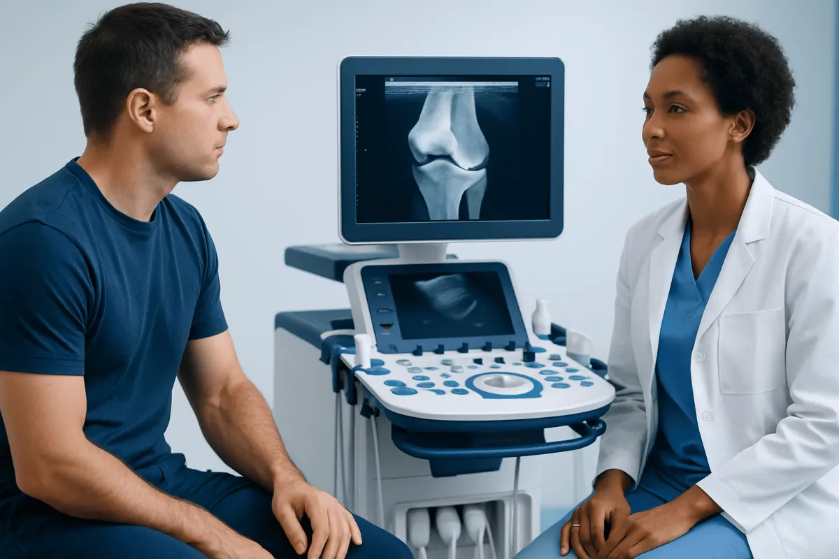
Fibrous cartilage, more commonly called fibrocartilage, is a particularly tough type of cartilage that helps stabilize body structures and absorb pressure. It is found in areas exposed to repeated compression and pulling forces, including the spinal discs, knee menisci and pubic symphysis.
What Is Fibrous Cartilage?
Fibrous cartilage, also known as fibrocartilage, is a specialized supportive tissue that provides both strength and cushioning. It is designed for parts of the body that must handle substantial mechanical stress, such as weight bearing, repeated movement, twisting and pressure. Its role is not simply to make movement smoother; it also helps distribute load and stabilize structures during everyday activity.
Cartilage is a firm but flexible connective tissue. Unlike bone, it does not contain a rich network of blood vessels. Fibrocartilage is especially dense and durable because it contains abundant bundles of type I collagen, a protein that gives connective tissues tensile strength. Cartilage cells, called chondrocytes, are positioned among these fibers in a firm, gel-like supporting material.
The term “fibrous cartilage” can sound as though it is a disease or an abnormal growth, but it describes normal anatomy. Problems occur when a fibrocartilage-containing structure is torn, degenerates over time, becomes inflamed nearby or no longer functions effectively. The symptoms and treatment depend on the location and the underlying cause rather than on the word fibrocartilage alone.
How Fibrocartilage Differs From Other Cartilage
The body has three main forms of cartilage: hyaline cartilage, elastic cartilage and fibrocartilage. Each has a different composition and purpose. Fibrocartilage is the most resistant to pulling and compression, making it suited to areas where tissues must withstand both body weight and movement-related forces.
Hyaline cartilage is smooth and glass-like in appearance. It covers the ends of many bones within joints, where it helps reduce friction, and it is also present in the nose, windpipe and rib connections. Elastic cartilage contains more elastic fibers and is flexible enough to maintain the shape of structures such as the outer ear and parts of the voice box.
In contrast, fibrocartilage is built more like a reinforced cushion. Its dense collagen fibers give it toughness, while its water-containing matrix helps it manage compression. This combination is valuable, but it does not make fibrocartilage indestructible. Aging, repetitive loading, trauma and some joint conditions can affect the structures that contain it.
- Hyaline cartilage: smooth joint-surface cushioning and structural support.
- Elastic cartilage: flexible shape support in structures such as the ear.
- Fibrocartilage: high-strength support and shock absorption where forces are greatest.
Where Is Fibrous Cartilage Found?
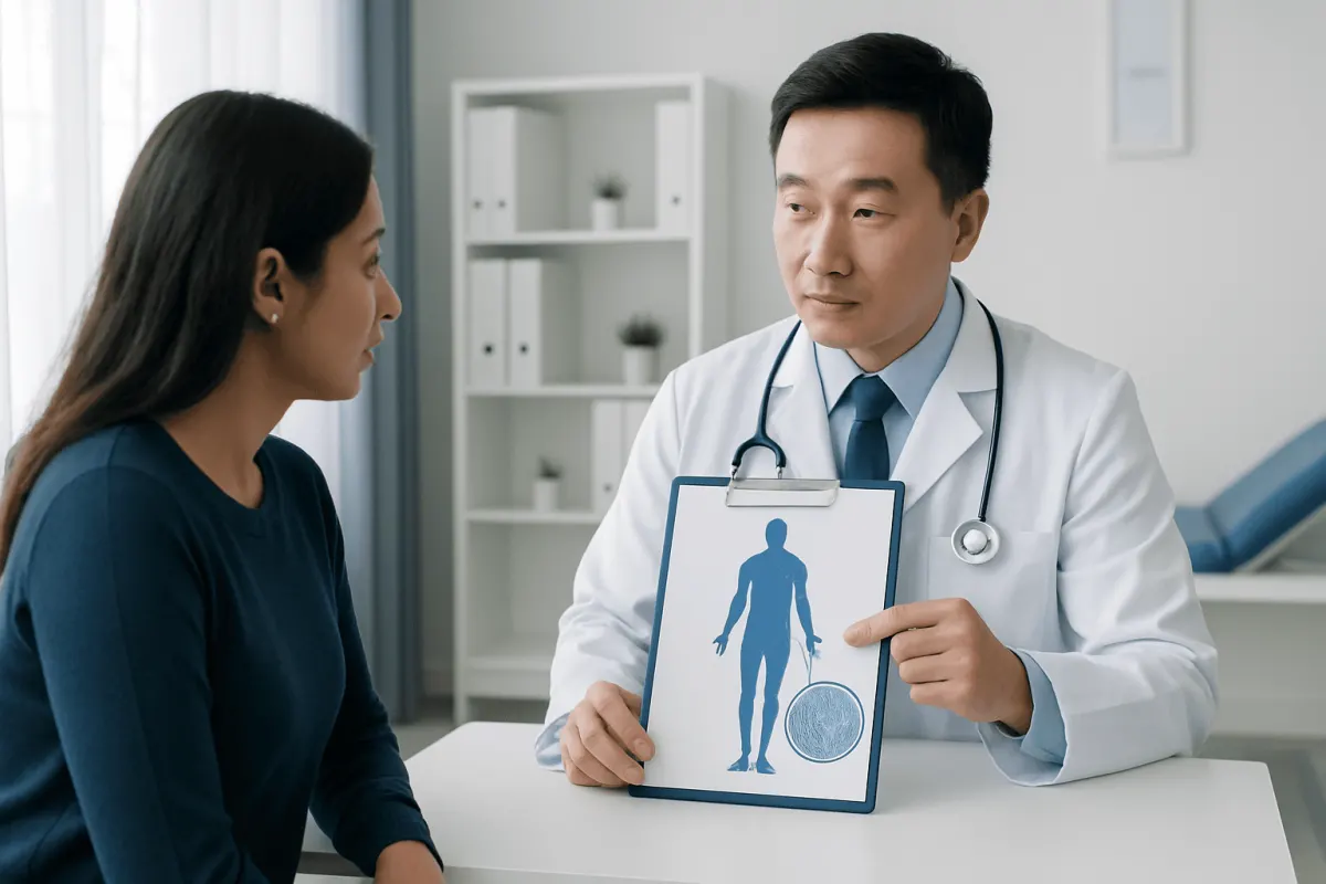
Fibrocartilage is found at several important load-bearing or stabilizing sites. A familiar example is the knee meniscus. Each knee has medial and lateral menisci, crescent-shaped structures that improve the fit between the thighbone and shinbone, spread forces across the joint and contribute to stability.
Another major example is the intervertebral disc. These discs sit between the bones of the spine. Their outer ring, called the annulus fibrosus, contains layers rich in fibrocartilage and collagen. This outer region helps contain the softer central portion of the disc and contributes to the spine’s ability to bend, rotate and bear load.
Fibrocartilage is also present in the pubic symphysis, the joint at the front of the pelvis where the two pubic bones meet. It supports the pelvis while allowing a very small amount of movement. Other examples include certain joint discs and labral structures, such as the disc in the jaw joint and the rings of tissue that deepen the shoulder and hip sockets.
Although these structures share fibrocartilage features, they are not identical. For example, a meniscus tear, a spinal disc problem and jaw-joint dysfunction involve different anatomy, symptoms and treatment decisions. It is therefore important not to apply advice for one location automatically to another.
Why Fibrocartilage Can Be Injured or Change Over Time
Fibrocartilage may be affected by a sudden injury, gradual wear-related changes or a combination of both. A forceful twist of a planted knee can tear a meniscus, particularly during sports or a fall. In the spine, repeated loading and age-related disc changes can contribute to small tears in the outer disc layers or to disc bulging. Not every structural change causes symptoms.
Risk varies by body part. Previous injury, physically demanding work, repetitive squatting or pivoting, high-impact activity, reduced muscle strength, poor movement control and some congenital or anatomical factors may all contribute. Age can also influence tissue resilience, as collagen and water content naturally change over time.
Body weight may increase the load placed on the knees, hips and spine, but it is only one of many influences. Genetics, activity patterns, overall health, smoking status and previous trauma can also matter. Inflammatory joint diseases may affect joints and nearby soft tissues, although they are not the same as a mechanical fibrocartilage injury.
A common misconception is that any pain in the knee, back, jaw or pelvis means the fibrocartilage has “worn away.” Pain can arise from muscles, tendons, ligaments, nerves, bone, joint lining and many other structures. A clinician considers the person’s symptoms, examination findings and, where appropriate, imaging before identifying the likely cause.
Possible Symptoms of Fibrocartilage Problems
Symptoms depend strongly on the affected area. A meniscal injury may cause pain along the joint line, swelling, catching, a feeling that the knee gives way or difficulty fully bending or straightening the knee. Some people with meniscal changes on imaging have little or no pain, especially when changes developed gradually with age.
When spinal discs are involved, symptoms may include local neck or back pain, stiffness or pain that worsens with certain movements. If a disc change irritates or compresses a nearby nerve, pain, tingling, numbness or weakness may travel into an arm or leg. These symptoms require careful assessment because their pattern helps guide management.
Fibrocartilage-related problems in the jaw may be associated with pain around the jaw joint, clicking, limited opening or discomfort when chewing. Pelvic symptoms related to the pubic symphysis can include pain at the front of the pelvis, especially with walking, turning in bed, climbing stairs or standing on one leg.
Clicking or cracking alone does not necessarily indicate damage. Many joints make sounds without causing pain or loss of function. More meaningful warning signs include persistent pain, swelling, reduced movement, locking, weakness or symptoms that interfere with normal daily activity.
How Doctors Assess and Treat These Conditions
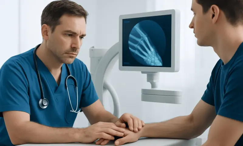
Assessment usually begins with a medical history and physical examination. A clinician may ask when symptoms began, whether there was an injury, which movements trigger symptoms, and whether there is swelling, numbness, weakness or loss of function. Examination may include checking posture, movement, muscle strength, joint stability, tenderness and nerve function.
Imaging is not always required immediately. X-rays are useful for evaluating bones, alignment and some forms of arthritis, but they do not show fibrocartilage clearly. Magnetic resonance imaging, or MRI, can provide more detail about menisci, discs, labral tissue and other soft tissues. Imaging findings are interpreted alongside symptoms because they do not always explain pain by themselves.
Treatment often starts with conservative measures. Depending on the diagnosis, this may include temporarily modifying aggravating activities, guided physiotherapy, exercises to improve strength and movement control, heat or cold when appropriate, and clinician-recommended pain relief. Gradual return to activity is often preferable to prolonged inactivity, but the plan should match the affected body part and severity of symptoms.
Some tears or structural problems may need specialist treatment, particularly when there is joint locking, significant instability, persistent disability, nerve involvement or failure to improve with appropriate nonsurgical care. Procedures can include repair, trimming of damaged tissue, injections in selected situations or surgery. The best option depends on tissue location, injury pattern, age, activity needs and the health of the surrounding joint.
Supporting Cartilage and Joint Health
There is no single food, supplement or exercise that can guarantee protection against all fibrocartilage injuries. However, practical habits can support the joints and muscles that share mechanical load. Regular physical activity, including strength, flexibility and balance work suited to the individual, can improve movement control and help maintain function.
When beginning a new sport or exercise routine, progression should be gradual. Warming up, learning appropriate technique and allowing time for recovery may reduce the chance of overload. Supportive footwear and appropriate equipment can be useful for some activities, although they cannot prevent every injury.
A balanced eating pattern that provides adequate protein, vitamins and minerals supports overall musculoskeletal health. Maintaining a weight that is appropriate for the individual may reduce stress on weight-bearing joints. Avoiding tobacco is also beneficial because smoking can impair circulation and may negatively affect tissue healing.
Rest after an injury can be helpful in the short term, but complete immobility for long periods may lead to stiffness and muscle weakness. A doctor or physiotherapist can advise which movements are safe and when normal activities can be resumed. Acıbadem Health Point’s multidisciplinary specialists and JCI-accredited hospitals assess musculoskeletal concerns and provide individualized care for international patients.
When to Seek Medical Care
Medical assessment is advisable for persistent or worsening joint, back, jaw or pelvic pain; swelling that does not settle; repeated catching or locking of a joint; or a noticeable reduction in movement or ability to use the affected area. Evaluation is also appropriate after an injury that causes difficulty bearing weight, instability or ongoing symptoms despite rest and activity adjustment.
Urgent medical care is important after significant trauma, especially if there is a visible deformity, inability to move a limb, severe swelling or suspected fracture. Prompt assessment is also needed for back or neck symptoms accompanied by new or worsening weakness, numbness in the groin or saddle area, or changes in bladder or bowel control.
Fever, a hot red swollen joint, unexplained weight loss or feeling generally unwell alongside joint pain should also be assessed urgently, as these features may point to a condition other than a simple mechanical injury. Early assessment can help clarify the cause and support an appropriate recovery plan.
Frequently asked questions
01Is fibrous cartilage the same as fibrocartilage?
Yes. Fibrous cartilage and fibrocartilage are terms used for the same type of cartilage. It is characterized by dense collagen fibers that give it substantial strength and resistance to pressure.
02What is the main function of fibrous cartilage?
Fibrocartilage helps absorb shock, distribute forces and stabilize structures that experience repeated stress. It is particularly important in the knee menisci, spinal discs and the joint at the front of the pelvis.
03Can fibrous cartilage heal after a tear?
Healing depends on the location, size and pattern of the injury, as well as the local blood supply. Some parts of structures such as the meniscus have better healing potential than others, while some tears may be managed without surgery.
04Does an MRI finding of fibrocartilage damage always need treatment?
No. MRI findings must be considered together with symptoms, examination results and daily function. Some structural changes are found in people who do not have pain and may not require specific treatment.
05Can exercise damage fibrocartilage?
Appropriately progressed exercise generally supports joint health, muscle strength and mobility. A sudden increase in training load, poor technique, trauma or repetitive high-force movements may increase injury risk in some people.
06Are supplements able to rebuild fibrous cartilage?
No supplement has been proven to reliably rebuild damaged fibrocartilage. A balanced diet can support general health, but persistent symptoms should be discussed with a qualified healthcare professional rather than managed with supplements alone.
This article is for general information only and is not a substitute for professional medical advice. Please consult a qualified doctor about your individual situation.
Joint and spine care in Turkey — expert assessment & treatment
JCI-accredited · board-certified surgeons · reply within 24h
Add us as a Preferred Source to see more of our trusted health content across Google Search, AI Overviews and Discover.