Excisional vs Punch Biopsy: Which Tissue Is Removed?
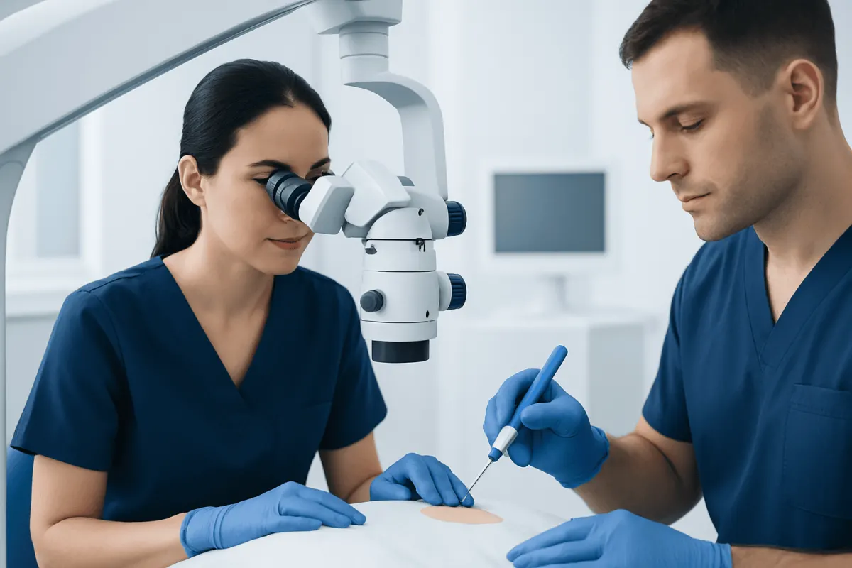
Excisional vs punch biopsy is primarily a question of whether a clinician needs to remove an entire skin lesion or take a small, representative tissue sample. The best option depends on the lesion’s size, location, appearance and the suspected diagnosis, particularly when melanoma is a concern.
Excisional vs Punch Biopsy: Side-by-Side Comparison
Excisional and punch biopsies are common procedures used to examine abnormal skin under a microscope. In an excisional biopsy, the clinician removes the entire visible lesion, often including a narrow rim of surrounding normal-looking skin. In a punch biopsy, a circular instrument removes a small cylinder of skin from one selected area.
The choice is not simply about which procedure is larger. It is about obtaining the tissue most likely to answer the clinical question while considering the lesion’s size, site, appearance and the potential cosmetic or functional effects of removal.
- Amount removed: Excisional biopsy removes the whole lesion; punch biopsy removes a sample.
- Typical use: Excision is often used for small suspicious pigmented lesions; punch biopsy is useful for widespread rashes, inflammatory disorders and lesions too large or difficult to remove initially.
- Information provided: Excision shows the lesion in its entirety; a punch shows tissue from one or more selected areas.
- Closure: Both may require stitches, although very small punch sites may heal without them.
- Procedure setting: Both are commonly performed with local anaesthetic in an outpatient setting.
How a Clinician Tells the Procedures Apart and Chooses
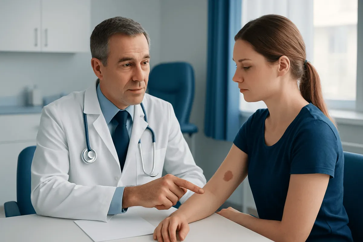
A clinician begins by examining the skin lesion and asking how long it has been present, whether it has changed, and whether it itches, bleeds, hurts or fails to heal. For pigmented spots, assessment may include dermoscopy, a handheld lighted device that helps reveal structures not visible to the naked eye. The clinician also considers personal and family history of skin cancer, sun exposure, immune suppression and previous biopsies.
Excisional biopsy is usually practical when a lesion is relatively small and can be fully removed without creating an unnecessarily large wound or affecting a sensitive area. It allows the pathologist to inspect the lesion’s overall architecture, borders and deepest point. This can be particularly important when evaluating a lesion that could be melanoma.
Punch biopsy may be selected when the condition covers a broad area, when several areas look different, or when complete initial removal would be difficult. For example, a punch sample can help diagnose inflammatory rashes, scarring hair loss, vasculitis or certain infections. More than one punch sample may be taken if different areas need comparison.
Why Is Excisional Biopsy Preferred?
Excisional biopsy is preferred in selected situations because it gives the pathologist the full lesion rather than a portion of it. Seeing the complete pattern of abnormal cells and their relationship to the surrounding skin may improve diagnostic confidence. If the lesion is benign, complete removal may also mean no further procedure is needed.
For a small lesion suspicious for melanoma, excisional biopsy with a narrow clinical margin is generally the preferred initial diagnostic approach when feasible. It enables accurate measurement of tumour thickness if melanoma is found, which is an important part of staging and treatment planning. The initial diagnostic removal is not the same as definitive wider surgery, which may be recommended later depending on the pathology report.
However, excision is not automatically the best choice for every lesion. On the face, hands, feet, ears, genital area or other functionally important locations, the clinician may choose a carefully planned partial biopsy first. A lesion that is very large, or one where surgical closure would be complex, may also need a different sampling strategy.
Which Biopsy Is More Accurate?
Neither biopsy is universally more accurate. Accuracy depends on whether the sample represents the most informative part of the lesion and whether it is sufficient for the suspected condition. An excisional biopsy can be more informative for a small, potentially cancerous pigmented lesion because it captures the whole lesion, including its edges and deepest portion.
A punch biopsy can be highly accurate for many skin disorders, especially when the clinician selects an active, representative site. In inflammatory or blistering diseases, the location and age of a lesion can strongly affect what appears under the microscope. Sometimes a pathologist may request an additional sample, special stains or direct immunofluorescence testing.
Partial sampling carries a small possibility that the sampled area does not show the most significant change within a varied lesion. If a pathology result does not fit the clinical appearance, the lesion continues changing, or concern remains, the clinician may recommend repeat biopsy or complete removal. This is a standard safety-focused step, not a sign that a diagnosis has already been made.
Which Biopsy Method Is Preferred for Melanoma Diagnosis?
When melanoma is suspected and the lesion can reasonably be removed, an excisional biopsy is generally preferred. The clinician removes the entire visible lesion with a narrow margin and sends it to a pathology laboratory. This approach helps the pathologist determine whether melanoma is present and, if it is, assess features such as thickness and ulceration.
A deep shave, saucerization or punch/incisional biopsy may be considered when complete excision is impractical because of lesion size or location. The priority is obtaining an adequate sample, including sufficient depth, without causing avoidable harm. A superficial sample that does not include the lesion’s full depth may make assessment more difficult.
People with a changing mole, an asymmetrical or unevenly coloured spot, or a lesion that bleeds or does not heal should arrange prompt clinical assessment. Evaluation may lead to testing for conditions such as melanoma, but only pathology can confirm the diagnosis.
Is an Excisional Biopsy Usually Indicative of Cancer?
No. An excisional biopsy is not usually indicative of cancer by itself. Clinicians remove lesions for many reasons, including a changing mole, a benign growth that is irritated, a cyst-like lesion, an unusual-looking spot, a recurrent skin problem or uncertainty about the diagnosis.
The purpose of biopsy is to replace uncertainty with a tissue diagnosis. Many biopsy results are benign, while others may identify inflammation, infection, precancerous change or a cancer that needs further management. The result should be discussed with the clinician who ordered the test, as its meaning depends on the pathology findings and the person’s overall health.
Waiting for results can understandably feel stressful. Processing time varies because some samples require additional laboratory techniques or review by a dermatopathologist. Patients should ask when and how they will receive results, and should contact the clinic if they have not heard back within the expected timeframe.
What to Expect, Recovery and Next Steps
Before either procedure, the area is cleaned and numbed with a local anaesthetic. Patients may feel a brief sting from the anaesthetic injection and pressure during the biopsy, but should not feel sharp pain. The tissue is placed in preservative and examined by a pathologist; the wound may be closed with stitches and covered with a dressing.
Aftercare usually includes keeping the site clean, following dressing instructions and avoiding activities that could pull on the wound until it has healed. Mild tenderness, bruising or a small amount of spotting can occur. A scar is possible with either procedure, and its size depends on the biopsy type, body site, wound tension and individual healing.
Patients should seek advice if they develop increasing redness, warmth, swelling, worsening pain, pus-like drainage, fever, persistent bleeding or a wound that opens. If cancer is identified, the next steps may include further excision, specialist review or additional assessment. Acıbadem Health Point’s multidisciplinary specialists and JCI-accredited hospitals diagnose and treat skin conditions for international patients, with care plans guided by pathology and individual needs.
When to Seek Medical Care
Medical assessment is advisable for a new or changing skin lesion, especially one that changes in size, shape, colour or thickness. Other reasons to seek care include a spot that repeatedly bleeds, crusts, ulcerates, causes persistent itching or pain, or does not heal as expected. A clinician can decide whether observation, photography, dermoscopy or biopsy is appropriate.
People at increased risk of skin cancer should be particularly attentive to new or changing lesions. This includes those with a previous skin cancer, a strong family history of melanoma, many moles, fair or sun-sensitive skin, substantial ultraviolet exposure, or a weakened immune system. Regular professional skin checks may be advised for some individuals.
Urgent emergency care is rarely needed for a skin lesion itself, but persistent bleeding after a biopsy that does not stop with firm, continuous pressure should be assessed promptly. For signs of a wound infection or a concerning pathology result, contacting the treating clinician is the appropriate next step.
Frequently asked questions
01What is the main difference between an excisional and punch biopsy?
An excisional biopsy removes the entire visible lesion, while a punch biopsy takes a small circular core from the lesion or rash. Both samples are sent to a pathology laboratory for microscopic examination. The appropriate method depends on what condition is suspected and where the abnormal skin is located.
02Does a punch biopsy remove all layers of skin?
A punch biopsy is designed to obtain a cylindrical sample that can extend through the epidermis and dermis and may include superficial fat. The depth depends on the punch instrument and the site being biopsied. It does not necessarily remove the entire skin lesion.
03How long do excisional and punch biopsy results take?
Results often take several days to a few weeks, depending on the laboratory and whether extra stains, expert review or further testing is needed. The clinic should explain the expected timeframe and how results will be communicated. A patient can contact the clinic if results are not received when expected.
04Will an excisional biopsy leave a scar?
Any procedure that cuts the skin can leave a scar. Excisional biopsies commonly require stitches and may leave a linear scar, while punch biopsies can leave a small round scar. Scar appearance varies by body location, skin type, wound care and individual healing.
05Can a punch biopsy miss melanoma?
A punch biopsy can miss the most diagnostically important area if a lesion is uneven and the sample is not representative. This is one reason excisional biopsy is generally preferred for a small lesion suspected to be melanoma when feasible. If clinical concern remains after a partial biopsy, further sampling or complete excision may be recommended.
06What should a person do after a skin biopsy?
They should follow their clinician’s wound-care instructions, keep the area clean and protect it from friction or pulling. They should ask when stitches need removal, if applicable, and when to expect pathology results. Increasing pain, spreading redness, drainage, fever or ongoing bleeding should be reported to a healthcare professional.
This article is for general information only and is not a substitute for professional medical advice. Please consult a qualified doctor about your individual situation.
Oncology care in Turkey — second opinion and treatment plan
JCI-accredited · board-certified surgeons · reply within 24h
Add us as a Preferred Source to see more of our trusted health content across Google Search, AI Overviews and Discover.