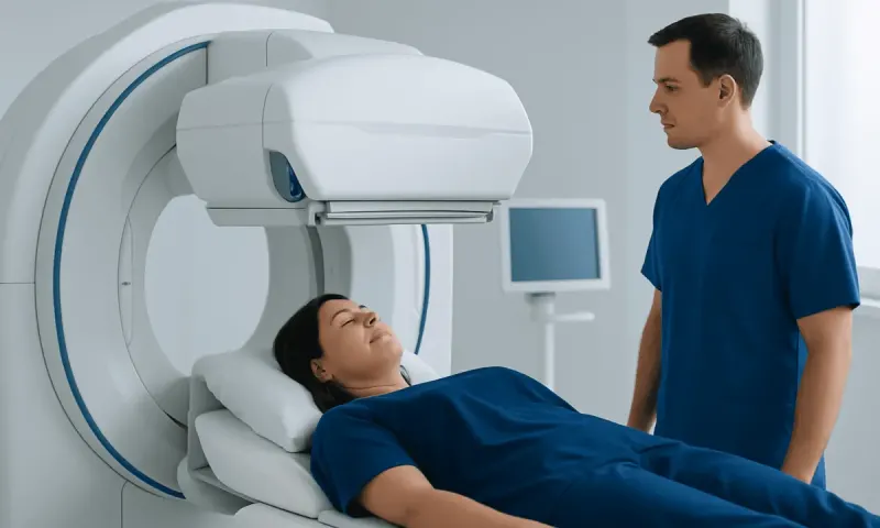
Echocardiogram Price: What Shapes Your Total Cost
An echocardiogram price cannot be estimated reliably from one figure alone because the final amount depends on the type of scan, where it is performed, professional interpretation, and insurance or public-health coverage. Patients can make costs clearer by asking for an itemized estimate before scheduling and confirming their expected out-of-pocket responsibility.
Echocardiogram Price: The Answer Depends on More Than the Scan
An echocardiogram price varies because it is not always the same test or billed in the same way. The total may reflect the ultrasound examination, the cardiologist or qualified clinician who interprets the images, the setting where it takes place, and any additional services needed. For this reason, the most useful question is often not simply “What does an echocardiogram cost?” but “What will this specific test cost me after my coverage is applied?”
An echocardiogram, often called an echo, uses sound waves to create moving images of the heart. It can help clinicians assess heart chambers, pumping strength, heart valves, blood flow and fluid around the heart. It does not use ionizing radiation and is commonly performed when a clinician needs more information after symptoms, a physical examination, an electrocardiogram, or another medical assessment.
Before booking, patients can ask the hospital, clinic or imaging center for an itemized estimate. It is helpful to ask whether the quoted amount includes technical imaging services, professional interpretation, facility charges, contrast if needed, sedation if applicable, and follow-up consultation. A health insurer or payer can also explain deductibles, copayments, coinsurance, network requirements and whether prior authorization is needed.
What Determines the Cost of an Echocardiogram?
The type of echocardiogram is one of the main cost considerations. A transthoracic echocardiogram (TTE), performed with an ultrasound probe moved over the chest, is the most common approach. A stress echocardiogram compares heart function at rest and during exercise or medicine-induced stress. A transesophageal echocardiogram (TEE) uses a probe passed through the mouth into the esophagus, allowing detailed images from close behind the heart; it generally requires more preparation and monitoring.
Where the test is performed can also affect the bill. A hospital outpatient department, independent imaging center and cardiology office may have different facility arrangements and pricing structures. The complexity of the study matters as well. Doppler measurements of blood flow, three-dimensional imaging, contrast enhancement, exercise supervision, medication administration, intravenous access, sedation and recovery observation may all add services beyond a basic scan.
Coverage is equally important. Private insurance and public programs may cover medically necessary echocardiography, but the patient’s share can depend on plan design and whether the clinician and facility are in network. A referral, diagnostic indication, or authorization may be required. Patients should request the procedure name or billing details from the ordering team and provide these to their insurer for the clearest personalized estimate.
- Ask whether both facility and professional fees apply.
- Confirm that the ordering clinician, imaging facility and interpreting specialist are covered by the plan.
- Ask about preauthorization, referral requirements and cost-sharing before the appointment.
- Request an explanation of any separate charges for contrast, stress testing, sedation or monitoring.
How an Echocardiogram Works and Who May Need One

During echocardiography, a device called a transducer sends harmless high-frequency sound waves into the chest. These waves echo back from the heart and are converted into images. Doppler ultrasound can show the direction and speed of blood flow, which is especially useful for evaluating valve function and estimating pressures within parts of the heart.
A clinician may recommend an echo for symptoms such as unexplained shortness of breath, chest discomfort, palpitations, fainting, leg swelling or a newly detected heart murmur. It may also be used to monitor known valve disease, heart muscle disease, congenital heart differences, heart failure, or the effects of certain treatments. An echo is selected based on an individual clinical question; it is not routinely needed for every person with cardiovascular risk factors alone.
Most people can have a transthoracic echocardiogram safely. A transesophageal echo may not be suitable for people with certain swallowing difficulties, esophageal conditions or recent upper gastrointestinal surgery, so the care team will review medical history carefully. People who are pregnant, take blood-thinning medicines, have diabetes, or have medication allergies should mention this when arranging a stress or transesophageal test, as preparation may need to be adjusted.
When an echo identifies or helps monitor a cardiac concern, it is usually considered alongside symptoms, examination findings and other tests. For example, it may contribute to the assessment of heart failure or valve-related problems, but it does not replace a full clinical evaluation.
What Happens During the Procedure and Recovery?
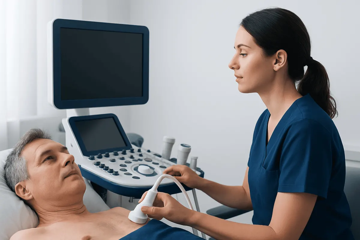
For a standard transthoracic echo, the patient usually removes clothing from the upper body and lies on an examination couch, often on the left side. Small electrodes may be placed on the chest to coordinate imaging with the heartbeat. Gel is applied to the skin, and the sonographer presses the transducer at different points on the chest. The probe pressure can feel firm but should not be painful. The test is generally completed without needles or anesthesia.
A stress echocardiogram includes images before and after the heart rate is increased through exercise or, when exercise is not appropriate, medication. Staff monitor heart rhythm, blood pressure and symptoms throughout. A transesophageal echo is more involved: fasting is usually required, the throat is numbed, and sedating medicine may be used. The patient is monitored while the probe is carefully placed in the esophagus to obtain images.
After a transthoracic echo, most people can return to usual activities straight away. After exercise stress testing, brief fatigue may occur. Following a transesophageal echo, the throat can feel mildly sore and driving should be avoided until the sedative has worn off and the care team confirms it is safe. The team will provide individualized discharge instructions, particularly when sedation or medicines have been used.
For patients needing a detailed cardiac assessment, echocardiography services may include the appropriate imaging approach based on the reason for testing and the person’s overall health.
Benefits, Limitations and Possible Risks
The main benefit of echocardiography is that it provides real-time information about how the heart is structured and working. It can help evaluate pumping function, chamber size, valve narrowing or leakage, fluid around the heart, and some congenital heart conditions. Results can guide decisions about further testing, monitoring, medications or referral to a heart specialist.
A standard transthoracic echocardiogram is noninvasive and generally very low risk. Some people experience minor discomfort from the probe being pressed against the chest or temporary skin irritation from electrodes. Contrast agents, when used, can rarely cause side effects or allergic reactions; the clinical team checks whether contrast is appropriate and monitors the patient.
Stress echocardiography carries the temporary physical demands of exercise or the effects of stress-inducing medication. Transesophageal echocardiography has additional uncommon risks, including throat irritation, dental injury, breathing problems related to sedation, bleeding or injury to the esophagus. These complications are uncommon, and the test is recommended only when its additional detail is likely to be clinically useful.
An echo also has limitations. Image quality can be affected by body shape, lung conditions or chest wall features. It may suggest problems that require confirmation through other testing, and a normal result does not rule out every possible cause of symptoms. A clinician interprets the results in the context of the patient’s complete medical history.
How Much Does Medicare Reimburse for an Echocardiogram?
Medicare reimbursement for an echocardiogram is not one fixed amount. It depends on the exact type of echo, the place where it is performed, regional payment rules, whether the service is provided in a hospital or office setting, and the billing components involved. The professional interpretation and technical imaging portion may also be billed separately in some circumstances.
For people enrolled in Medicare, coverage generally depends on whether the test is medically necessary and ordered appropriately by a qualified clinician. What the patient pays can vary according to Original Medicare versus a Medicare Advantage plan, supplemental coverage, deductibles, coinsurance, network rules and prior authorization policies. The most reliable source of an individual estimate is the plan administrator together with the facility that will perform the test.
Patients may wish to ask the ordering office for the planned test description and relevant billing information, then contact Medicare or their Medicare Advantage plan before the appointment. They should also ask whether the interpreting physician and testing facility participate in their coverage network.
Can an Echo Show Clogged Arteries? Why Can It Be Expensive?
An echocardiogram cannot directly show clogged coronary arteries in the way that coronary angiography or coronary CT angiography can. However, it may show indirect signs that blood flow to part of the heart muscle has been affected, such as an area of the heart wall that does not move normally. A stress echocardiogram can sometimes identify changes in heart muscle movement that raise concern for reduced blood flow during exertion.
When coronary artery disease is suspected, a clinician chooses further tests based on symptoms, risk factors, examination findings and the urgency of the situation. These may include an electrocardiogram, blood tests, stress testing, coronary CT angiography, or invasive coronary angiography. An echo is therefore an important but limited part of cardiovascular assessment, not a direct test for every type of artery blockage.
Patients may feel that an echocardiogram is expensive because the invoice can combine specialized equipment, trained sonography staff, image analysis, physician interpretation, facility operations and additional components such as Doppler studies, contrast, stress testing or sedation. Insurance deductibles and out-of-network services can further increase the amount a person sees on a bill. Requesting an itemized estimate beforehand and reviewing an explanation of benefits afterward can clarify each part of the charge.
Is an Echocardiogram Worth Having? When to Seek Medical Care
An echocardiogram is worth having when it is likely to answer an important clinical question and influence care. For someone with a heart murmur, persistent breathlessness, suspected valve disease, symptoms of possible heart failure, or a known condition requiring monitoring, the information may be particularly useful. The decision should be based on a discussion with a clinician about the expected benefit, alternatives, limitations and financial considerations.
People should seek prompt medical assessment for new or worsening chest pressure, severe shortness of breath, fainting, a rapid or irregular heartbeat with feeling unwell, or sudden swelling accompanied by breathing difficulty. Emergency services should be contacted for severe chest pain, trouble breathing, collapse, or symptoms that could suggest a heart attack or stroke. These symptoms need urgent evaluation and should not be managed by waiting for a routine outpatient echo.
For nonurgent concerns, patients can speak with their primary care clinician or cardiologist about whether echocardiography is appropriate. Acıbadem Health Point’s multidisciplinary specialists and JCI-accredited hospitals diagnose and treat cardiovascular conditions for international patients, including through coordinated cardiac imaging and follow-up care.
Frequently asked questions
01How much does an echocardiogram cost without insurance?
The price without insurance varies widely by country, city, facility type and the kind of echocardiogram performed. A standard transthoracic study is generally less complex than a stress or transesophageal echo. Patients can ask the facility for a self-pay, itemized estimate that includes interpretation and any possible additional charges.
02How much does Medicare reimburse for an echocardiogram?
Medicare reimbursement depends on the specific procedure, location of service and local payment rules, so there is no single reimbursement figure. Patient responsibility also depends on coverage type, deductible, coinsurance and supplemental or Medicare Advantage benefits. Checking with the plan and testing facility before scheduling provides the most individualized information.
03Can an echo show clogged arteries?
An echocardiogram does not directly visualize coronary artery blockages. It may show indirect effects of reduced blood supply to the heart muscle, particularly during a stress echocardiogram. Other tests may be needed when coronary artery disease is suspected.
04Why is my echocardiogram so expensive?
The total charge may include image acquisition, specialized equipment, sonographer services, physician interpretation and facility fees. More complex testing, such as stress imaging, contrast use, sedation or transesophageal echocardiography, can involve additional services. Insurance deductibles, coinsurance and network status also influence the amount billed to the patient.
05Is an echocardiogram worth having?
It can be very worthwhile when it helps explain symptoms, evaluate a heart murmur, monitor known heart disease or guide treatment decisions. The test is noninvasive in its standard form and provides useful information about heart structure and function. A clinician can explain whether the likely benefit is meaningful for an individual situation.
06How long does an echocardiogram take?
A standard transthoracic echocardiogram commonly takes less than an hour, although appointment times vary by facility and the complexity of imaging needed. Stress and transesophageal echocardiograms usually require more time for preparation, monitoring and recovery. The testing center can provide instructions and an expected visit length in advance.
This article is for general information only and is not a substitute for professional medical advice. Please consult a qualified doctor about your individual situation.
Explore treatments in Turkey — costs, top hospitals & a free quote
JCI-accredited · board-certified surgeons · reply within 24h
Add us as a Preferred Source to see more of our trusted health content across Google Search, AI Overviews and Discover.