Double Vision After a Stroke: Why It Happens and When Neuro-Ophthalmology Helps
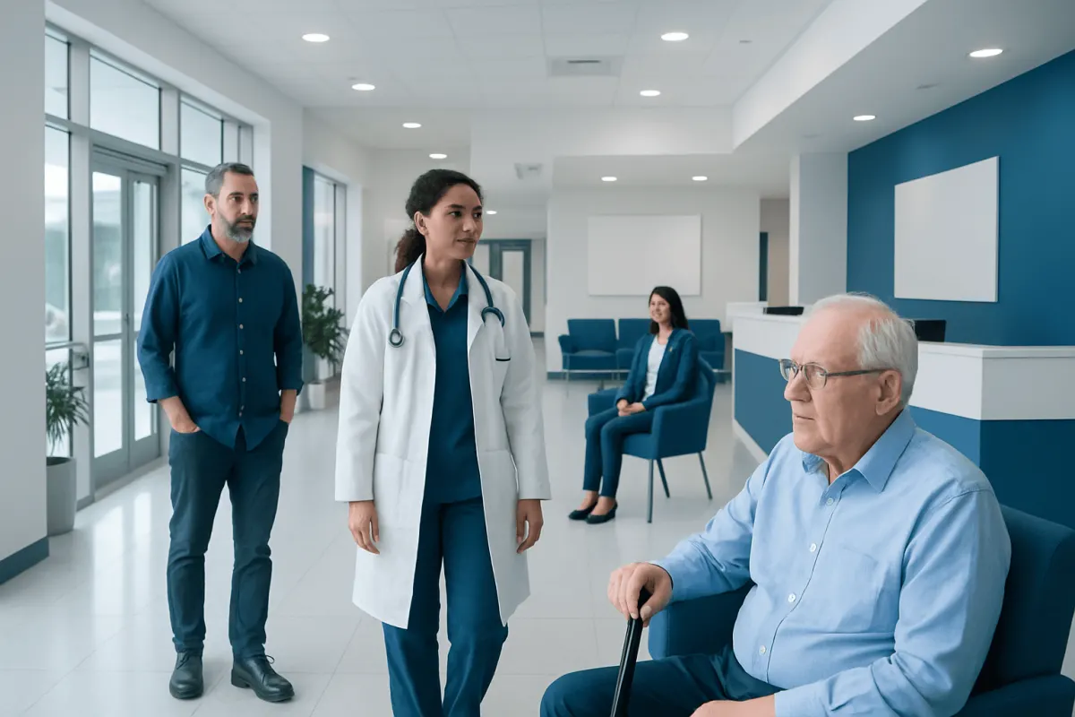
Double vision after a stroke is a common visual symptom that can occur when the brain pathways controlling eye movements or visual processing are injured. Careful assessment by stroke and neuro-ophthalmology specialists can identify the cause and help guide recovery, symptom relief, and rehabilitation.
Overview
Double vision, also called diplopia, means seeing one object as two. After a stroke, this can happen if the areas of the brain that control eye movements, eye alignment, or visual pathways are affected. Some people notice side-by-side images, while others see one image above the other. The symptom may be constant or may appear only when looking in certain directions.
A stroke can interrupt the signals that keep both eyes working together as a team. Even if the eyes themselves are healthy, the brain may have difficulty coordinating them. This is why some people develop double vision as part of stroke-related visual problems.
For many patients, double vision improves during recovery as the brain heals and adapts. In other cases, symptoms last longer and need targeted care. A structured evaluation can help explain what is happening and what Neuromodulation" class="ahp-ilk">treatment options may improve comfort, safety, and daily function.
Why a Stroke Can Cause Double Vision
To produce a single clear image, the brain must accurately control the muscles that move each eye and combine visual information from both sides. A stroke may disturb this system at several levels. It can affect the brainstem, where important eye movement centers and cranial nerve pathways are located, or it can involve higher brain regions that help coordinate gaze and visual attention.
Double vision is especially likely when a stroke affects the nerves or pathways linked to eye movement. These include the third, fourth, and sixth cranial nerves, which control most eye muscles. If one eye does not move in the same direction or at the same speed as the other, the two images no longer match, and diplopia can result.
Some people also develop related visual symptoms rather than true double vision, such as blurred vision, difficulty focusing, loss of part of the visual field, or a tilted sense of the world. A neuro-ophthalmology assessment helps separate these problems and identify the exact cause. This can be especially useful when symptoms are subtle, fluctuate, or overlap with balance and dizziness complaints.
Symptoms and Related Signs

The main symptom is seeing two images of a single object. The images may be horizontal, vertical, or diagonal. Some people notice that double vision becomes worse when looking to one side, reading, walking, or feeling tired. Others may find that closing one eye makes the symptom disappear, which often suggests a problem with eye alignment rather than a surface problem of the eye.
Post-stroke double vision may occur together with other neurological or visual symptoms. These can include dizziness, nausea, imbalance, headache, drooping of one eyelid, difficulty tracking moving objects, or trouble judging distance. In some cases, people turn or tilt their head without realizing it because this position reduces the double vision.
Practical effects can be significant. Reading may become slow, stairs may feel unsafe, and driving may not be possible until symptoms are fully assessed. People recovering from a stroke may also be at higher risk of falls if vision and balance are both affected. Recognizing these symptoms early can help the care team plan safer recovery and rehabilitation.
- Seeing two images side by side or one above the other
- Worse symptoms when looking in certain directions
- Eye strain, headache, or fatigue while reading
- Head tilt or face turn to compensate
- Dizziness, unsteadiness, or depth-perception problems
How Neuro-Ophthalmology Helps
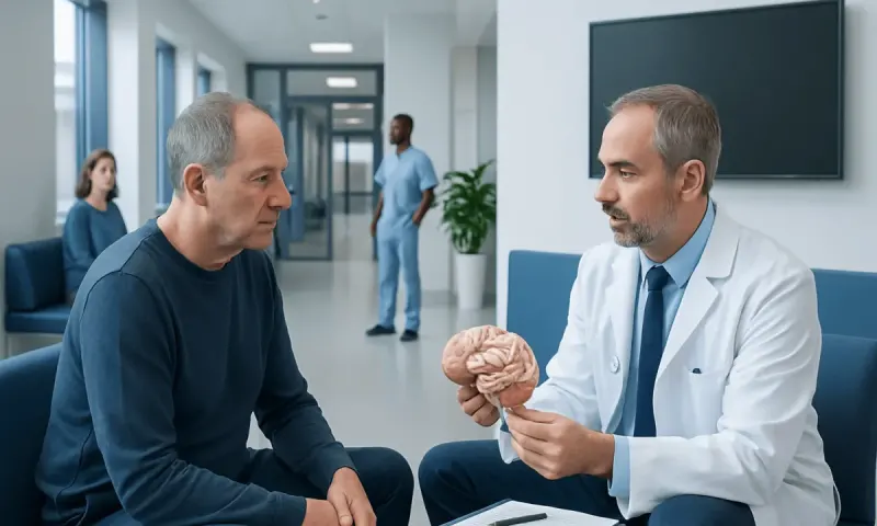
Neuro-ophthalmology focuses on vision problems related to the brain, optic nerves, and eye movement control systems. This specialty is especially helpful when double vision happens after a neurological event such as a stroke. The goal is not only to confirm that diplopia is present, but also to identify exactly which pathway has been affected.
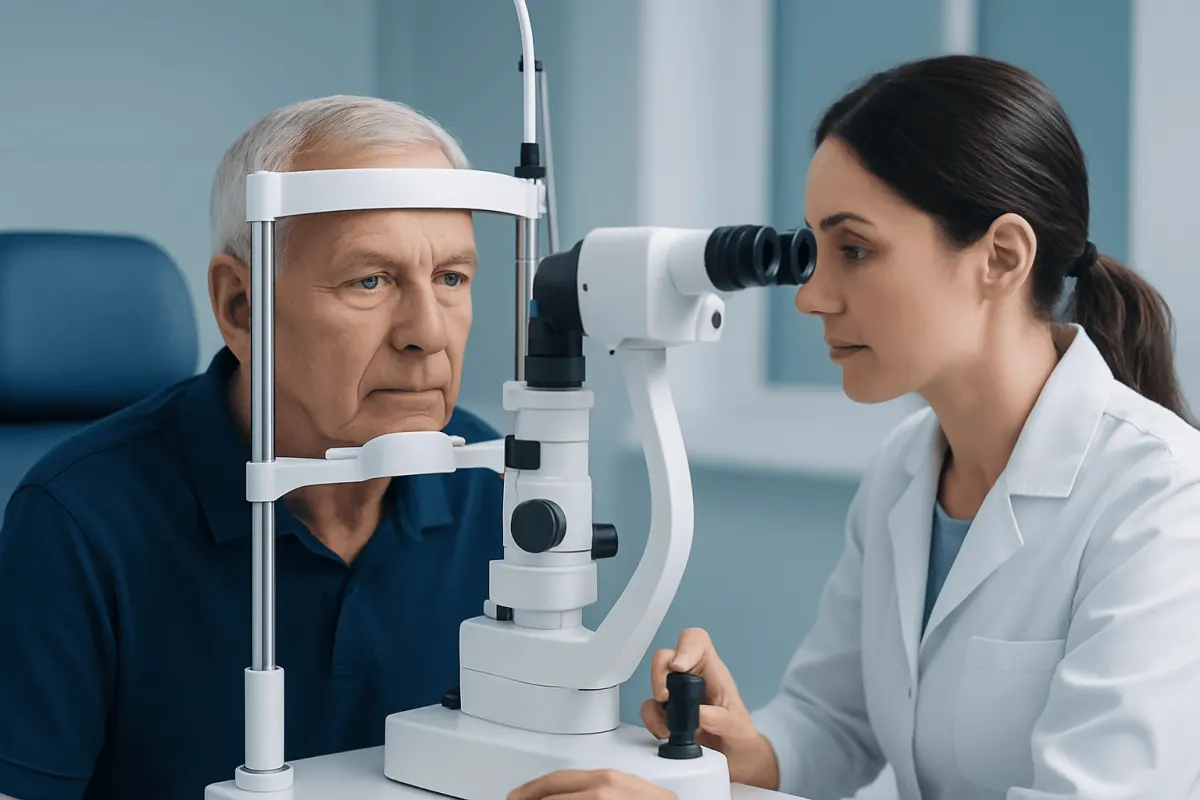
During the evaluation, the specialist may assess eye alignment, eye movements in different directions, eyelid position, pupils, visual fields, and the way both eyes work together. This can show whether the problem is due to a cranial nerve palsy, a gaze disorder, skew deviation, internuclear ophthalmoplegia, or another stroke-related change. That level of detail helps guide treatment and gives a better sense of likely recovery.
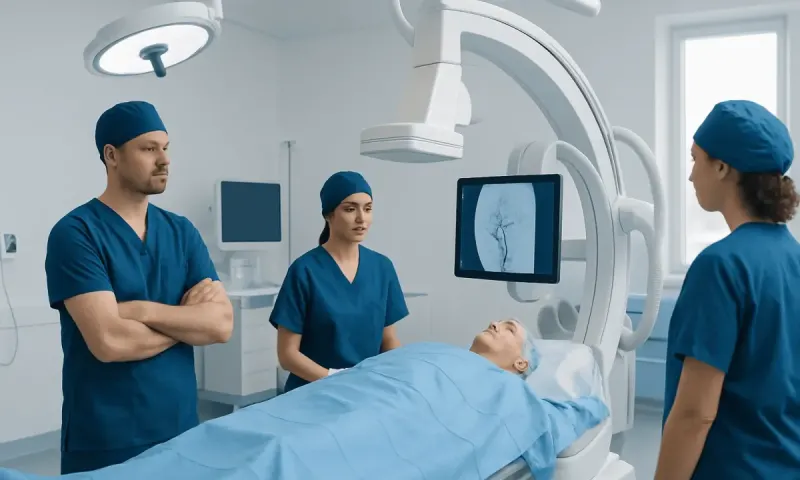
Imaging and neurological review are often part of the process. Brain imaging, vascular studies, and other tests may already have been done during initial stroke care, but they may be reviewed again in the context of the visual symptoms. In a multidisciplinary setting, specialists in stroke medicine, neuro-ophthalmology, and neuroradiology can work together to build a clearer picture of the problem.
Diagnosis and Evaluation
Diagnosis begins with a careful history. Doctors usually ask when the double vision started, whether it was sudden or gradual, whether it affects one or both eyes, and whether it changes with direction of gaze. They also ask about other symptoms such as limb weakness, facial drooping, speech changes, numbness, imbalance, or a recent transient ischemic attack.
The examination may include tests of visual acuity, eye alignment, pupil reactions, and eye movements. Cover testing, prism measurements, and bedside neurological examination can reveal subtle misalignment that the patient may not be able to describe clearly. If needed, blood tests or additional scans may be ordered to rule out other conditions that can mimic stroke-related diplopia.
Imaging is important when the cause is uncertain, symptoms are new, or there are signs of ongoing vascular risk. Depending on the situation, a patient may need MRI, CT, or vascular imaging to evaluate the brain and blood vessels. Patients with persistent symptoms may also benefit from assessment in vascular neurology or, when procedures are needed for the underlying blood vessel problem, interventional neuroradiology.
Treatment Options
Treatment depends on the cause, severity, and stage of recovery. The first priority is always proper stroke management and prevention of another event. This may include medicines prescribed by the doctor, rehabilitation, and treatment of risk factors such as high blood pressure, diabetes, irregular heart rhythm, or cholesterol problems.
For the double vision itself, doctors may recommend temporary measures while the nervous system heals. These can include patching one eye for comfort, applying a translucent filter to one lens, or using prism glasses to bring the two images closer together. These approaches do not treat the stroke directly, but they can reduce symptoms and make daily activities easier.
If the double vision continues, further options may include visual rehabilitation, orthoptic exercises in selected cases, botulinum toxin for certain eye muscle imbalances, or eye muscle surgery after the condition has stabilized. Not every patient needs all of these treatments, and timing matters. Many specialists prefer to wait for the pattern of recovery to become clearer before considering permanent interventions.
Recovery often benefits from a team approach. Neurologists, neuro-ophthalmologists, rehabilitation specialists, and optometrists or orthoptists may all contribute. Near the end of the care pathway, if symptoms remain disruptive, patients may discuss tailored rehabilitation plans at centers such as Acıbadem Health Point, where multidisciplinary specialists in JCI-accredited hospitals diagnose and treat neurological and visual complications for international patients.
Prevention, Self-Care, and Recovery
Although a person cannot reverse a stroke on their own, healthy recovery habits can support rehabilitation and lower the risk of another stroke. Following the prescribed treatment plan, taking medicines as directed, attending follow-up appointments, and participating in physical and occupational therapy are all important parts of care. If reading, walking, or navigating busy spaces is difficult, practical adjustments at home can also help.
Simple strategies may reduce discomfort from double vision during recovery. Good lighting, larger print, frequent rest breaks, and avoiding visually crowded environments can ease strain. Patients should ask their clinician before driving, returning to work, or using stairs alone if vision remains affected.
Long-term prevention focuses on stroke risk reduction. This may include controlling blood pressure, managing diabetes, not smoking, staying physically active within medical guidance, eating a balanced diet, and treating conditions such as carotid artery stenosis when present. Families and caregivers can play an important role by helping monitor symptoms and encouraging safe, steady recovery.
When to See a Doctor
Sudden double vision should always be taken seriously. If it starts abruptly, especially with weakness, numbness, difficulty speaking, severe headache, loss of balance, or facial drooping, urgent medical assessment is needed. These signs can suggest a stroke or another neurological emergency.
Even after a stroke has already been diagnosed, new or worsening visual symptoms deserve prompt review. Double vision may change during recovery, but it should not simply be ignored. Early assessment can improve comfort, reduce fall risk, and help identify complications that need treatment.
A doctor should also review persistent symptoms that interfere with reading, walking, work, or daily activities. If vision problems continue beyond the early recovery phase, referral to neuro-ophthalmology may provide more specific answers and practical strategies for rehabilitation.
Frequently asked questions
01Is double vision common after a stroke?
It can be a relatively common visual problem after a stroke, especially when the brainstem or eye movement pathways are involved. Some people notice it immediately, while others become more aware of it during recovery activities such as reading or walking.
02Can double vision after a stroke go away?
Yes, it may improve over time, particularly in the first weeks to months after the stroke. Recovery depends on the location and extent of the brain injury, as well as the person's overall rehabilitation progress.
03What is the difference between an eye problem and stroke-related double vision?
An eye problem may come from the surface of the eye, the lens, or the retina, while stroke-related double vision usually comes from disrupted brain control of eye alignment or visual pathways. A specialist examination helps distinguish between these causes because treatment can differ significantly.
04What does a neuro-ophthalmologist do for post-stroke double vision?
A neuro-ophthalmologist evaluates how the eyes, optic nerves, and brain work together. The specialist helps identify the exact cause of the double vision and can recommend options such as prisms, patching, rehabilitation, or referral for additional neurological care.
05Should someone drive with double vision after a stroke?
In general, driving should be avoided until the condition has been assessed and a doctor says it is safe. Double vision can affect depth perception, reaction time, and awareness of surroundings, which may make driving unsafe.
06When is double vision an emergency?
It is an emergency when it starts suddenly, especially if it comes with weakness, facial drooping, speech problems, severe headache, or difficulty walking. These symptoms need immediate medical attention because they can signal a stroke or another urgent neurological condition.
This article is for general information only and is not a substitute for professional medical advice. Please consult a qualified doctor about your individual situation.
Explore treatments in Turkey — costs, top hospitals & a free quote
JCI-accredited · board-certified surgeons · reply within 24h
Add us as a Preferred Source to see more of our trusted health content across Google Search, AI Overviews and Discover.