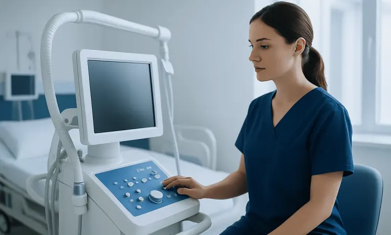
Congenital Nevi: Birthmark Monitoring, Biopsy, and Removal Decisions
Congenital nevi are moles or pigmented birthmarks that are present at birth or appear soon afterward. Most are harmless, but careful monitoring and individualized decisions about biopsy or removal help protect skin health and support comfort, function, and appearance.
Overview
Congenital nevi are moles or birthmarks caused by clusters of pigment-producing cells in the skin. They are present at birth or become noticeable during the first months of life. These lesions may be flat or raised, light brown to black, smooth or pebbled, and sometimes covered with thicker hair. Their appearance can change gradually as a child grows, because the skin itself is growing.
Doctors often classify congenital melanocytic nevi by their expected adult size: small, medium, large, or giant. This classification is important because size, location, and appearance help guide follow-up and treatment decisions. Small and medium congenital nevi are common and usually do not cause medical problems. Large and giant nevi are less common and need closer monitoring because they carry a higher lifetime risk of melanoma than smaller lesions.
For many families, the main questions are practical: Is the birthmark safe? Should it be photographed or checked regularly? Is biopsy needed? Would removal reduce risk or improve appearance? The answers are individualized. A dermatologist, pediatric dermatologist, plastic surgeon, or other specialist can assess the nevus and help families choose a plan that balances medical safety, cosmetic outcomes, and the child’s well-being.
What Congenital Nevi Can Look and Feel Like
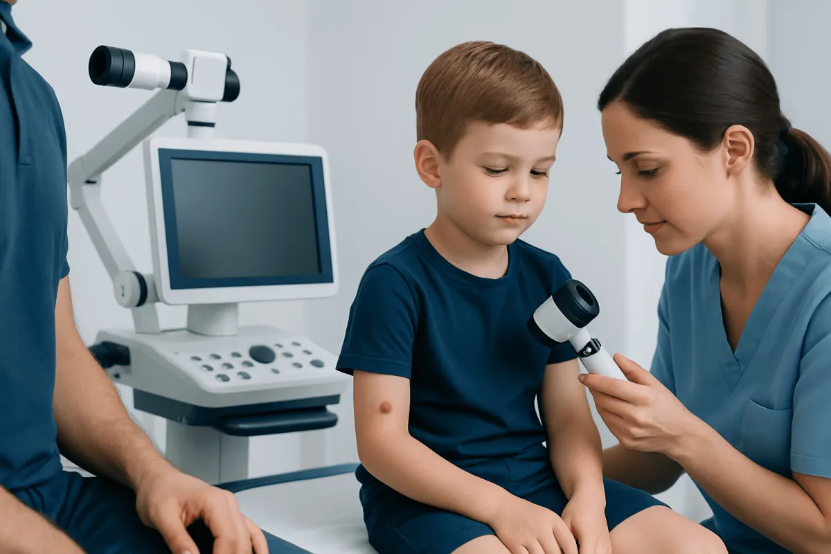
Congenital nevi can look very different from one person to another. Some are small, round, evenly colored moles. Others are larger patches with mixed shades of tan, brown, blue-gray, or black. The surface may be smooth, slightly bumpy, thickened, or wart-like. Hair growth within the nevus is common and does not by itself mean the lesion is dangerous.
As children grow, congenital nevi often enlarge in proportion to the body. They may become more raised or textured over time, especially during childhood and puberty. Mild color variation can be normal, but sudden or uneven changes should be assessed. Parents may also notice small darker spots or nodules within a larger lesion; many are benign, but new or rapidly changing nodules deserve medical evaluation.
Features that are especially useful to track include size, border shape, color pattern, thickness, surface texture, hair growth, and symptoms. A simple monitoring routine may include:
- Taking clear photographs with consistent lighting and a ruler for scale.
- Recording the date and any symptoms such as itching, tenderness, crusting, or bleeding.
- Checking areas that are hard to see, such as the scalp, back, buttocks, or behind the ears.
- Keeping follow-up appointments recommended by the clinician.
Causes and Risk Factors
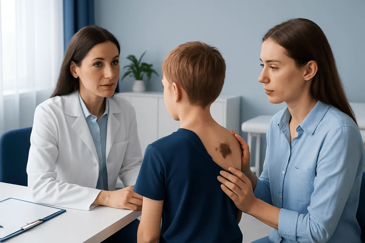
Congenital nevi form during fetal development when melanocytes, the cells that produce skin pigment, grow in clusters. In most cases, this happens sporadically and is not caused by anything parents did or did not do during pregnancy. Congenital nevi are usually not inherited in a simple family pattern, although genetic changes within the nevus cells can contribute to their development.
The most important risk factor for later complications is size. Small and medium congenital nevi have a low risk of becoming melanoma. Large and giant congenital melanocytic nevi carry a higher risk, especially when they cover a large surface area, involve the trunk, or are accompanied by multiple smaller satellite nevi. Risk assessment should be discussed with a dermatologist because estimates vary by lesion type and individual features.
In some children with large or giant congenital melanocytic nevi, doctors may consider the possibility of neurocutaneous melanosis, a rare condition in which melanocytic cells are also present in the brain or spinal cord coverings. Not every child with a large nevus needs imaging, but neurological symptoms, very extensive lesions, or multiple satellite lesions may prompt referral. Decisions about MRI or other evaluation are made case by case.
Monitoring and Diagnosis
Diagnosis is usually made by clinical examination. A dermatologist examines the lesion’s size, color, borders, surface, and distribution. Dermoscopy, a noninvasive tool that magnifies skin patterns, can help distinguish expected congenital nevus features from changes that need closer attention. In children, clinicians also consider how the lesion has evolved over time and whether changes match normal growth.
Monitoring schedules vary. A small, stable nevus may only need routine skin checks during general health visits or periodic dermatology review. A large, giant, irregular, or difficult-to-monitor nevus may need more structured follow-up. Medical photography can be very helpful because it allows subtle changes to be compared over months or years, rather than relying on memory.
Families are often taught to watch for the ABCDE warning signs used for moles: asymmetry, border irregularity, color variation, diameter or enlargement, and evolution. For congenital nevi, evolution must be interpreted carefully because growth with the child can be normal. More concerning signs include rapid change out of proportion to body growth, a new firm lump, persistent bleeding, ulceration, unexplained pain, or a color change that is very different from the rest of the lesion.
When Biopsy May Be Recommended
A biopsy means removing a small sample or sometimes the entire suspicious area so it can be examined under a microscope by a pathologist. It is not needed for every congenital nevus. In many cases, careful clinical monitoring is enough. A biopsy is considered when the doctor sees a feature that cannot be confidently explained by normal development or irritation.
Possible reasons for biopsy include a new or rapidly enlarging nodule, persistent crusting or ulceration, repeated bleeding without injury, a sharply different color area, pain that does not settle, or a change that appears clinically suspicious. In large or giant nevi, small nodules can be benign proliferative nodules, especially in infants, but evaluation is still important because the appearance can overlap with more serious conditions.
The type of biopsy depends on the lesion’s size, depth, and location. Options may include a punch biopsy, shave biopsy, incisional biopsy, or excisional biopsy. The doctor will consider comfort, scarring, diagnostic accuracy, and whether local anesthesia or sedation is appropriate. Families should ask what the biopsy is meant to answer, how results will be reported, and whether further treatment might be needed afterward.
Removal Decisions and Treatment Options
Removal of a congenital nevus may be considered for medical, functional, cosmetic, or emotional reasons. For small and medium nevi, complete surgical excision can sometimes remove the lesion in one procedure, depending on location and skin laxity. For larger lesions, staged excision may be needed, meaning the nevus is removed gradually over more than one operation. The likely scar, healing time, and need for anesthesia are important parts of the decision.
Large or giant congenital nevi may require reconstructive planning. Techniques can include staged excision, tissue expansion, skin grafting, local flaps, or a combination of methods. Tissue expansion uses a temporary device placed under nearby normal skin to create extra skin for reconstruction. When nevi involve the scalp, hairline, eyelids, or face, planning must consider both appearance and function; selected patients may benefit from procedures related to scalp reconstruction and aesthetic scalp surgery or oculoplastic care for eyelid and periocular lesions.
Laser treatment, dermabrasion, or other surface treatments may lighten or smooth some lesions, but they may not remove all nevus cells and may make future monitoring more difficult in selected cases. For this reason, these approaches require careful counseling. Some families choose observation rather than removal, especially when surgical risks, scarring, or anesthesia concerns outweigh expected benefits. The best plan is one that is medically sound and respectful of the patient’s age, preferences, and quality of life.
For international patients seeking assessment, Acıbadem Health Point’s multidisciplinary specialists and JCI-accredited hospitals can evaluate congenital nevi and discuss dermatology, pediatric, plastic surgery, and reconstructive options when appropriate. Decisions should always be made after an in-person examination and a clear explanation of benefits, limitations, and alternatives.
Prevention, Self-care, and Skin Protection
Congenital nevi cannot be prevented because they develop before birth. However, good skin protection is helpful for overall skin health and may reduce avoidable sun damage. Sunburn should be avoided, especially in children. Protective clothing, hats, shade, and broad-spectrum sunscreen are practical measures for exposed skin, including skin surrounding the nevus.
Daily care should be gentle. The nevus can usually be washed like normal skin unless a doctor gives different instructions after a procedure. Avoid picking, scratching, or repeatedly shaving over raised areas that bleed easily. If hair growth bothers the patient, the safest hair-removal method depends on the nevus location and texture; a dermatologist can advise on trimming, shaving, laser hair removal, or other options.
Emotional well-being also matters. Visible birthmarks can attract questions or unwanted attention, especially during school years and adolescence. Families can help by using simple, confident language to explain the birthmark and by involving older children in decisions about monitoring or treatment. If a child feels distressed, support from a pediatrician, dermatologist, counselor, or patient support group may be useful.
When to See a Doctor
A medical evaluation is recommended for any congenital nevus that is large, changing quickly, difficult to monitor, symptomatic, or located in a sensitive area such as the eyelid, mouth, genitals, hands, feet, or scalp. Babies with large or multiple congenital nevi should be assessed early so the family understands monitoring, possible imaging needs, and future treatment options.
Prompt review is also important if a nevus develops persistent bleeding, ulceration, a new firm lump, sudden color change, increasing pain, or rapid growth that seems different from normal body growth. Most changes are not cancer, especially in children, but timely assessment allows the clinician to decide whether reassurance, photographs, dermoscopy, biopsy, or removal is appropriate.
Adults with congenital nevi should continue skin awareness throughout life. A dermatologist can help determine how often professional skin checks are needed, particularly if the person has many moles, a personal or family history of melanoma, fair skin, extensive sun exposure, or a large congenital nevus. Regular care helps patients make calm, informed choices rather than reacting only when a change becomes obvious.
Frequently asked questions
01Are congenital nevi dangerous?
Most small and medium congenital nevi are benign and remain harmless. Large and giant congenital nevi have a higher melanoma risk than smaller lesions, so they need individualized follow-up. A dermatologist can estimate risk based on size, location, appearance, and associated findings.
02Does every congenital nevus need to be removed?
No. Many congenital nevi can be safely monitored without removal. Surgery may be considered for suspicious changes, repeated irritation, functional problems, cosmetic concerns, or risk reduction in selected larger lesions. The decision should include discussion of scarring, anesthesia, age, and realistic outcomes.
03How often should a congenital nevus be checked?
The schedule depends on the nevus. A small, stable lesion may need only periodic routine checks, while a large, irregular, or hard-to-see lesion may need closer dermatology follow-up. Photographs taken with the same lighting and scale can make monitoring more accurate.
04What changes should prompt a doctor visit?
Medical review is recommended for rapid growth, a new firm lump, persistent bleeding, ulceration, unexplained pain, or a sudden color or texture change. In children, some change can be normal as the body grows, but changes that are sudden or different from the rest of the nevus should be assessed.
05Will removing a congenital nevus eliminate melanoma risk?
Removal may reduce the number of nevus cells in the treated area, but it may not always eliminate risk completely, especially for very large or deep lesions. Some cells can remain in deeper tissues or nearby areas, and melanoma can also arise elsewhere on the skin. Continued skin awareness is still important after surgery.
06Is biopsy painful for children?
A biopsy is usually performed with local anesthesia to numb the area, and comfort measures are used to reduce anxiety. For very young children, large lesions, or sensitive locations, sedation or a planned operating-room procedure may be considered. The clinician will explain the safest approach for the child’s age and lesion location.
This article is for general information only and is not a substitute for professional medical advice. Please consult a qualified doctor about your individual situation.
Hair transplant in Turkey — costs, graft counts & a free assessment
JCI-accredited · board-certified surgeons · reply within 24h
Add us as a Preferred Source to see more of our trusted health content across Google Search, AI Overviews and Discover.