Breast Tumors Pictures: What Images Can and Cannot Show
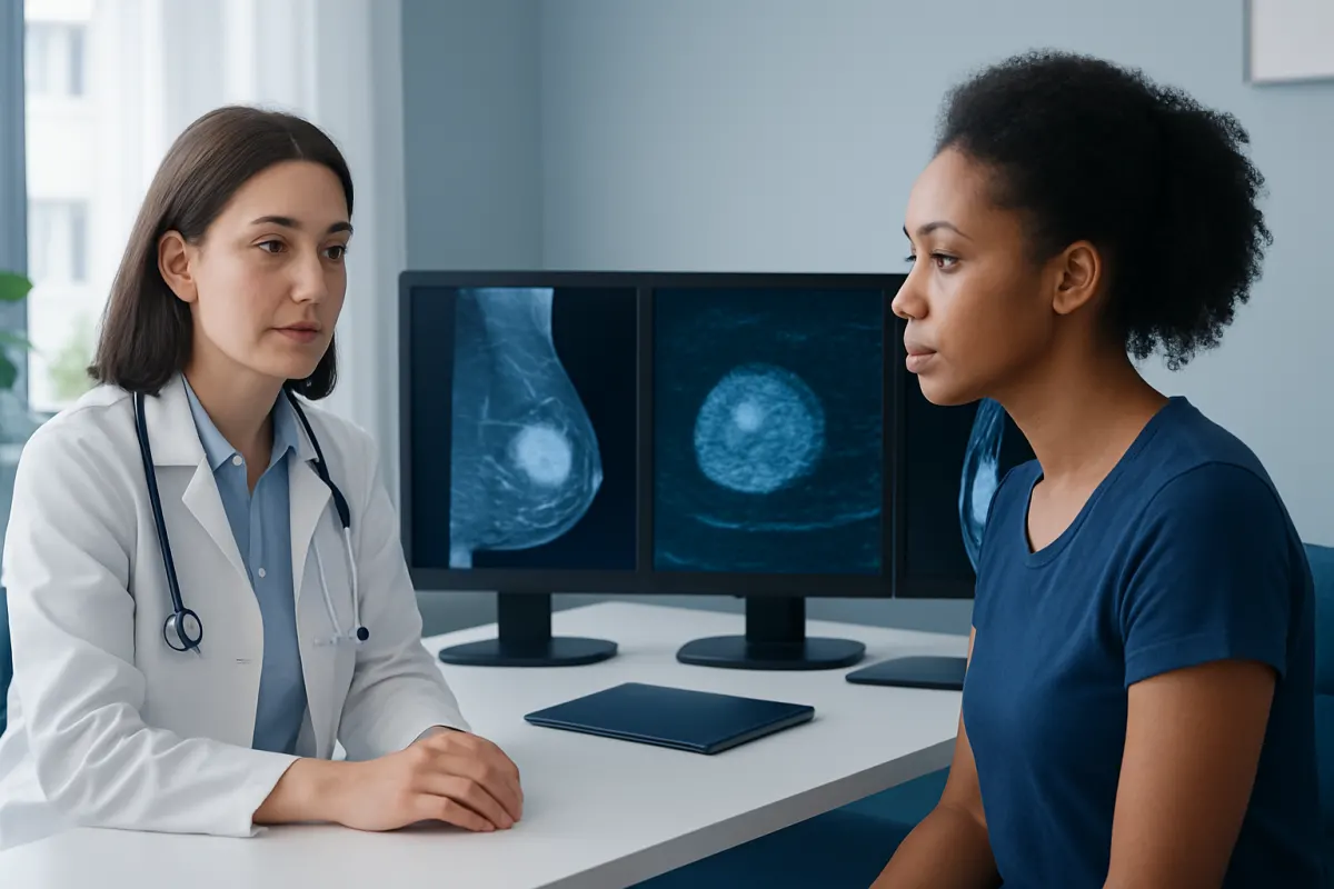
Breast tumors pictures may help patients understand what changes can look like on the skin or on medical imaging, but they cannot confirm whether a lump is benign or cancerous. A new breast change should be assessed by a qualified clinician, even when it does not resemble images seen online.
What breast tumors pictures can show
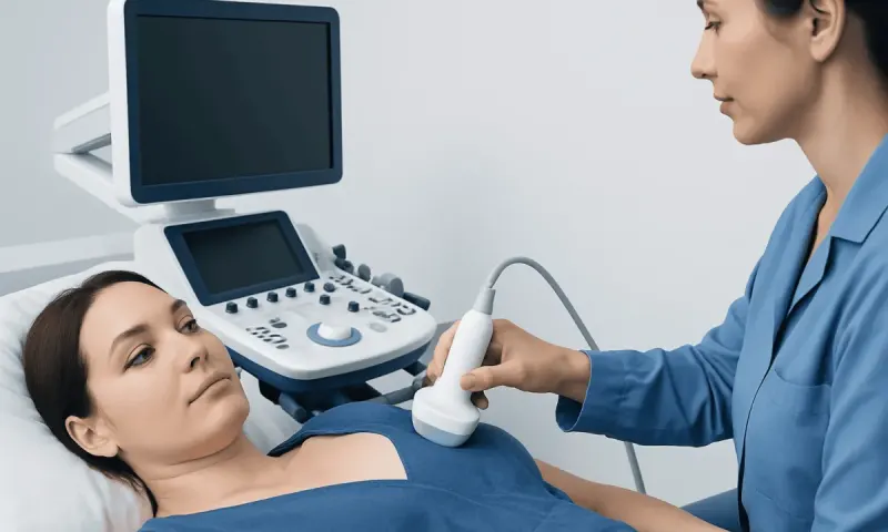
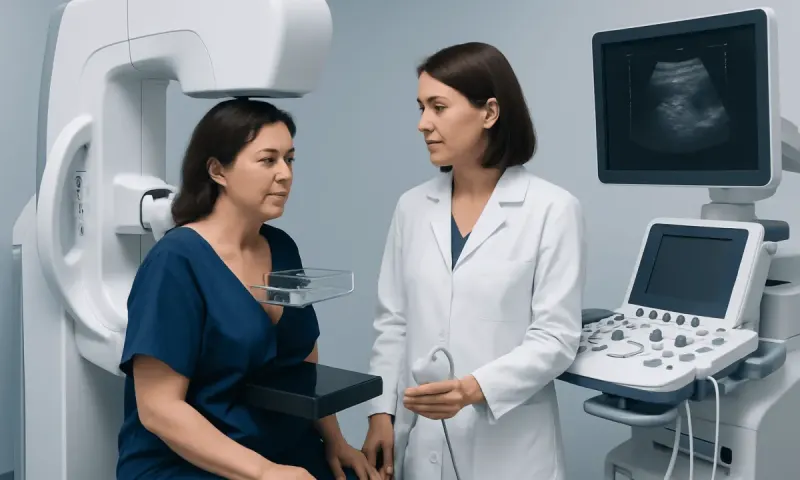
Breast tumors pictures can refer to photographs of visible breast changes, as well as images produced by mammography, ultrasound, or magnetic resonance imaging (MRI). These images can be useful for explaining why a clinician recommends further assessment. However, no online photograph or scan image should be used to decide whether a breast lump is cancerous.
Many breast tumors do not cause an obvious change in appearance. A lump may be felt but not seen, and some breast cancers are found on screening mammograms before a person notices any symptom. Conversely, redness, swelling, dimpling, or nipple changes can have non-cancer causes, such as infection, inflammation, injury, or normal hormonal variation.
Images are interpreted in context. A radiologist considers the person’s age, symptoms, breast density, prior imaging, family history, and the detailed appearance of the finding. If an area looks uncertain or suspicious, a biopsy may be needed to identify the cells and provide a definite diagnosis.
Visible breast changes: what photographs may illustrate
Clinical photographs may show outward changes that deserve medical attention, but they are not a reliable way to identify a particular condition. A breast tumor can sometimes alter the contour of the breast, create a localized area of thickening, or pull on nearby skin or the nipple. These changes can be subtle and may be easier to notice when comparing both breasts in a mirror.
Possible visible changes include persistent dimpling or puckering of the skin, a newly inverted nipple, scaling or a rash around the nipple that does not settle, unexplained swelling, or a change in breast size or shape. Skin that appears thickened or has a texture sometimes described as similar to an orange peel also needs timely assessment.
It is important not to interpret these signs in isolation. Breast appearance naturally varies between individuals, and mild asymmetry is common. What matters most is a change that is new, continues, or becomes more noticeable. A clinician can examine the breast and decide whether imaging is appropriate.
- Photographs cannot show cells, tissue type, or whether a mass has spread.
- A breast lump can be present without any skin or nipple change.
- Not all visible changes are caused by a tumor, but persistent changes should be checked.
How breast tumors appear on mammogram, ultrasound, and MRI
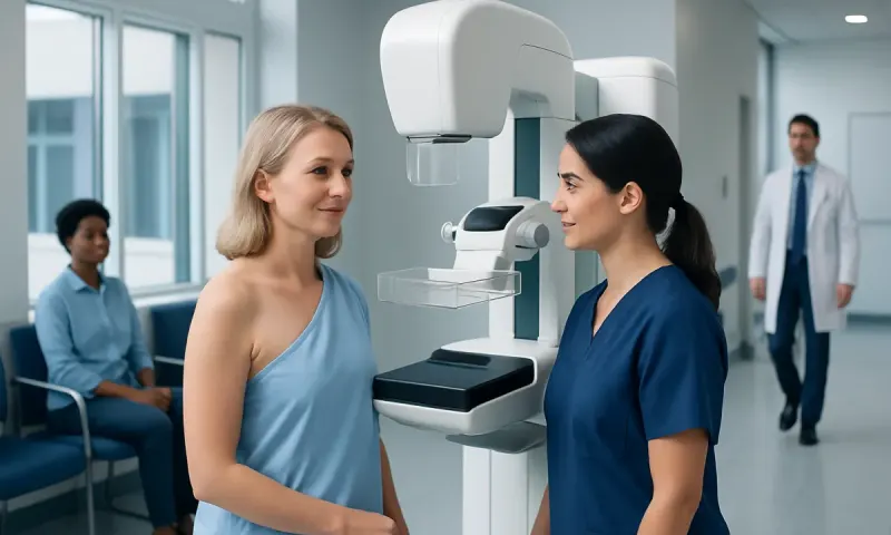
A mammogram is a low-dose X-ray of the breast and is commonly used for screening and evaluating symptoms. On a mammogram, a tumor may appear as a dense mass, an area of distortion in the normal tissue pattern, or a group of tiny calcium deposits called calcifications. Some calcifications are harmless, while certain patterns may require further investigation.
Ultrasound uses sound waves and is especially helpful for assessing a lump that can be felt or an area seen on a mammogram. It can often distinguish a fluid-filled cyst from a solid mass. Features such as the shape, edges, orientation, and internal appearance of a solid mass help the radiologist assess how likely it is to be benign or whether tissue sampling is advisable.
Breast MRI uses contrast material and provides detailed images of breast tissue and blood flow. It may be recommended for selected people, including those at higher inherited risk of breast cancer, those with unclear findings after other tests, or those needing further evaluation after a cancer diagnosis. MRI is sensitive but can identify findings that are ultimately benign, so results are interpreted carefully.
Radiologists may use the Breast Imaging Reporting and Data System, known as BI-RADS, to communicate the level of concern and the recommended next step. This may range from routine screening to short-interval follow-up, additional imaging, or biopsy. A BI-RADS result is not, by itself, a final diagnosis.
Common benign and malignant breast tumors
Breast tumors are growths or abnormal areas in breast tissue. They can be benign, meaning non-cancerous, or malignant, meaning cancerous. Benign lumps are common and do not spread to distant parts of the body. Nevertheless, they may still need assessment, monitoring, or treatment depending on their type, size, symptoms, and imaging appearance.
Examples of benign breast findings include simple cysts, fibroadenomas, lipomas, and some papillomas. Cysts are fluid-filled sacs that may become tender around menstrual periods. Fibroadenomas are usually smooth, firm, mobile lumps and are more common in younger adults. Their appearance may suggest a benign diagnosis, but clinicians may still recommend imaging or biopsy to be certain.
Breast cancer includes several types that begin in different breast structures. The most common forms arise in the milk ducts or lobules. Some cancers form a distinct mass, whereas others present as calcifications, tissue distortion, skin changes, or a change visible only on imaging. This is why a normal-looking breast photograph cannot exclude cancer.
For broader information about symptoms, testing, and care, patients can read about breast cancer. A diagnosis should always be based on a clinician’s examination and appropriate tests rather than comparisons with images found online.
How a breast lump or suspicious image is diagnosed
Assessment usually begins with a medical history and breast examination. The clinician may ask when a lump or visible change was first noticed, whether it varies with the menstrual cycle, whether there is pain or nipple discharge, and whether there is a personal or family history of breast or ovarian cancer. They will also check the breast tissue and nearby lymph nodes.
For a new breast concern, diagnostic mammography and targeted ultrasound are commonly used. The choice of tests depends on the person’s age, symptoms, pregnancy status, breast density, and previous screening results. Additional views or repeat imaging do not necessarily mean cancer is likely; they often help clarify an area that was not fully visible or was difficult to interpret.
If imaging cannot confidently show that a finding is benign, a needle biopsy may be advised. In this procedure, a specialist removes small samples of tissue, often using ultrasound, mammography, or MRI guidance. A pathologist examines the cells under a microscope. This is the most reliable way to determine whether a suspicious lesion is benign, malignant, or requires another form of follow-up.
Waiting for results can be stressful. Patients may find it helpful to ask what test is being performed, when results are expected, and what the possible next steps might be. Clear communication with the breast care team can make the diagnostic process easier to understand.
Treatment and follow-up depend on the diagnosis
Not every breast tumor requires active treatment. A simple cyst or a typical fibroadenoma may only need observation, particularly when it is small and not causing discomfort. A clinician may recommend follow-up imaging to confirm that a probably benign finding remains stable over time.
Some benign lumps are removed if they are growing, painful, large, uncertain on biopsy, or causing significant concern. The method may include a minimally invasive procedure or surgery, depending on the lesion. Decisions should be individualized after discussing the expected benefits, possible risks, and alternatives.
When breast cancer is diagnosed, treatment is planned according to the cancer type, size, grade, stage, hormone receptor status, HER2 status, and the person’s general health and preferences. Options may include surgery, radiation therapy, chemotherapy, hormone therapy, targeted therapy, or immunotherapy. Many people receive a combination of treatments coordinated by a multidisciplinary team.
Acıbadem Health Point’s multidisciplinary specialists and JCI-accredited hospitals diagnose and treat breast conditions for international patients. Any treatment plan should be discussed with a qualified breast surgeon, oncologist, radiologist, and other relevant specialists as needed.
Breast awareness, screening, and when to seek medical care
Breast awareness means knowing what is usual for one’s own breasts and reporting changes promptly. It does not require a formal, intensive self-examination routine. Looking and feeling for changes during normal activities, such as bathing or dressing, can help a person notice a new lump, persistent thickening, or skin or nipple change.
Regular screening mammography can detect breast cancer before symptoms develop, but the recommended starting age and interval vary by country, individual risk, and professional guidance. People with a strong family history, known inherited gene changes, prior chest radiation, or certain previous breast findings may need earlier or additional screening. A clinician can help create a personalized screening plan.
Seek medical care promptly for a new breast or underarm lump; a persistent focal area of thickening; bloody or clear discharge from one nipple; new nipple inversion; unexplained breast swelling; skin dimpling, redness, or thickening that does not improve; or a breast change that continues after a menstrual cycle. Breast pain alone is often not caused by cancer, but persistent pain in one specific area should also be discussed with a clinician.
Most breast changes have explanations other than cancer, and obtaining an assessment is a practical step toward reassurance or timely treatment. Avoid delaying care because an image online appears different from, or similar to, a personal symptom.
Frequently asked questions
01Can breast tumors be diagnosed from pictures?
No. Photographs of the breast and images viewed online cannot diagnose a breast tumor. Mammograms, ultrasound, and MRI must be interpreted by trained clinicians, and a biopsy may be needed to confirm the diagnosis.
02What does a breast cancer lump look like?
A breast cancer lump may feel firm, irregular, or fixed, but cancer can also feel smooth or mobile. Many non-cancerous lumps can feel similar, so the feel of a lump alone cannot determine its cause. Any new or changing lump should be assessed by a healthcare professional.
03Can a mammogram tell whether a breast tumor is cancerous?
A mammogram can identify features that appear benign, probably benign, or suspicious, but it cannot always provide a definite diagnosis. Ultrasound or additional mammogram views may clarify the finding. If concern remains, a biopsy provides the most reliable answer.
04Are most breast lumps cancerous?
No, many breast lumps are benign. Cysts, fibroadenomas, hormonal tissue changes, and other non-cancer causes are common. Still, a new lump should be evaluated because early assessment helps distinguish benign conditions from those requiring treatment.
05Can breast cancer occur without a lump?
Yes. Some breast cancers are detected as abnormal calcifications or tissue distortion on a screening mammogram before a lump can be felt. Others may cause skin, nipple, or breast-shape changes rather than a distinct mass.
06When should a person see a doctor for a breast change?
A person should arrange medical assessment for a new lump, nipple discharge, persistent focal pain, skin dimpling, nipple inversion, swelling, or a change in breast shape. Urgent evaluation is particularly appropriate for rapidly increasing redness or swelling, especially if accompanied by fever or feeling unwell.
This article is for general information only and is not a substitute for professional medical advice. Please consult a qualified doctor about your individual situation.
Oncology care in Turkey — second opinion and treatment plan
JCI-accredited · board-certified surgeons · reply within 24h
Add us as a Preferred Source to see more of our trusted health content across Google Search, AI Overviews and Discover.